Citation: Sukhwinder K. Bhullar, Harpal S. Buttar. Perspectives on nanofiber dressings for the localized delivery of botanical remedies in wound healing[J]. AIMS Materials Science, 2017, 4(2): 370-382. doi: 10.3934/matersci.2017.2.370
[1] | Misato Kuroyanagi, Yoshimitsu Kuroyanagi . Tissue-engineered products capable of enhancing wound healing. AIMS Materials Science, 2017, 4(3): 561-581. doi: 10.3934/matersci.2017.3.561 |
[2] | Daria Wehlage, Robin Böttjer, Timo Grothe, Andrea Ehrmann . Electrospinning water-soluble/insoluble polymer blends. AIMS Materials Science, 2018, 5(2): 190-200. doi: 10.3934/matersci.2018.2.190 |
[3] | Huilin Yang, Amy M. Peterson . Inkjet printed drug-releasing polyelectrolyte multilayers for wound dressings. AIMS Materials Science, 2017, 4(2): 452-469. doi: 10.3934/matersci.2017.2.452 |
[4] | Elena Kossovich . Theoretical study of chitosan-graphene and other chitosan-based nanocomposites stability. AIMS Materials Science, 2017, 4(2): 317-327. doi: 10.3934/matersci.2017.2.317 |
[5] | Dorel Feldman . Poly(vinyl alcohol) nanocomposites: Recent contributions to engineering and medicine. AIMS Materials Science, 2021, 8(1): 119-129. doi: 10.3934/matersci.2021008 |
[6] | Zhenguo Yu, Dong Wang, Zhentan Lu . Nanocomposite hydrogel fibers in the field of diagnosis and treatment. AIMS Materials Science, 2023, 10(6): 1004-1033. doi: 10.3934/matersci.2023054 |
[7] | Raffaele Conte, Anna Di Salle, Francesco Riccitiello, Orsolina Petillo, Gianfranco Peluso, Anna Calarco . Biodegradable polymers in dental tissue engineering and regeneration. AIMS Materials Science, 2018, 5(6): 1073-1101. doi: 10.3934/matersci.2018.6.1073 |
[8] | João Otávio Donizette Malafatti, Thamara Machado de Oliveira Ruellas, Camila Rodrigues Sciena, Elaine Cristina Paris . PLA/starch biodegradable fibers obtained by the electrospinning method for micronutrient mineral release. AIMS Materials Science, 2023, 10(2): 200-212. doi: 10.3934/matersci.2023011 |
[9] | Chiara Emma Campiglio, Chiara Marcolin, Lorenza Draghi . Electrospun ECM macromolecules as biomimetic scaffold for regenerative medicine: challenges for preserving conformation and bioactivity. AIMS Materials Science, 2017, 4(3): 638-669. doi: 10.3934/matersci.2017.3.638 |
[10] | DavidWood, Dale K. Hensley, Nicholas Roberts . Enhanced thermal conductance of polymer composites through embeddingaligned carbon nanofibers. AIMS Materials Science, 2016, 3(3): 851-861. doi: 10.3934/matersci.2016.3.851 |
Nonwoven nano-and micro-fibrous structures are becoming popular for a wide variety of applications such as biomedical devices, water filtration, protective garment material, electrical and optical applications, sensors and nanofibers reinforced composites [1]. Nanoscale materials also offer potential usages for creating surgical devices like dressings and mats, cardiovascular stents, targeted-delivery of therapeutic agents, smart nanofibrous textiles for the management of lymphedema in cancer survivors. Nanofibres are known to mimic collagen fibrils in the extracellular matrix. Due to their smaller size in the range of 1-100 nm, nanoparticles can easily penetrate through cellular and microbial membranes and seem to exhibit differential uptake efficiency in the target cells and injured tissue over normal cells. Theseattributes of nanoparticles not only enhance their permeation and retention effect in diseased organs and tissues but also improve therapeutic potency, resulting in lower toxicity to normal cells or tissues [2]. The multifold properties of nanofiber devices, namely large surface-area-to-volume ratio, high porosity, improved cell adherence, cellular proliferation and migration, as well as controlled in vivo biodegradation rate promote their candidature for many biomedical applications as illustrated in Figure 1, including, scaffolds, drug delivery systems, implants, prosthesis and wound care [3,4,5,6,7].
Traditional dressings consisted of cotton swabs and gauze dressings for managing the chronic and highly exuding wounds over centuries. This practice subsequently led to the development of advanced wound and burn care dressings for the localised delivery of therapeutic products and the modern nonwoven polymeric wound care scaffolds. Advanced antimicrobial wound care dressings or bioactive dressings are comprised of a wide variety of materials such as sodium alginate, ionic silver, chitosan, hydrocolloid, foam, gel or paste, molecular iodine, and have been marketed for several years. The fabrication of modern and novel biocompatible dressings which incorporate bioactive wound healing materials like growth factors have been reported in [7]. Growth factors help to repair the damaged tissues and promote healthy cellular growth. Wounds often provide a favourable environment for the colonization of microbes which delay healing. Incorporated anti-septic and anti-inflammatory agents and growth factors in modern dressings are slowly released at the wound surface and consequently minimize the opportunity for infection and promote healing. Because of its effectiveness against a broad range of micro-organisms, silver is included in many wound and healthcare products. Silver nanoparticles release silver ions in sustainable form to maintain desired concentration for antimicrobial, anti-inflammatory, and wound healing activity, while minimizing the toxic effect of silver. Silver accelerates healing of injured tissue through anti-microbial, anti-inflammatory, and anti-oxidant effect. The emergence of bacterial resistance to silver and its potential to induce cross-resistance to antibiotics has also been reported [6]. In cell culture experiments done with human mesenchymal stem cells, silver ions were found to be much more toxic than silver nanoparticles [7]. However, despite of these risks, the use of silver-containing dressings (e.g., Hydrofiber dressing, polyurethane foams and gauzes) is increasing in wound and burn care products. Currently, the development of a variety of biocompatible dressings is the focus of attention of biomedical researchers [8,9,10]. These dressings serve as vehicle for the promising delivery of wound or burn care ingredients or even allogenic cells which may provide a specific wound healing benefit. Further, the dressing acts to maintain a locally moist environment needed for wound healing.
Currently, the health care professionals encounter many problems arising from patients suffering from injuries related to wounds, diabetic ulcers and burns [11]. Some wounds and burns damage the underlying structures like bone, muscle, tendon, arteries and nerves, which are deeper and need medical supervision to prevent infection and loss of function [12]. There are many ointments, topical gels and creams available in the market for treating wounds, but the most common disadvantage is the easily washing off or removal of the medicament from the application site, which ultimately results in therapeutic failure. In addition, patient compliance is also a matter of concern, which can result in therapy failure. Several clinical studies and animal experiments demonstrate that nanofibers with diameter range of 50-1000 nm offer a great potential in biomedical applications due to their large size area, high porosity and small pore size. Nonwoven nanofibrous scaffolds or patches for wound care have shown to produce skin substitutes with optimal cellular organization, tissue proliferation and to reduce wound contraction [13].
Non-woven nanofibrous membranes of both biopolymers and synthetic polymers are useful for making wound dressings. Such materials decrease infection and morbidity, since they fit well with pathophysiological and biochemical environment of wound healing [5,14,15,16,17]. A number of polymeric nanfibrous membranes including polycaprolactone (PCL), poly(L-lactic acid) (PLA), polyvinyl alcohol (PVA), collagen, gelatin, chitosan, cellulous, alginate and silk fibroin blended natural or synthetic active agent containing silver nanoparticles and silver salts, vitamins, antibiotics, curcumin, growth factors have been prepared [18,19,20,21,22,23,24,25]. The nanofibrouswound care dressings which promote cellular proliferation and subsequent healing have been reported in the literature [18,19,20,21,22,23,24,25]. Silver containing polymeric nanofiber dressings have been studied by several investigators [16,20,21,22,25]. Also, it has been reported by the present authors that the chitosan/sericin composite nanofibers have a promising potential for making wound care dressings due to their antibacterial properties [26].
In the beginning of 19th century allopathic or synthetic medicines became the primary mode of pharmacotherapy. Fortunately in the 21st century, there are many choices for treating communicable and non-communicable diseases with synthetic pharmaceuticals, alternative remedies, natural health products (NHPs), nutraceuticals, and dietary supplements. In spite of the enormously rapid development of new drug discoveries made in the field of allopathy, plant-derived products, Ayurvedic remedies, and Chinese medicines still remain the major source of therapy in the developing countries. Many botanical products have shown anti-bacterial, anti-cancer, anti-oxidant, and anti-inflammatory activities. Emerging evidence suggests that herbal remedies containing flavonoids, carotenoids, anti-oxidants and anti-inflammatory agents not only decrease oxidative stress by scavenging free radicals, but also modulate gene and protein expression and thereby modify endogenous metabolic pathways and homeostasis, and consequently reduce the risk of cardiovascular and chronic diseases multifactorial in origin. Self-medication with NHPs and plant-derived therapies is becoming popular globally among the lay population. According to the WHO studies of 2008 [27], around 80% population in developing countries relies on traditional plant-derived remedies, and such traditional medicines make around 25% market share of the entire pharmacotherapy cache. In view of the high cost of synthetic drugs, the developing countries are desperately looking for cost-effective alternative therapies for treating communicable and non-communicable diseases, including wounds and burns as well as skin disorders like dermatitis and eczema. However, the safety and efficacy remain critical issues for botanical healing agents used for treating communicable and non-communicable diseases, wounds, burns and diabetic foot ulcers.
Biocompatible and biodegradable polymer micro- and nano-fiber devices fabricated from nanofiber materials with sizes less than 1 µm are especially useful in the field of medicine because these nanomaterials tend to replicate the molecular components of in vivo cellular and bimolecular environment. As mentioned earlier, the nanofiberous devices are beneficial for burn and wound healing due to their large surface-area-to-volume ratio, high porosity, improved cell adherence, cellular proliferation and migration, as well as controlled in vivo biodegradation rates. The large surface area of polymer nanofiber mats not only allows increased close interaction of therapeutic agents and exchange of O2 and CO2 with tissues but also provides a mechanism for sustained release and localised delivery of plant-derived remedies, analgesics, antibiotics, and growth factors needed for burn and wound healing. In addition, the high porosity of nanofiber mats and dressings permits diffusion of nutrients and removal of waste products from the application site. With all these attributes and functions, nanofiber devices promote wound and burn healing. Owing to their multifacet properties, the nanofiber mats/dressings created from both natural and synthetic polymers have attracted the attention of surgeons, physicians, biomedical researchers, and industry. Their envisioned potential applications are due to the optically transparent functional materials and nano-composites required for making scaffolds to grow stem cells, wound healing dressings and mats, transdermal patches, targeted drug-delivery systems, tissue compatibility and biodegradability, improved cell adherence, and relatively lower manufacturing cost [28,29,30,31]. Furthermore, as illustrated in Figure 2, tissue engineering techniques, namely nanofibrous polymer scaffolds used for stem cell growth and regeneration and of cells have proven successful in the wound and burn healing process [2]. To enhance the therapeutic efficiency, poorly soluble synthetic drugs and plant-derived materials can be incorporated into nanofiber devices during their fabrication. On the basis of large surface-area-to-volume ratio and high porosity, the nanofiber mats or dressings provide mechanisms for the localized sustained release of herbal products and plant extracts in their nano-to-micro forms to treat burns and wounds as well as some inflammation-related chronic skin diseases, e.g., dermatitis and eczema. Several examples can be cited from the published literature where nanofiber materials have been utilised for the localised delivery of plant-based remedies for wound care. For instance, crude bark extract of Tecomella undulata blended with nanofibers of synthetic polymers PCL/PVP with solvents chloroform/methanol has been reported for wound healing [32]. Bionanocellulose-based dressings are used for treating burns and diabetic ulcers [33]. Aloe vera with biocompatible and biodegradable polymer PCL with chloroform solvent was investigated for wound dressing [34]. Nanofibers of PCL and butanol, dichloromethane, hexane and methanol loaded with extraction of Centella Asiatica were utilized for topical drug administration and wound healing [35]. Shikonin loaded nanofibers fabricated with polymer PCL with dichloromethane (DCM) and N, N-dimethylformamide (DMF) have been tried for wound cure and/or atopic dermatitis [36]. Also, PCL and chloroform-methanol nanofibers impregnated with Indigofera aspalathoides, Azadirachta indica, Memecylon edule, and Myristica andamanica was reported for skin tissue engineering [37].
Furthermore, silk nanofiberous dressing fabricated from biomaterial silk have shown promising approach in the treatment of skin wounds [38,39,40]. A comparative study was done to evaluate the safety and efficacy of colostrum powder dressing with conventional dressing in the management of deep wounds. It was found that the colostrum dressing group not only required less number of dressing changes, but also showed rapid healing, short healing time, and decreased pain compared to the conventional dressing group. The results suggest that colostrum powder dressings are safe and effective for healing deep wounds, and may be used as an adjunct therapy for the management of deep wounds [41]. Biopolymers cellulose is one of the most abundant natural product that has attracted the attention of basic researchers involved in the biomedical applications of nanomaterials [42,43,44,45,46,47]. Studies have shown that cellulose nanofibers derived from wood and plants have widespread applications in pharmaceutical industry and biomedical areas due to their biocompatibility, physical and mechanical properties. These materials can be useful for drug delivery, make foams and aerogels as well as cell carriers, biomaterial substitutes, and scaffold synthesis [48,49,50,51]. In addition, cellulose nanofibers are highly hydrophilic in nature and owing to their large surface area have a great potential for making wound dressings [45].
Fabrication of polymeric nano/micro fibrous structures is an increasingly emerging tissue engineering area that offers potential applications in biomedical sciences and biotechnology industry, as well as for making scaffolds, stents, targeted drug delivery systems, implants and prosthesis. To produce these sophisticated devices, electrospinning technique is a widely accepted method for making micro- to nano-scale structures using high voltage to create an electrically charged jet of polymer solution or melt from the nozzle or needle [46,47,48,49,50,51]. Since 1930, nanofibers have been produced from a variety of polymers, both natural and synthetic [52]. It has been reported that compared to mechanical spinning processes, electrospininning is simple, economical and most versatile technique for producing nanofibres [53,54,55,56]. Several methods of electrospinning, namely direct-dispersed electrospinning, gas-solid reaction, in situ photoreduction, sol-gel processes, emulsions, co-evaporation, and co-axial electrospinning have been reported by a number of investigators [5,6,14,57,58,59,60]. Fabrication of nanofiber structures is illustrated in Figure 3.
Several authors have pointed out that a variety of nanofiber structures can be tailored by electrospinning multicomponent mixtures and post-modification of fabricated nanofibrous membranes. At present, electrostatic spinning process is used for producing nanofibers with diameters in the range of 10-100 nanometers. Similar techniques have been described in the literature by several investigators. Melt- or solvent-(s)-based polymer solution can be used for the fabrication of nano- and micro-fibers. The machine set up used for electrospinning and the fabrication of nanofibers is shown in Figure 4.
Furthermore, the various steps involved from polymer solution to fabrication of nanofibers by the electrospinning technique are illustrated in Figure 5. With the application of high voltage, the polymer suspended in the solution or melt form results in the formation of charged polymer called meniscus, and consequently the high voltage electric field produces a Taylor cone meniscus that is formed during electrospinning process [61,62]. Further, the high electric field is strong enough to overcome the surface tension of Taylor cone to a moving jet and the resulting jet is solidified into micro- to nano-fibers after solvent evaporation.
The nano- and micro-fiber structures offer a great potential for the localised delivery of herbal products alone or in combination with synthetic drugs. For systemic delivery of products administered orally, the extremely high surface area of nanofibers can enhance the absorption and bioavailability of poorly soluble xenobiotics because the large surface area leads to increased dissolution rate in the gastrointestinal tract [63,64]. The in vitro release of drugs from the electrospun nanofibers composed of water-insoluble and nonbiodegradable polymer was found to be dependent on the drug: polymer ratio and fiber diameter [65]. There are a number of technical conditions that usually affect the nanofiber diameter, viz., viscosity of polymeric solution, voltage strength, distance between nozzle and fiber collector plate, flow rate of pump, nozzle diameter and fiber diameter [66]. The relationship between fiber diameter and equipment nozzle diameter calculated from experimental and numerical data is discussed in [67].
Another technique used for the fabrication of polymer-based micro- and nano-fiber structers is called airbrushing method shown in Figure 6. In this technique, the polymer solution is injected using compressed air to make nanofibers of a wide range of regular and irregular shapes onto a collector [68,69,70].
The airbrushing method is not only comparatively cost effective, but also is more safer than other electrospining techniques, since fabrication can be accomplished without using any type of high voltage equipment [71,72,73]. Furthermore, it’s a easily applicable technique for creating the highly porous and lower modulus aligned and loosely packed bundles of nanofibers using a larger array of polymer solutions at the higher injection rates. It has been reported that airbrushing method can be applied for direct nanofiber deposition onto different target sites for surgical homeostasis, as surgical sealant, or for tissue reconstruction [74,75]. It therefore appears that nanofiber dressings loaded with herbal products or plant-derived extracts could be potentially used for the localised delivery of botanical remedies for wound and burn healing.
Sometimes, the wounds may get infected and the infection may spread systemically. Under such circumstances, the topical application of a therapeutic agent alone may not be effective for wound treatment and the topical therapy may need to be supplemented with alternate routes of drug administration (e.g., oral, intravenous, intramuscular, etc.). This may constitute one potential limitation of the localized delivery of antimicrials and/or botanical remedies in wound healing.
Limited numbers of studies have been done for the localised delivery of plant-based remedies for wound and burn care as well as atopic dermatitis using biocompatible and biodegradable dressings. Considering the meagre amount of work done in this area and the promising biomedical applications of drug-coated nanofiber structures, further well designed clinical studies are needed to determine the localised and controlled delivery of botanical and synthetic products for wound and burn healing. The unique physico-chemical properties of nanofiber dressings and mats render them highly useful for several novel biomedical applications. The findings of research studies discussed in our review paper open a range of possibilities for further mechanistic studies of the localised and controlled delivery of botanical extracts as well as for creating possible alternative treatment of wound and burn healing. Plant-derived remedies and herbal products have been used since ancient times for wound and burn cure as well as for treating chronic skin diseases like dermatitis and eczema. In view of the high cost of synthetic drugs, the developing countries are desperately looking for cost-effective alternative therapies for wound and burn care as well as dermatitis and eczema.
The authors declare that there is no conflict of interest regarding the publication of this manuscript.
[1] | Chirayil C, Mathew L, Thomas S (2014) Review of recent research in nano cellulose preparation from different lignocellulosic fibers. Rev Adv Mater Sci 37: 20–28. |
[2] |
Zhang L, Webster T (2009) Nanotechnology and nanomaterials: promises for improved tissue regeneration. Nano Today 4: 66–80. doi: 10.1016/j.nantod.2008.10.014
![]() |
[3] |
Sill TJ, von Recum HA (2008) Electrospinning: Applications in drug delivery and tissue engineering. Biomaterials 29: 1989–2006. doi: 10.1016/j.biomaterials.2008.01.011
![]() |
[4] |
Li WJ, Laurencin CT, Caterson EJ, et al. (2002) Electrospun nanofibrous structure: A novel scaffold for tissue engineering. J Biomed Mater Res A 60: 613–621. doi: 10.1002/jbm.10167
![]() |
[5] | Zahedi P, Rezaeian I, Ranaei‐Siadat SO, et al. (2010) A review on wound dressings with an emphasis on electrospun nanofibrous polymeric bandages. Polym Advan Technol 21: 77–95. |
[6] | Harcup JW, Saul PA (1986) A Study of The Effect of Cadexomer Iodine in The Treatment of Venous Leg Ulcers. Br J Clin Pract 40: 360–364. |
[7] | Harding KG, Jones V, Price P (2000) Topical treatment: which dressing to choose. Diabetes Metab Res Rev 16: S47–S50. |
[8] |
Kittler S, Greulich C, Diendorf J, et al. (2010) Toxicity of silver nanoparticles increases during storage because of slow dissolution under release of silver ions. Chem Mater 22: 4548–4554. doi: 10.1021/cm100023p
![]() |
[9] |
Ormiston MC, Seymour MT, Venn GE, et al. (1985) Controlled Trial of Iodosorb in Chronic Venous Ulcers. Br Med J (Clin Res Ed) 291: 308–310. doi: 10.1136/bmj.291.6491.308
![]() |
[10] | Percival SL, Bowler PG, Russell D (2005) Bacterial resistance to silver in wound care. J Hosp Infect 40: 1–7. |
[11] | Nguyen DT, Orgill DP, Murphy GF (2009) The Pathophysiologic Basis for Wound Healing and Cutaneous Regeneration. In: Orgill DP, Blanco C, Biomaterials For Treating Skin Loss, Woodhead Publishing (UK/Europe) & CRC Press (US), Cambridge/Boca Raton, 25–57. |
[12] |
Powell HM, Supp DM, Boyce ST (2008) Influence of electrospun collagen on wound contraction of engineered skin substitutes. Biomaterials 29: 834–843. doi: 10.1016/j.biomaterials.2007.10.036
![]() |
[13] | Wound Care Market by Product (Advanced (Foam, Alginate, NPWT, Active), Surgical, Traditional), Wound Type (Chronic (DFU, Pressure Ulcer), Acute (Burn)), End User (Hospital (Inpatient, Outpatient), Long-Term Care, Home Healthcare)-Global Forecast to 2021, 2016. Available from: http://www.marketsandmarkets.com/PressReleases/wound-care.asp. |
[14] |
Lu X, Wang C, Wei Y (2009) One-Dimensional Composite Nanomaterials: Synthesis by Electrospinning and Their Applications. Small 5: 2349–2370. doi: 10.1002/smll.200900445
![]() |
[15] |
Boland ED, Wnek GE, Simpson DG, et al. (2001) Tailoring Tissue Engineering Scaffolds Using Electrostatic Processing Techniques: A Study of Poly(Glycolic Acid) Electrospinning. J Macromol Sci A 38: 1231–1243. doi: 10.1081/MA-100108380
![]() |
[16] | Khil MS, Cha DI, Kim HY, et al. (2003) Electrospun Nanofibrous Polyurethane Membrane as Wound Dressing. J Biomed Mater Res B 67: 675–679. |
[17] |
Duan Y, Jia J, Wang S, et al. (2007) Preparation of antimicrobial poly(γ-caprolactone) electrospun nanofibers containing silver-loaded zirconium phosphate nanoparticles. J Appl Polym Sci 106: 1208–1214. doi: 10.1002/app.26786
![]() |
[18] |
Kriegel C, Kit KM, McClements DJ, et al. (2009) Electrospinning of chitosan-poly(ethylene oxide) blend nanofibers in the presence of micellar surfactant solutions. Polymer 50: 189–200. doi: 10.1016/j.polymer.2008.09.041
![]() |
[19] |
Au HT, Pham LN, Vu THT, et al. (2012) Fabrication of an antibacterial non-woven mat of a poly(lactic acid)/chitosan blend by electrospinning. Macromol Res 20: 51–58. doi: 10.1007/s13233-012-0010-9
![]() |
[20] |
Van der Schueren L, Steyaert I, De Schoenmaker B, et al. (2012) Polycaprolactone/chitosan blend nanofibres electrospun from an acetic acid/formic acid solvent system. Carbohyd Polym 88: 1221–1226. doi: 10.1016/j.carbpol.2012.01.085
![]() |
[21] |
Sundaramurthi D, Vasanthan KS, Kuppan P, et al. (2012) Electrospun nanostructured chitosan-poly(vinyl alcohol) scaffolds: a biomimetic extracellular matrix as dermal substitute. Biomed Mater 7: 045005. doi: 10.1088/1748-6041/7/4/045005
![]() |
[22] | Chen JP, Chang GY, Chen JK (2008) Electrospun collagen/chitosan nanofibrous membrane as wound dressing. Colloid Surface A 313: 183–188. |
[23] | Dhandayuthapani B, Krishnan UM, Sethuraman S (2010) Fabrication and characterization of chitosan-gelatin blend nanofibers for skin tissue engineering. J Biomed Mater Res B 94: 264–272. |
[24] |
Cai Z, Mo X, Zhang K, et al. (2010) Fabrication of Chitosan/Silk Fibroin Composite Nanofibers for Wound-dressing Applications. Int J Mol Sci 11: 3529–3539. doi: 10.3390/ijms11093529
![]() |
[25] |
Wang R, Wang Z, Lin S, et al. (2015) Green fabrication of antibacterial polymer/silver nanoparticle nanohybrids by dual-spinneret electrospinning. RSC Adv 5: 40141–40147. doi: 10.1039/C5RA03288A
![]() |
[26] |
Zhao R, Li X, Sun B, et al. (2014) Electrospun chitosan/sericin composite nanofibers with antibacterialproperty as potential wound dressings. Int J Biol Macromol 68: 92–97. doi: 10.1016/j.ijbiomac.2014.04.029
![]() |
[27] | Gupta RC (2016) Nutraceuticals: Efficacy, Safety and Toxicity. |
[28] |
Habibi Y, Lucia LA, Rojas OJ (2010) Cellulose nanocrystals: Chemistry, selfassembly, and applications. Chem Rev 110: 3479–3500. doi: 10.1021/cr900339w
![]() |
[29] | Siro I, Plackett D (2010) Microfibrillated cellulose and new nanocomposite materials: a review. Cellulose 17:459–494. |
[30] |
Visakh PM, Thomas S (2010) Preparation of Bionanomaterials and their Polymer Nanocomposites. Waste Biomass Valouri 1: 121–134. doi: 10.1007/s12649-010-9009-7
![]() |
[31] |
Klemm D, Heublein B, Fink HP, et al. (2005) Cellulose: Fascinating biopolymer and sustainable raw material. Angew Chem Int Edit 44: 3358–3393. doi: 10.1002/anie.200460587
![]() |
[32] |
Suganya S, Senthil Ram T, Lakshmi BS, et al. (2011) Herbal Drug Incorporated Antibacterial Nanofibrous Mat Fabricated by Electrospinning: An Excellent Matrix For Wound Dressings. J Appl Polym Sci 121: 2893–2899. doi: 10.1002/app.33915
![]() |
[33] |
Sikareepaisan P, Suksamrarn A, Supaphol P (2008) Electrospun gelatin fiber mats containing a herbal-Centellaasiatica-extract and release characteristic of asiaticoside. Nanotechnology 19: 015102. doi: 10.1088/0957-4484/19/01/015102
![]() |
[34] | Han J, Zhang HT, Zhu LM, et al. (2009) Electrospun biodegradable nanofiber mats for controlled release of herbal medicine. 3rd International Conference on Bioinformatics and Biomedical Engineering. |
[35] |
Agnes Mary S, Giri Dev VR (2015) Electrospun herbal nanofibrous wound dressings for skin tissue engineering. J Text I 106: 886–895. doi: 10.1080/00405000.2014.951247
![]() |
[36] | DeMario MD, Ratain MJ (1998) Oral chemotherapy: rationale and future directions. J Clin Oncol 16: 2557–2567. |
[37] |
Schneider A, Wang XY, Kaplan DL, et al. (2009) Biofunctionalized electrospun silk mats as a topical bioactive dressing for accelerated wound healing. Acta Biomater 5: 2570–2578. doi: 10.1016/j.actbio.2008.12.013
![]() |
[38] |
Gil ES, Panilaitis B, Bellas E, et al. (2013) Functionalized silk biomaterials for wound healing. Adv Healthc Mater 2: 206–217. doi: 10.1002/adhm.201200192
![]() |
[39] |
Navone SE, Pascucci L, Dossena M, et al. (2014) Decellularized silk fibroin scaffold primed with adipose mesenchymal stromal cells improves wound healing in diabetic mice. Stem Cell Res Ther 5: 7–17. doi: 10.1186/scrt396
![]() |
[40] | Kshirsagar AY, Vekariya MA, Gupta V, et al. (2015) A Comparative Study of Colostrum Dressing Versus Conventional Dressing in Deep Wounds. J Clin Diagn Res 9: PC01–PC04. |
[41] |
Borges AC, Eyholzer C, Duc F, et al. (2011) Nanofibrillated cellulose composite hydrogel for the replacement of the nucleus pulposus. Acta Biomater 7: 3412–3421. doi: 10.1016/j.actbio.2011.05.029
![]() |
[42] | Mathew AP, Oksman K, Pierron D, et al. (2011) Crosslinked fibrous composites based on cellulose nanofibers and collagen with in situ pH induced fibrillation. Cellulose 19: 139–150. |
[43] |
Valo H, Arola S, Laaksonen P, et al. (2013) Drug release from nanoparticles embedded in four different nanofibrillar cellulose aerogels. Eur J Pharm Sci 50: 69–77. doi: 10.1016/j.ejps.2013.02.023
![]() |
[44] |
Laurén P, Lou YR, Raki M, et al. (2014) Technetium-99 mlabeled nanofibrillar cellulose hydrogel for in vivo drug release. Eur J Pharm Sci 65: 79–88. doi: 10.1016/j.ejps.2014.09.013
![]() |
[45] |
Sakai K, Kobayashi Y, Saito T, et al. (2016) Partitioned airs at microscale and nanoscale: thermal diffusivity in ultrahigh porosity solids of nanocellulose. Sci Rep 6: 20434. doi: 10.1038/srep20434
![]() |
[46] |
Mertaniemi H, Escobedo-Lucea C, Sanz-Garcia A, et al. (2016) Human stem cell decorated nanocellulose threads for biomedical applications. Biomaterials 82: 208–220. doi: 10.1016/j.biomaterials.2015.12.020
![]() |
[47] |
Chang C, Zhang L (2011) Cellulose-based hydrogels: present status and application prospects. Carbohydr Polym 84: 40–53. doi: 10.1016/j.carbpol.2010.12.023
![]() |
[48] | El-Newehy MH, Al-Deyab SS, Kenawy ER, et al. (2011) Nanospider Technology for the Production of Nylon-6 Nanofibers for Biomedical Applications. J Nanomater 2011. |
[49] |
Zhang S, Shim WS, Kim J (2009) Design of ultra-fine nonwovens via electrospinning of Nylon 6: spinning parameters and filtration efficiency. Mater Design 30: 3659–3666. doi: 10.1016/j.matdes.2009.02.017
![]() |
[50] | Pedicini A, Farris RJ (2004) Thermally induced color change in electrospun fiber mats. J Polym Sci Pol Phys 42: 752–757. |
[51] | Deitzel JM, Kosik W, McKnight SH, et al. (2001) Electrospinning of polymer nanofibers with specific surface chemistry. Polymer 43: 1025–1029. |
[52] |
Nirmala R, Navamathavan R, El-Newehy MH, et al. (2011) Preparation and electrical characterization of polyamide-6/chitosan composite nanofibers via electrospinning. Mater Lett 65: 493–496. doi: 10.1016/j.matlet.2010.10.066
![]() |
[53] | Zachariades AE, Porter RS, Doshi J, et al. (1995) High modulus polymers. A novel electrospinning process. Polym News 20: 206–207. |
[54] |
Reneker DH, Yarin AL, Fong H, et al. (2000) Bending instability of electrically charged liquid jets of polymer solutions in Electrospinning. J Appl Phys 87: 4531–4547. doi: 10.1063/1.373532
![]() |
[55] | Fong H, Reneker DH (1999) Elastomeric nanofibers of styren–butadiene–styrene triblock copolymer. J Polym Sci Polym Phys 37: 3488–3493. |
[56] | Teo WE, Ramakrishna S (2006) A Review on Electrospinning Design and Nanofibre Assemblies. Nanotechnology 17: 89–106. |
[57] |
Wan LS, Wu J, Xu ZK (2006) Porphyrinated Nanofibers via Copolymerization and Electrospinning. Macromol Rapid Comm 27: 1533–1538. doi: 10.1002/marc.200600381
![]() |
[58] | Wahl DA, Sachlos E, Liu C, et al. (2007) Controlling the processing of collagen hydroxyapatite scaffolds for bone tissue engineering. J Mater Sci-Mater M 18: 201–209. |
[59] |
Jayakumar R, Prabaharan M, Kumar PTS, et al. (2011) Biomaterials based on chitin and chitosan in wound dressing applications. Biotechnol Adv 29: 322–337. doi: 10.1016/j.biotechadv.2011.01.005
![]() |
[60] |
Hu WW, Yu HN (2013) Co-electrospinning of chitosan/alginate fibers by dual-jet system for modulating material surfaces. Carbohydr Polym 95: 716–727. doi: 10.1016/j.carbpol.2013.02.083
![]() |
[61] | Lyons J, Ko FK (2004) Nanofibers. Encycl Nanosci Nanotech 6: 727–738. |
[62] |
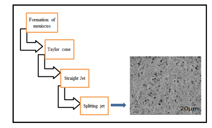
Yarin AL, Koombhongse S, Reneker DH (2001) Taylor cone and jetting from liquid droplets in electrospinning of nanofibers. J Appl Phys 90: 4836–4846. doi: 10.1063/1.1408260
![]() |
[63] |
Stanger J, Tucker N, Kirwan K, et al. (2009) Effect of charge density on the Taylor cone in electrospinning. Int J Mod Phys B 23: 1956–1961. doi: 10.1142/S0217979209061895
![]() |
[64] | Anton F (1938) Method and apparatus for the production of fibers. US Patent 2116942, 1938-5-10. |
[65] |
Ignatious F, Sun L, Lee CP, et al. (2010) Electrospun nanofibers in oral drug delivery. Pharm Res 27: 576–588. doi: 10.1007/s11095-010-0061-6
![]() |
[66] |
Verreck G, Chun I, Rosenblatt J, et al. (2003) Incorporation of drugs in an amorphous state into electrospun nanofibers composed of a water-insoluble nonbiodegradable polymer. J Control Release 92: 349–360. doi: 10.1016/S0168-3659(03)00342-0
![]() |
[67] |
Ko J, Bhullar SK, Mohtaram NK, et al. (2014) Using mathematical modeling to control topographical properties of poly (ε-caprolactone) melt electrospun scaffolds. J Micromech Microeng 24: 065009. doi: 10.1088/0960-1317/24/6/065009
![]() |
[68] |
Denn MM (1980) Continuous Drawing of Liquids to Form Fibers. Annu Rev Fluid Mech 12: 365–387. doi: 10.1146/annurev.fl.12.010180.002053
![]() |
[69] | Feng L, Li S, Li H, et al. (2002) Super-Hydrophobic Surface of Aligned Polyacrylonitrile Nanofibers. Angew Chem 141: 1269–1271. |
[70] | Ramakrishna S, Fujihara K, Teo W, et al. (2005) An introduction to electrospinning and nanofibers, Singapore: World Scientific Publishing Company. |
[71] |
Ellison CJ, Phatak A, Giles DW, et al. (2007) Melt blown nanofibers: fiber diameter distributions and onset of fiber breakup. Polymer 48: 3306–3316. doi: 10.1016/j.polymer.2007.04.005
![]() |
[72] | Grafe T, Graham K (2003) Nanofibers and Nanofiber Webs: A New Class of Nonwovens. Nonwoven Technol Rev 12: 51–55. |
[73] |
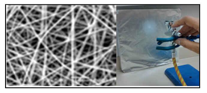
Medeiros ES, Glenn GM, Klamczynski AP, et al. (2009) Solution blow spinning: A new method to produce micro‐and nanofibers from polymer solutions. J Appl Polym Sci 113: 2322–2330. doi: 10.1002/app.30275
![]() |
[74] |
Srinivasan S, Chhatre SS, Mabry JM, et al. (2011) Solution spraying of poly(methyl methacrylate) blends to fabricate microtextured, superoleophobic surfaces. Polymer 52: 3209–3218. doi: 10.1016/j.polymer.2011.05.008
![]() |
[75] |
Behrens AM, Casey BJ, Sikorski MJ, et al. (2014) In situ deposition of PLGA nanofibers via solution blow spinning. ACS Macro Lett 3: 249–254. doi: 10.1021/mz500049x
![]() |
1. | Cláudia Mouro, Raul Fangueiro, Isabel C. Gouveia, Preparation and Characterization of Electrospun Double-layered Nanocomposites Membranes as a Carrier for Centella asiatica (L.), 2020, 12, 2073-4360, 2653, 10.3390/polym12112653 | |
2. | Homa Maleki, Rouhollah Semnani Rahbar, Mohammad Mahdi Saadatmand, Hossein Barani, Physical and morphological characterisation of poly(L-lactide) acid-based electrospun fibrous structures: tunning solution properties, 2018, 47, 1465-8011, 438, 10.1080/14658011.2018.1532672 | |
3. | Ravikumar Rramaswamy, Ganesh Mani, Senthil Venkatachalam, Ramesh Venkata Yasam, J. C. Bose Rajendran, Jang Hyun Tae, Preparation and Characterization of Tetrahydrocurcumin-Loaded Cellulose Acetate Phthalate/Polyethylene Glycol Electrospun Nanofibers, 2018, 19, 1530-9932, 3000, 10.1208/s12249-018-1122-0 | |
4. | Kirandeep Kaur, Atamjit Singh, Hamayal Sharma, Sanha Punj, Neena Bedi, Formulation Strategies and Therapeutic Applications of Shikonin and Related Derivatives, 2022, 16, 26673878, 55, 10.2174/2667387816666220302112201 | |
5. | Biruk Fentahun Adamu, Jing Gao, Shaojie Tan, Esubalew K Gebeyehu, Comparison of antibacterial property of herbal plant–based bio-active extract loaded polymer electrospun nanofibrous mat wound dressings, 2022, 51, 1528-0837, 1793S, 10.1177/15280837221086899 | |
6. | Cláudia Mouro, Isabel C. Gouveia, Electrospun wound dressings with antibacterial function: a critical review of plant extract and essential oil incorporation, 2023, 0738-8551, 1, 10.1080/07388551.2023.2193859 |