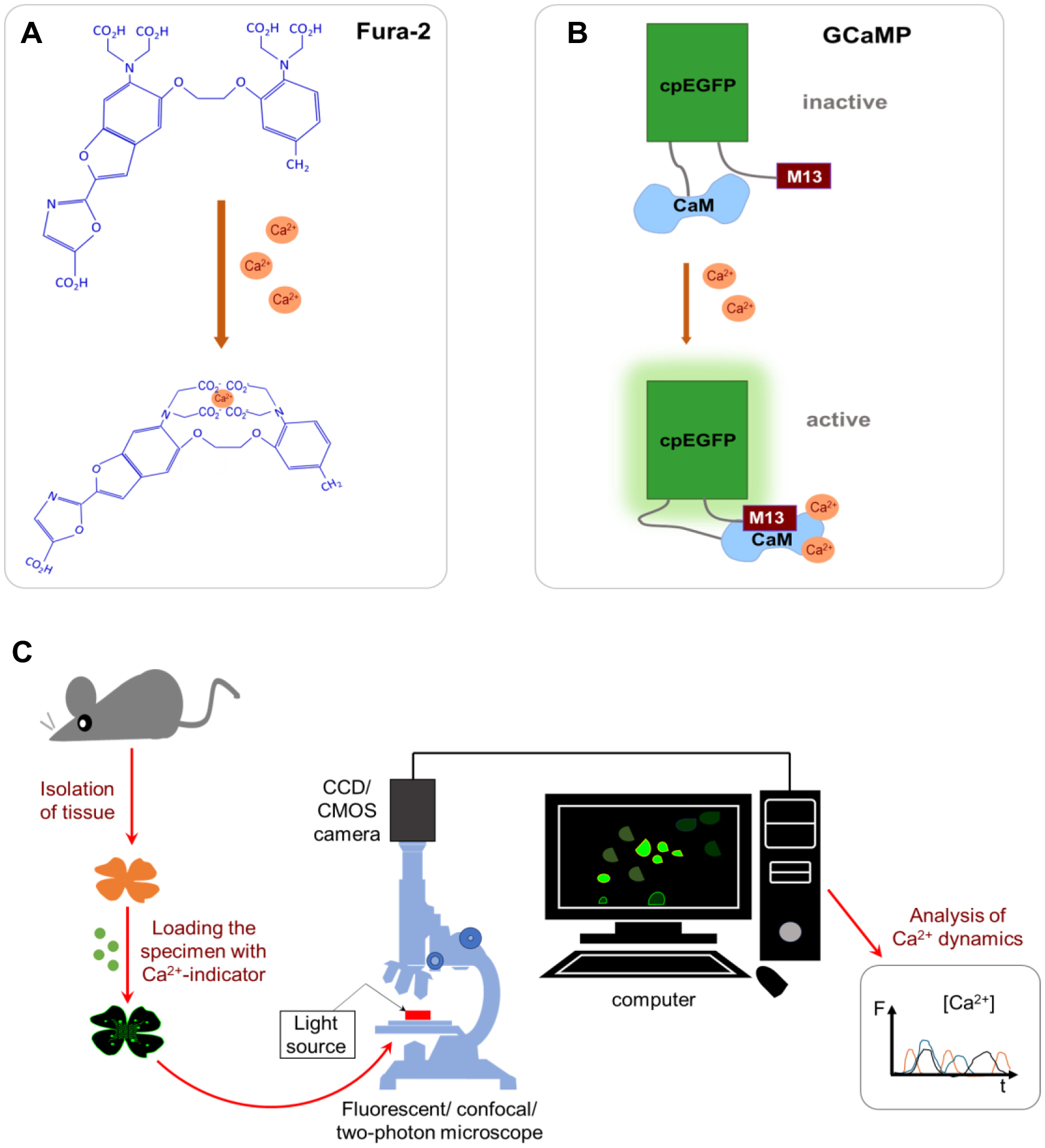
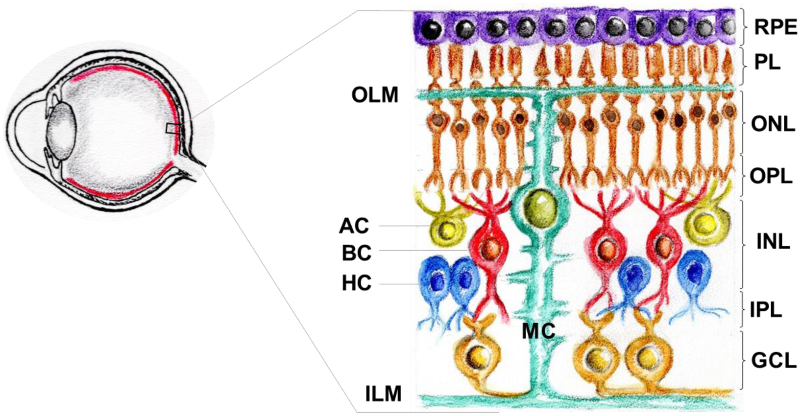
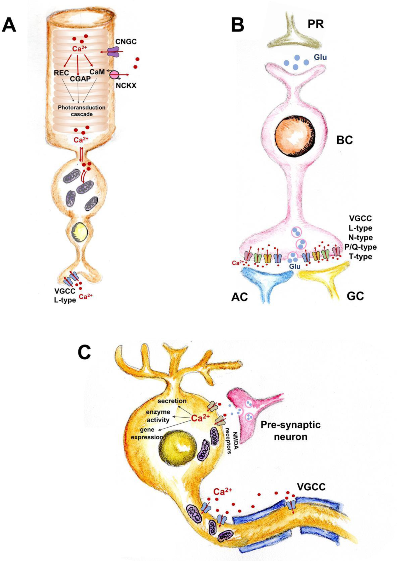
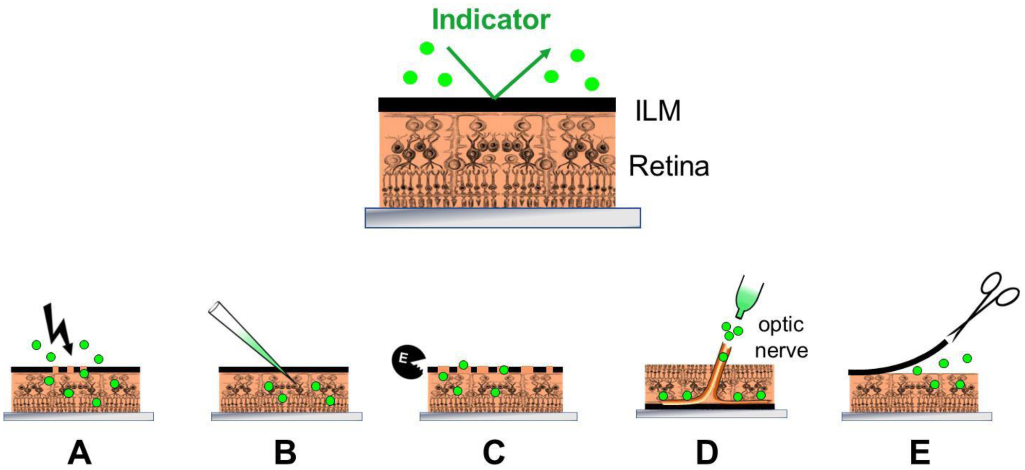
Calcium ions are universal signaling particles used alongside other signaling systems in animal cells. Changes in intracellular calcium concentration trigger vital reactions in different types of eukaryotic cells by acting through molecular calcium sensors. Over several decades, calcium imaging has been developed to study calcium-based signaling pathways. This method visualizes changes in intracellular free calcium using special fluorescent indicators. The retina, a type of nervous tissue in the eye, is responsible for perception, primary processing, and transmission of visual information to the brain. Signal cascades within retinal cells and synaptic transmission between cells play a crucial role in implementing these functions. Calcium plays a significant and diverse role in the functioning of the retina, in both normal and pathological conditions. Studying the fundamental processes of visual perception at the retinal level requires the visualization of changes in intracellular calcium concentration at different time scales, including very rapid changes. Consequently, the calcium imaging method, which was originally developed and used for studying other tissues, has now entered the field of visual neuroscience. While there are currently many examples of calcium imaging being used to study the functioning principles of all major types of retinal cells, adapting this method to the study of this tissue presents a number of difficulties. This review discussed these problems and how to solve them.
Citation: Yakov S. Veselov, Luba A. Astakhova. Calcium imaging in retinal research: challenges and prospects for its application[J]. AIMS Biophysics, 2025, 12(4): 510-543. doi: 10.3934/biophy.2025025
Calcium ions are universal signaling particles used alongside other signaling systems in animal cells. Changes in intracellular calcium concentration trigger vital reactions in different types of eukaryotic cells by acting through molecular calcium sensors. Over several decades, calcium imaging has been developed to study calcium-based signaling pathways. This method visualizes changes in intracellular free calcium using special fluorescent indicators. The retina, a type of nervous tissue in the eye, is responsible for perception, primary processing, and transmission of visual information to the brain. Signal cascades within retinal cells and synaptic transmission between cells play a crucial role in implementing these functions. Calcium plays a significant and diverse role in the functioning of the retina, in both normal and pathological conditions. Studying the fundamental processes of visual perception at the retinal level requires the visualization of changes in intracellular calcium concentration at different time scales, including very rapid changes. Consequently, the calcium imaging method, which was originally developed and used for studying other tissues, has now entered the field of visual neuroscience. While there are currently many examples of calcium imaging being used to study the functioning principles of all major types of retinal cells, adapting this method to the study of this tissue presents a number of difficulties. This review discussed these problems and how to solve them.

| [1] |
Kahl CR, Means AR (2003) Regulation of cell cycle progression by calcium/calmodulin-dependent pathways. Endocr Rev 24: 719-736. https://doi.org/10.1210/er.2003-0008 
|
| [2] |
Hammad AS, Machaca K (2021) Store operated calcium entry in cell migration and cancer metastasis. Cells 10: 1246. https://doi.org/10.3390/cells10051246
|
| [3] |
Short AD, Bian J, Ghosh TK, et al. (1993) Intracellular Ca2+ pool content is linked to control of cell growth. P Natl A Sci 90: 4986-4990. https://doi.org/10.1073/pnas.90.11.4986
|
| [4] |
Smaili SS, Pereira G, Costa MM, et al. (2012) The role of calcium stores in apoptosis and autophagy. Curr Mol Med 13: 252-265. https://doi.org/10.2174/156652413804810772
|
| [5] |
Sukumaran P, Schaar A, Sun Y, et al. (2016) Functional role of TRP channels in modulating ER stress and autophagy. Cell Calcium 60: 123-132. https://doi.org/10.1016/j.ceca.2016.02.012
|
| [6] |
Ashley CC (1978) Calcium ion regulation in barnacle muscle fibers and its relation to force development. Ann NY Acad Sci 307: 308-329. https://doi.org/10.1111/j.1749-6632.1978.tb41959.x
|
| [7] |
Marban E, Rink TJ, Tsien RW, et al. (1980) Free calcium in heart muscle at rest and during contraction measured with Ca2+-sensitive microelectrodes. Nature 286: 845-850. https://doi.org/10.1038/286845a0
|
| [8] |
Williams DA, Fogarty KE, Tsien RY, et al. (1985) Calcium gradients in single smooth muscle cells revealed by the digital imaging microscope using Fura-2. Nature 318: 558-561. https://doi.org/10.1038/318558a0
|
| [9] |
Bennett LL, Curry DL, Grodsky GM (1969) Calcium-magnesium antagonism in insulin secretion by the perfused rat pancreas. Endocrinology 85: 594-596. https://doi.org/10.1210/endo-85-3-594
|
| [10] |
Milner RDG, Hales CN (1967) The role of calcium and magnesium in insulin secretion from rabbit pancreas studied in vitro. Diabetologia 3: 47-49. https://doi.org/10.1007/bf01269910
|
| [11] |
Augustine GJ, Adler EM, Charltonc MP (1991) The calcium signal for transmitter secretion from presynaptic nerve terminals. Ann NY Acad Sci 635: 365-381. http://dx.doi.org/10.1111/j.1749-6632.1991.tb36505.x
|
| [12] | Goutman JD, Glowatzki E (2007) Time course and calcium dependence of transmitter release at a single ribbon synapse. Ann NY Acad Sci 104: 16341-16346. https://doi.org/10.1073/pnas.0705756104 |
| [13] |
Castellano-Muñoz M, Schnee ME, Ricci AJ (2016) Calcium-induced calcium release supports recruitment of synaptic vesicles in auditory hair cells. J Neurophysiol 115: 226-239. https://doi.org/10.1152/jn.00559.2015
|
| [14] |
Liu M, Chen TY, Ahamed B, et al. (1994) Calcium-calmodulin modulation of the olfactory cyclic nucleotide-gated cation channel. Science 266: 1348-1354. https://doi.org/10.1126/science.266.5189.1348
|
| [15] |
Nakatani K, Yau KW (1988) Calcium and magnesium fluxes across the plasma membrane of the toad rod outer segment. J Physiol 395: 695-729. https://doi.org/10.1113/jphysiol.1988.sp016942
|
| [16] |
Luan S, Wang C (2021) Calcium signaling mechanisms across kingdoms. Annu Rev Cell Dev Bi 37: 311-340. https://doi.org/10.1146/annurev-cellbio-120219-035210
|
| [17] |
Berridge MJ, Bootman MD, Roderick HL (2003) Calcium signalling: dynamics, homeostasis and remodelling. Nat Rev Mol Cell Bio 4: 517-529. https://doi.org/10.1038/nrm1155
|
| [18] |
Ghosh S, Dahiya M, Kumar A, et al. (2023) Calcium imaging: a technique to monitor calcium dynamics in biological systems. Physiol Mol Biol Pla 29: 1777-1811. https://doi.org/10.1007/s12298-023-01405-6
|
| [19] |
Campbell AK (2014) Intracellular Calcium. Chichester: John Wiley & Sons.
|
| [20] |
Brini M (2009) Plasma membrane Ca2+-ATPase: from a housekeeping function to a versatile signaling role. Pflüg Arch-Eur J Phy 457: 657-664. https://doi.org/10.1007/s00424-008-0505-6
|
| [21] | Nicoll DA, Ottolia M, Goldhaber JI, et al. (2013) 20 Years from NCX Purification and Cloning: Milestones. Sodium Calcium Exchange: A Growing Spectrum of Pathophysiological Implications, Advances in Experimental Medicine and Biology, 961 . New York: Springer 17-23. https://doi.org/10.1007/978-1-4614-4756-6_2 |
| [22] |
Giorgi C, Marchi S, Pinton P (2018) The machineries, regulation and cellular functions of mitochondrial calcium. Nat Rev Mol Cell Biol 19: 713-730. https://doi.org/10.1038/s41580-018-0052-8
|
| [23] |
Loncke J, Kaasik A, Bezprozvanny I, et al. (2021) Balancing ER-mitochondrial Ca2+ fluxes in health and disease. Trends Cell Biol 31: 598-612. https://doi.org/10.1016/j.tcb.2021.02.003
|
| [24] |
Kovacs G, Reimer L, Jensen PH (2021) Endoplasmic reticulum-based calcium dysfunctions in synucleinopathies. Front Neurol 12: 742625. https://doi.org/10.3389/fneur.2021.742625
|
| [25] |
Giorgi C, Marchi S, Pinton P (2018) The machineries, regulation and cellular functions of mitochondrial calcium. Nat Rev Mol Cell Biol 19: 713-730. https://doi.org/10.1038/s41580-018-0052-8
|
| [26] |
Luan S, Kudla J, Rodriguez-Concepcion M, et al. (2002) Calmodulins and calcineurin B–like proteins: Calcium sensors for specific signal response coupling in plants. Plant Cell 14: S389-S400. https://doi.org/10.1105/tpc.001115
|
| [27] |
Corbalan-Garcia S, Gómez-Fernández JC (2014) Signaling through C2 domains: more than one lipid target. BBA–Biomembranes 1838: 1536-1547. https://doi.org/10.1016/j.bbamem.2014.01.008
|
| [28] |
Coussens L, Parker PJ, Rhee L, et al. (1986) Multiple, distinct forms of bovine and human protein kinase C suggest diversity in cellular signaling pathways. Science 233: 859-866. https://doi.org/10.1126/science.3755548
|
| [29] |
Akopian A, Witkovsky P (2002) Calcium and retinal function. Mol Neurobiol 25: 113-132. https://doi.org/10.1385/MN:25:2:113
|
| [30] |
Bruton J, Cheng AJ, Westerblad H (2020) Measuring Ca2+ in Living Cells. Calcium Signaling, Advances in Experimental Medicine and Biology . Cham: Springer 7-26. https://doi.org/10.1007/978-3-030-12457-1_2
|
| [31] | Ammann D, Meier P, Simon W (1979) Design and use of calcium-selective microelectrodes. Detection and Measurement of Free Ca2+ in Cells : 117-129. |
| [32] |
Tsien RY (1980) Liquid sensors for ion-selective microelectrodes. Trends Neurosci 3: 219-221.
|
| [33] |
Ashley CC, Campbell AK (1979) Detection and Measurement of Free Ca2+ in Cells. Amsterdam: Elsevier/North-Holland Biomedical Press 1-461. |
| [34] |
Merchant FA, Bartels KA, Bovik AC, et al. (2005) Confocal Microscopy. Handbook of Image and Video Processing, Communications, Networking and Multimedia . Academic Press 1291-1309. http://dx.doi.org/10.1016/B978-012119792-6/50135-2
|
| [35] |
Elliott AD (2019) Confocal microscopy: principles and modern practices. Curr Protoc Cytom 92: e68. https://doi.org/10.1002/cpcy.68
|
| [36] |
Denk W, Strickler JH, Webb WW (1990) Two-photon laser scanning fluorescence microscopy. Science 248: 73-76. https://doi.org/10.1126/science.2321027
|
| [37] |
Ashley CC, Ridgway EB (1968) Simultaneous recording of membrane potential, calcium transient and tension in single muscle fibres. Nature 219: 1168-1169. https://doi.org/10.1038/2191168a0
|
| [38] |
Paredes RM, Etzler JC, Watts LT, et al. (2008) Chemical calcium indicators. Methods 46: 143-151. https://doi.org/10.1016/j.ymeth.2008.09.025
|
| [39] | Oheim M, van 't Hoff M, Feltz A, et al. (2014) New red-fluorescent calcium indicators for optogenetics, photoactivation and multi-color imaging. BBA–Mol Cell Res 1843: 2284-2306. https://doi.org/10.1016/j.bbamcr.2014.03.010 |
| [40] |
Kao JPY (1994) Practical aspects of measuring [Ca2+] with fluorescent indicators. Methods Cell Biol 40: 155-181. https://doi.org/10.1016/S0091-679X(08)61114-0
|
| [41] |
Grienberger C, Konnerth A (2012) Imaging calcium in neurons. Neuron 73: 862-885. https://doi.org/10.1016/j.neuron.2012.02.011
|
| [42] |
Iseppon F, Linley JE, Wood JN (2022) Calcium imaging for analgesic drug discovery. Neurobiol Pain 11: 100083. https://doi.org/10.1016/j.ynpai.2021.100083
|
| [43] |
Barnett LM, Hughes TE, Drobizhev M (2017) Deciphering the molecular mechanism responsible for GCaMP6m's Ca2+-dependent change in fluorescence. Plos One 12: e0170934. https://doi.org/10.1371/journal.pone.0170934
|
| [44] |
McGregor JE, Godat T, Dhakal KR, et al. (2020) Optogenetic restoration of retinal ganglion cell activity in the living primate. Nat Commun 11: 1703. https://doi.org/10.1038/s41467-020-15317-6
|
| [45] |
Nimpf S, Kaplan HS, Nordmann GC, et al. (2024) Long-term, high-resolution in vivo calcium imaging in pigeons. Cell Rep Methods 4: 100711. https://doi.org/10.1016/j.crmeth.2024.100711
|
| [46] |
Franke K, Berens P, Schubert T, et al. (2017) Inhibition decorrelates visual feature representations in the inner retina. Nature 542: 439-444. https://doi.org/10.1038/nature21394
|
| [47] |
Weitz AC, Behrend MR, Lee NS, et al. (2013) Imaging the response of the retina to electrical stimulation with genetically encoded calcium indicators. J Neurophysiol 109: 1979-1988. https://doi.org/10.1152/jn.00852.2012
|
| [48] |
Zimmermann MJY, Nevala NE, Yoshimatsu T, et al. (2018) Zebrafish differentially process color across visual space to match natural scenes. Curr Biol 28: 2018-2032.e5. https://doi.org/10.1016/j.cub.2018.04.075
|
| [49] |
Dombeck DA, Khabbaz AN, Collman F, et al. (2007) Imaging large-scale neural activity with cellular resolution in awake, mobile mice. Neuron 56: 43-57. https://doi.org/10.1016/j.neuron.2007.08.003
|
| [50] |
Ghosh KK, Burns LD, Cocker ED, et al. (2011) Miniaturized integration of a fluorescence microscope. Nat Methods 8: 871-878. https://doi.org/10.1038/nmeth.1694
|
| [51] |
Chia TH, Levene MJ (2009) Microprisms for in vivo multilayer cortical imaging. J Neurophysiol 102: 1310-1314. https://doi.org/10.1152/jn.91208.2008
|
| [52] |
Murayama M, Larkum ME (2009) In vivo dendritic calcium imaging with a fiberoptic periscope system. Nat Protoc 4: 1551-1559. https://doi.org/10.1038/nprot.2009.142
|
| [53] |
Svoboda K, Yasuda R (2006) Principles of two-photon excitation microscopy and its applications to neuroscience. Neuron 50: 823-839. https://doi.org/10.1016/j.neuron.2006.05.019
|
| [54] | Helmchen F (2011) Calibration of fluorescent calcium indicators. Cold Spring Harbor Protoc 2011: 923-930. https://doi.org/10.1101/pdb.top120 |
| [55] |
Romano SA, Pérez-Schuster V, Jouary A, et al. (2017) An integrated calcium imaging processing toolbox for the analysis of neuronal population dynamics. Plos Comput Biol 13: e1005526. https://doi.org/10.1371/journal.pcbi.1005526
|
| [56] |
Giovannucci A, Friedrich J, Gunn P, et al. (2019) CaImAn an open source tool for scalable calcium imaging data analysis. eLife 8: e38173. https://doi.org/10.7554/elife.38173
|
| [57] |
Dursun G, Bijelić D, Ayşit N, et al. (2023) Combined segmentation and classification-based approach to automated analysis of biomedical signals obtained from calcium imaging. Plos One 18: e0281236. https://doi.org/10.1371/journal.pone.0281236
|
| [58] |
Tchito Tchapga C, Mih TA, Tchagna Kouanou A, et al. (2021) Biomedical image classification in a big data architecture using machine learning algorithms. J Healthc Eng 2021: 9998819. https://doi.org/10.1155/2021/9998819
|
| [59] |
Tippani M, Pattie EA, Davis BA, et al. (2022) CaPTure: Calcium PeakToolbox for analysis of in vitro calcium imaging data. BMC Neurosci 23: 71. https://doi.org/10.1186/s12868-022-00751-7
|
| [60] |
Rupprecht P, Carta S, Hoffmann A, et al. (2021) A database and deep learning toolbox for noise-optimized, generalized spike inference from calcium imaging. Nat Neurosci 24: 1324-1337. https://doi.org/10.1038/s41593-021-00895-5
|
| [61] |
Aseyev N, Borodinova A, Pavlova S, et al. (2024) CADENCE — Neuroinformatics tool for supervised calcium events detection. Neuroinformatics 22: 379-387. https://doi.org/10.1007/s12021-024-09677-3
|
| [62] |
Zhou Z, Yip HM, Tsimring K, et al. (2023) Effective and efficient neural networks for spike inference from in vivo calcium imaging. Cell Rep Methods 3: 100462. https://doi.org/10.1016/j.crmeth.2023.100462
|
| [63] |
Sebastian J, Sur M, Murthy HA, et al. (2021) Signal-to-signal neural networks for improved spike estimation from calcium imaging data. Plos Comput Biol 17: e1007921. https://doi.org/10.1371/journal.pcbi.1007921
|
| [64] |
Ku RY, Bansal A, Dutta DJ, et al. (2024) Evaluating chemical effects on human neural cells through calcium imaging and deep learning. iScience 27: 111298. https://doi.org/10.1016/j.isci.2024.111298
|
| [65] |
Kolb H (2003) How the retina works. Am Sci 91: 28. http://dx.doi.org/10.1511/2003.11.28
|
| [66] |
Szikra T, Križaj D (2006) The dynamic range and domain-specific signals of intracellular calcium in photoreceptors. Neuroscience 141: 143-155. https://doi.org/10.1016/j.neuroscience.2006.03.054
|
| [67] |
Perkins BD, Fadool JM (2010) Photoreceptor structure and development: analyses using GFP transgenes. Methods in Cell Biology . New York: Academic Press 205-218. https://doi.org/10.1016/b978-0-12-384892-5.00007-4
|
| [68] |
Joselevitch C, Kamermans M (2007) Interaction between rod and cone inputs in mixed-input bipolar cells in goldfish retina. J Neurosci Research 85: 1579-1591. https://doi.org/10.1002/jnr.21249
|
| [69] |
Protti DA, Flores-Herr N, von Gersdorff H (2000) Light evokes Ca2+ spikes in the axon terminal of a retinal bipolar cell. Neuron 25: 215-227. https://doi.org/10.1016/s0896-6273(00)80884-3
|
| [70] |
Baden T, Berens P, Bethge M, et al. (2013) Spikes in mammalian bipolar cells support temporal layering of the inner retina. Curr Biol 23: 48-52. https://doi.org/10.1016/j.cub.2012.11.006
|
| [71] |
Saszik S, DeVries SH (2012) A mammalian retinal bipolar cell uses both graded changes in membrane voltage and all-or-nothing Na+ spikes to encode light. J Neurosci 32: 297-307. http://dx.doi.org/10.1523/JNEUROSCI.2739-08.2012
|
| [72] |
Babai N, Thoreson WB (2009) Horizontal cell feedback regulates calcium currents and intracellular calcium levels in rod photoreceptors of salamander and mouse retina. J Physiol 587: 2353-2364. https://doi.org/10.1113/jphysiol.2009.169656
|
| [73] |
Yoshimatsu T, Bartel P, Schröder C, et al. (2021) Ancestral circuits for vertebrate color vision emerge at the first retinal synapse. Sci Adv 7: eabj6815. https://doi.org/10.1126/sciadv.abj6815
|
| [74] |
Thoreson WB, Dacey DM (2019) Diverse cell types, circuits, and mechanisms for color vision in the vertebrate retina. Physiol Rev 99: 1527-1573. https://doi.org/10.1152/physrev.00027.2018
|
| [75] |
Kim US, Mahroo OA, Mollon JD, et al. (2021) Retinal ganglion cells—diversity of cell types and clinical relevance. Front Neurol 12: 661938. https://doi.org/10.3389/fneur.2021.661938
|
| [76] |
Aranda ML, Schmidt TM (2021) Diversity of intrinsically photosensitive retinal ganglion cells: circuits and functions. Cell Mol Life Sci 78: 889-907. https://doi.org/10.1007/s00018-020-03641-5
|
| [77] |
Raja S, Milosavljevic N, Allen AE, et al. (2023) Burning the candle at both ends: Intraretinal signaling of intrinsically photosensitive retinal ganglion cells. Front Cell Neurosci 16: 1095787. https://doi.org/10.3389/fncel.2022.1095787
|
| [78] |
Silverman SM, Wong WT (2018) Microglia in the retina: roles in development, maturity, and disease. Annu Rev Vis Sci 4: 45-77. https://doi.org/10.1146/annurev-vision-091517-034425
|
| [79] |
Halfter W, Reckhaus W, Kroger S (1987) Nondirected axonal growth on basal lamina from avian embryonic neural retina. J Neurosci 7: 3712-3722. https://doi.org/10.1523/jneurosci.07-11-03712.1987
|
| [80] | Lee JE, Byon IS, Park SW (2021) Internal Limiting Membrane Surgery. Singapore: Springer Nature 1-105. https://doi.org/10.1007/978-981-15-9403-8 |
| [81] |
Zhang KY, Tuffy C, Mertz JL, et al. (2021) Role of the internal limiting membrane in structural engraftment and topographic spacing of transplanted human stem cell-derived retinal ganglion cells. Stem Cell Rep 16: 149-167. https://doi.org/10.1016/j.stemcr.2020.12.001
|
| [82] |
Fain GL, Lamb TD, Matthews HR, et al. (1989) Cytoplasmic calcium as the messenger for light adaptation in salamander rods. J Physiol 416: 215-243. https://doi.org/10.1113/jphysiol.1989.sp017757
|
| [83] |
Sampath AP, Matthews HR, Cornwall MC, et al. (1999) Light-dependent changes in outer segment free-Ca2+ concentration in salamander cone photoreceptors. J Gen Physiol 113: 267-277. https://doi.org/10.1085/jgp.113.2.267
|
| [84] |
Ratto G, Payne R, Owen W, et al. (1988) The concentration of cytosolic free calcium in vertebrate rod outer segments measured with fura-2. J Neurosci 8: 3240-3246. http://dx.doi.org/10.1523/JNEUROSCI.08-09-03240.1988
|
| [85] |
Gray-Keller MP, Detwiler PB (1994) The calcium feedback signal in the phototransduction cascade of vertebrate rods. Neuron 13: 849-861. https://doi.org/10.1016/0896-6273(94)90251-8
|
| [86] |
Fain GL, Matthews HR, Cornwall MC, et al. (2001) Adaptation in vertebrate photoreceptors. Physiol Rev 81: 117-151. https://doi.org/10.1152/physrev.2001.81.1.117
|
| [87] |
Sampath AP, Matthews HR, Cornwall MC, et al. (1998) Bleached pigment produces a maintained decrease in outer segment Ca2+ in salamander rods. J Gen Physiol 111: 53-64. https://doi.org/10.1085/jgp.111.1.53
|
| [88] |
Yamagata K, Goto K, Kuo CH, et al. (1990) Visinin: A novel calcium binding protein expressed in retinal cone cells. Neuron 4: 469-476. https://doi.org/10.1016/0896-6273(90)90059-O
|
| [89] |
Ames JB, Dizhoor AM, Ikura M, et al. (1999) Three-dimensional structure of guanylyl cyclase activating protein-2, a calcium-sensitive modulator of photoreceptor guanylyl cyclases. J Biol Chem 274: 19329-19337. https://doi.org/10.1074/jbc.274.27.19329
|
| [90] |
Lim S, Peshenko I, Dizhoor A, et al. (2009) Effects of Ca2+, Mg2+, and myristoylation on guanylyl cyclase activating protein 1 structure and stability. Biochemistry 48: 850-862. https://doi.org/10.1021/bi801897p
|
| [91] |
Muresan Z, Besharse JC (1993) D2-like dopamine receptors in amphibian retina: localization with fluorescent ligands. J Comp Neurol 331: 149-160. https://doi.org/10.1002/cne.903310202
|
| [92] |
Van Hook MJ, Nawy S, Thoreson WB (2019) Voltage- and calcium-gated ion channels of neurons in the vertebrate retina. Prog Retin Eye Res 72: 100760. https://doi.org/10.1016/j.preteyeres.2019.05.001
|
| [93] |
Zhang G, Liu JB, Yuan HL, et al. (2022) Multiple calcium channel types with unique expression patterns mediate retinal sgnaling at bipolar cell ribbon synapses. J Neurosci 42: 6487-6505. https://doi.org/10.1523/JNEUROSCI.0183-22.2022
|
| [94] |
Williams B, Maddox JW, Lee A (2022) Calcium channels inretinal function and disease. Annu Rev Vis Sci 8: 53-77. https://doi.org/10.1146/annurev-vision-012121-111325
|
| [95] |
Sargoy A, Sun X, Barnes S, et al. (2014) Differential calcium sgnaling mediated by voltage-gated calcium channels in rat retinal ganglion cells and their unmyelinated axons. Plos One 9: e84507. https://doi.org/10.1371/journal.pone.0084507
|
| [96] |
Do MTH (2019) Melanopsin and the intrinsically photosensitive retinal ganglion cells: biophysics to behavior. Neuron 104: 205-226. https://doi.org/10.1016/j.neuron.2019.07.016
|
| [97] |
Do MTH, Yau K-W (2013) Adaptation to steady light by intrinsically photosensitive retinal ganglion cells. P Natl Acad Sci 110: 7470-7475. https://doi.org/10.1073/pnas.1304039110
|
| [98] |
Lee YH, Kothmann WW, Lin YP, et al. (2023) Sources of calcium at connexin 36 gap junctions in the retina. eNeuro 10: ENEURO.0493-22.2023. https://doi.org/10.1523/eneuro.0493-22.2023
|
| [99] |
Kothmann WW, Trexler EB, Whitaker CM, et al. (2012) Nonsynaptic NMDA receptors mediate activity-dependent plasticity of gap junctional coupling in the AII amacrine cell network. J Neurosci 32: 6747-6759. https://doi.org/10.1523/JNEUROSCI.5087-11.2012
|
| [100] |
Newman EA (2005) Calcium increases in retinal glial cells evoked by light-induced neuronal activity. J Neurosci 25: 5502-5510. https://doi.org/10.1523/jneurosci.1354-05.2005
|
| [101] |
Keirstead SA, Miller RF (1997) Metabotropic glutamate receptor agonists evoke calcium waves in isolated Müller cells. Glia 21: 194-203. https://doi.org/10.1002/(SICI)1098-1136(199710)21:2<194::AID-GLIA3>3.0.CO;2-7Citations:26
|
| [102] |
Wurm A, Erdmann I, Bringmann A, et al. (2009) Expression and function of P2Y receptors on Müller cells of the postnatal rat retina. Glia 57: 1680-1690. https://doi.org/10.1002/glia.20883
|
| [103] |
Newman EA, Zahs KR (1997) Calcium waves in retinal glial cells. Science 275: 844-847. https://doi.org/10.1126/science.275.5301.844
|
| [104] |
Agte S, Pannicke T, Ulbricht E, et al. (2017) Two different mechanosensitive calcium responses in Müller glial cells of the guinea pig retina: differential dependence on purinergic receptor signaling. Glia 65: 62-74. https://doi.org/10.1002/glia.23054
|
| [105] |
Reichenbach A, Bringmann A (2016) Purinergic signaling in retinal degeneration and regeneration. Neuropharmacology 104: 194-211. https://doi.org/10.1016/j.neuropharm.2015.05.005
|
| [106] |
Euler T, Franke K, Baden T (2019) Studying a light sensor with light: multiphoton imaging in the retina. Multiphoton Microscopy, Neuromethods . New York: Humana 225-250. https://doi.org/10.1007/978-1-4939-9702-2_10
|
| [107] |
Denk W, Detwiler PB (1999) Optical recording of light-evoked calcium signals in the functionally intact retina. P Natl Acad Sci 96: 7035-7040. https://doi.org/10.1073/pnas.96.12.7035
|
| [108] |
Wei T, Schubert T, Paquet-Durand F, et al. (2012) Light-driven calcium signals in mouse cone photoreceptors. J Neurosci 32: 6981-6994. https://doi.org/10.1523/jneurosci.6432-11.2012
|
| [109] |
Griffin DR, Hubbard R, Wald G (1947) The sensitivity of the human eye to infra-red radiation. J Opt Soc Am 37: 546. https://doi.org/10.1364/josa.37.000546
|
| [110] |
Palczewska G, Vinberg F, Stremplewski P, et al. (2014) Human infrared vision is triggered by two-photon chromophore isomerization. P Natl Acad Sci 111: E5445-E5454. https://doi.org/10.1073/pnas.1410162111
|
| [111] |
Roy S, Wang D, Rudzite AM, et al. (2023) Large-scale interrogation of retinal cell functions by 1-photon light-sheet microscopy. Cell Rep Methods 3: 100453. https://doi.org/10.1016/j.crmeth.2023.100453
|
| [112] |
Shemetov AA, Monakhov MV, Zhang Q, et al. (2021) A near-infrared genetically encoded calcium indicator for in vivo imaging. Nat Biotechnol 39: 368-377. https://doi.org/10.1038/s41587-020-0710-1
|
| [113] | Euler T, Detwiler PB, Margolis DJ, et al. (2006) Eyecup scope – optophysiological recordings of light–stimulus evoked fluorescence signals in the retina. Invest Ophth Vis Sci 47: 5394. |
| [114] |
Franke K, Maia CA, Zhao Z, et al. (2019) An arbitrary-spectrum spatial visual stimulator for vision research. eLife 8: e48779. https://doi.org/10.7554/eLife.48779
|
| [115] |
Pitkänen L, Pelkonen J, Ruponen M, et al. (2004) Neural retina limits the nonviral gene transfer to retinal pigment epithelium in an in vitro bovine eye model. AAPS J 6: 72-80. https://doi.org/10.1208/aapsj060325
|
| [116] |
del Amo EM, Rimpelä AK, Heikkinen E, et al. (2017) Pharmacokinetic aspects of retinal drug delivery. Prog Retin Eye Res 57: 134-185. https://doi.org/10.1016/j.preteyeres.2016.12.001
|
| [117] |
Jackson TL, Antcliff RJ, Hillenkamp J, et al. (2003) Human retinal molecular weight exclusion limit and estimate of species variation. Invest Ophth Vis Sci 44: 2141-2146. https://doi.org/10.1167/iovs.02-1027
|
| [118] |
Behrend MR, Ahuja AK, Humayun MS, et al. (2009) Selective labeling of retinal ganglion cells with calcium indicators by retrograde loading in vitro. J Neurosci Methods 179: 166-172. https://doi.org/10.1016/j.jneumeth.2009.01.019
|
| [119] |
Gray DC, Merigan W, Wolfing JI, et al. (2006) In vivo fluorescence imaging of primate retinal ganglion cells and retinal pigment epithelial cells. Opt Express 14: 7144-7158. https://doi.org/10.1364/oe.14.007144
|
| [120] | Sargoy A, Barnes S, Brecha NC, et al. (2014) Immunohistochemical and calcium imaging methods in wholemount rat retina. J Vis Exp 92: e51396. https://doi.org/10.3791/51396 |
| [121] | Ivanova E, Toychiev AH, Yee CW, et al. (2013) Optimized protocol for retinal wholemount preparation for imaging and immunohistochemistry. J Vis Exp 82: 51018. https://doi.org/10.3791/51018 |
| [122] |
Blankenship AG, Ford KJ, Johnson J, et al. (2009) Synaptic and extrasynaptic factors governing glutamatergic retinal waves. Neuron 62: 230-241. https://doi.org/10.1016/j.neuron.2009.03.015
|
| [123] | Cameron MA, Kekesi O, Morley JW, et al. (2017) Prolonged incubation of acute neuronal tissue for electrophysiology and calcium-imaging. J Vis Exp 120: 55396. https://doi.org/10.3791/55396 |
| [124] |
Briggman KL, Euler T (2011) Bulk electroporation and population calcium imaging in the adult mammalian retina. J Neurophysiol 105: 2601-2609. https://doi.org/10.1152/jn.00722.2010
|
| [125] |
Weaver JC (1993) Electroporation: a general phenomenon for manipulating cells and tissues. J Cell Biochem 51: 426-435. https://doi.org/10.1002/jcb.2400510407
|
| [126] | Reberšek M, Miklavčič D (2010) Concepts of electroporation pulse generation and overview of electric pulse generators for cell and tissue electroporation. Advanced Electroporation Techniques in Biology and Medicine . Boca Raton: CRC Press 343-360. https://doi.org/10.1201/EBK1439819067 |
| [127] | Neu WK, Neu JC (2009) Theory of electroporation. Cardiac Bioelectric Therapy . Boston: Springer US 133-161. http://dx.doi.org/10.1007/978-0-387-79403-7_7 |
| [128] | Kandušer M, Miklavčič D (2008) Electroporation in biological cell and tissue: an overview. Food Engineering Series . New York: Springer New York 1-37. https://doi.org/10.1007/978-0-387-79374-0_1 |
| [129] |
Szarka G, Ganczer A, Balogh M, et al. (2024) Gap junctions fine-tune ganglion cell signals to equalize response kinetics within a given electrically coupled array. iScience 27: 110099. https://doi.org/10.1016/j.isci.2024.110099
|
| [130] |
Gonschorek D, Goldin MA, Oesterle J, et al. (2025) Nitric oxide modulates contrast suppression in a subset of mouse retinal ganglion cells. eLife 13: RP98742. https://doi.org/10.7554/eLife.98742.3
|
| [131] |
Baden T, Berens P, Franke K, et al. (2016) The functional diversity of retinal ganglion cells in the mouse. Nature 529: 345-350. https://doi.org/10.1038/nature16468
|
| [132] |
Chapot CA, Behrens C, Rogerson LE, et al. (2017) Local signals in mouse horizontal cell dendrites. Curr Biol 27: 3603-3615.e5. https://doi.org/10.1016/j.cub.2017.10.050
|
| [133] |
Euler T, Detwiler PB, Denk W (2002) Directionally selective calcium signals in dendrites of starburst amacrine cells. Nature 418: 845-852. https://doi.org/10.1038/nature00931
|
| [134] |
Hausselt SE, Euler T, Detwiler PB, et al. (2007) A dendrite-autonomous mechanism for direction selectivity in retinal starburst amacrine cells. Plos Biol 5: e185. https://doi.org/10.1371/journal.pbio.0050185
|
| [135] |
Dyszkant N, Oesterle J, Qiu Y, et al. (2025) Photoreceptor degeneration has heterogeneous effects on functional retinal ganglion cell types. J Physiol 603: 6599-6621. https://doi.org/10.1113/jp287643
|
| [136] | Palanker D (2023) Electronic retinal prostheses. CSH Perspect Med 13: a041525. https://doi.org/10.1101/cshperspect.a041525 |
| [137] |
Haq W, Dietter J, Zrenner E (2018) Electrical activation of degenerated photoreceptors in blind mouse retina elicited network-mediated responses in different types of ganglion cells. Sci Rep 8: 16998. https://doi.org/10.1038/s41598-018-35296-5
|
| [138] |
Azrad Leibovitch T, Farah N, Markus A, et al. (2024) A novel GCaMP6f-RCS rat model for studying electrical stimulation in the degenerated retina. Front Cell Dev Biol 12: 1386141. https://doi.org/10.3389/fcell.2024.1386141
|
| [139] |
Yin L, Geng Y, Osakada F, et al. (2013) Imaging light responses of retinal ganglion cells in the living mouse eye. J Neurophysiol 109: 2415-2421. https://doi.org/10.1152/jn.01043.2012
|
| [140] |
Cheong SK, Xiong W, Strazzeri JM, et al. (2018) In vivo functional imaging of retinal neurons using red and green fluorescent calcium indicators. Advances in Experimental Medicine and Biology . Cham: Springer International Publishing 135-144. https://doi.org/10.1007/978-3-319-75402-4_17
|
| [141] |
Yin L, Masella B, Dalkara D, et al. (2014) Imaging light responses of foveal ganglion cells in the living macaque eye. J Neurosci 34: 6596-6605. https://doi.org/10.1523/jneurosci.4438-13.2014
|
| [142] |
Baez HC, LaPorta JM, Walker AD, et al. (2025) Inner limiting membrane peel extends in vivo calcium imaging of retinal ganglion cell activity beyond the fovea in non-human primate. Invest Ophth Vis Sci 66: 25. https://doi.org/10.1101/2024.06.02.597041
|
| [143] |
Astakhova LA, Rotov AYu, Chernetsov NS (2023) The relationship between the magnetic compass and vision in birds: in search of receptor cells. Neurosci Behav Physiol 53: 1014-1024. https://doi.org/10.1007/s11055-023-01495-5
|
| [144] |
Wiltschko R, Nießner C, Wiltschko W (2021) The magnetic compass of birds: the role of cryptochrome. Front Physiol 12: 667000. https://doi.org/10.3389/fphys.2021.667000
|
| [145] |
Hore PJ, Mouritsen H (2016) The radical-pair mechanism of magnetoreception. Annu Rev Biophys 45: 299-344. https://doi.org/10.1146/annurev-biophys-032116-094545
|
Figures(4) / Tables(1)

Yakov S. Veselov, Luba A. Astakhova. Calcium imaging in retinal research: challenges and prospects for its application[J]. AIMS Biophysics, 2025, 12(4): 510-543. doi: 10.3934/biophy.2025025


 DownLoad: