In the last decade, complementary metal-oxide semi-conductor (CMOS) cameras became the state-of-the-art technology in many biological applications, and the ability to reach high acquisition rates represents one of the characteristics that outperform previous technologies. In this review, I concentrated on neuronal functional imaging (voltage and/or ions) that requires recording fluorescence from multiples sites of a neuron or of a network at kHz rates to sample signals associated with neuronal excitability. After introducing the physical constrains of this type of imaging and reviewing the technologies used in the past, I analysed how CMOS can address the challenge of neuronal functional imaging. I focused on the characteristics of two CMOS cameras that are in use in my laboratory: DaVinci2K and Kinetix. DaVinci2K achieves high acquisitions rates at 14-bit depth by using parallel processing from 16 sub-sensors whereas Kinetix achieves higher spatiotemporal resolution by sampling fluorescence at 8-bit depth, but at the cost of decreasing the dynamic range which represents a limitation in several experimental scenarios. I present comparable membrane potential imaging recordings of action potentials from the axon initial segment, which were achieved at 20 kHz with the two cameras. Finally, I conclude the review with some perspective considerations on future availability of CMOS cameras that may overcome the performance of present devices and the limitations in developing optimal devices for biological and biomedical applications.
Citation: Marco Canepari. CMOS cameras: state-of-the-art technology for neuronal functional imaging with high spatiotemporal resolution[J]. AIMS Biophysics, 2025, 12(4): 499-509. doi: 10.3934/biophy.2025024
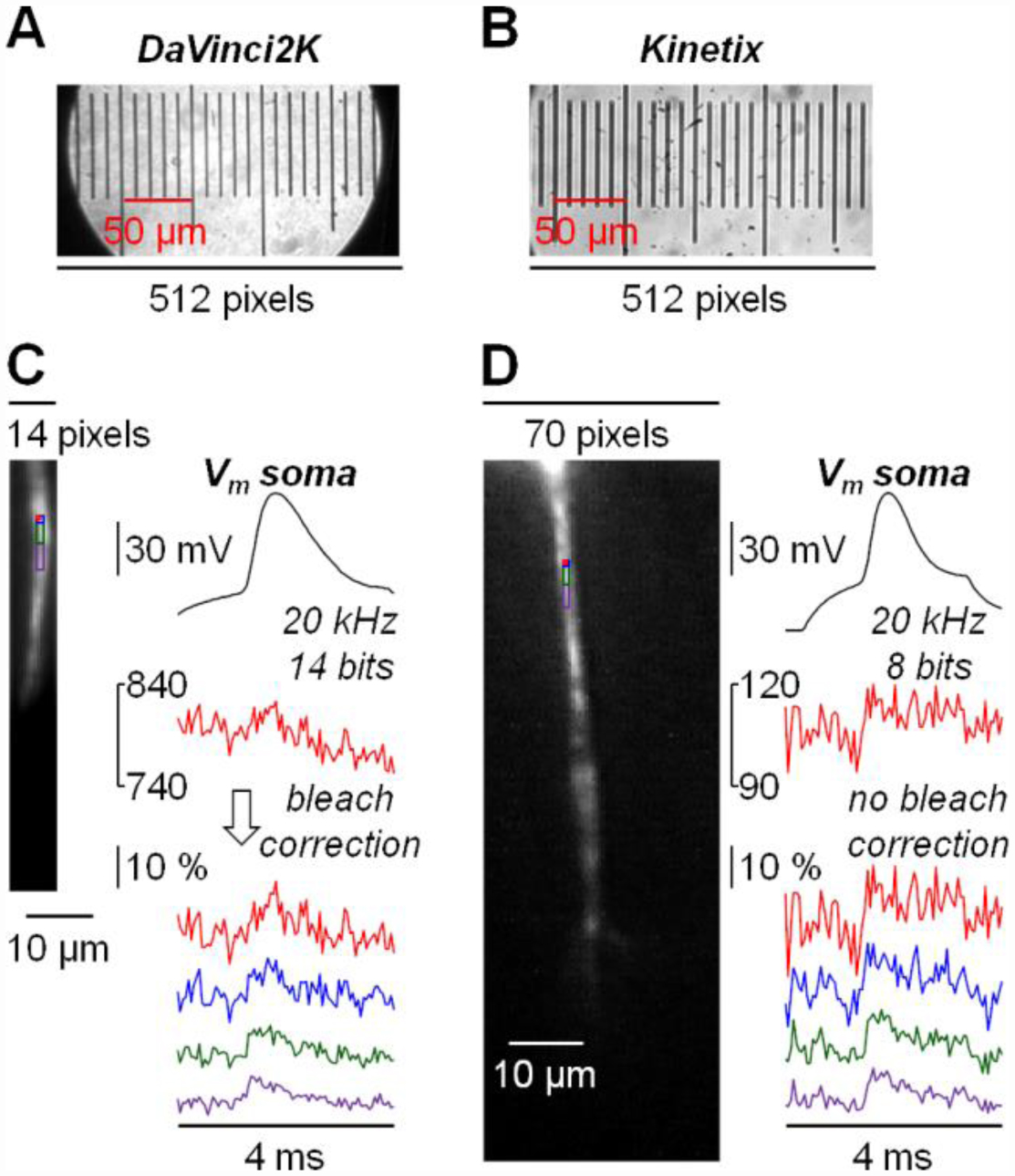
In the last decade, complementary metal-oxide semi-conductor (CMOS) cameras became the state-of-the-art technology in many biological applications, and the ability to reach high acquisition rates represents one of the characteristics that outperform previous technologies. In this review, I concentrated on neuronal functional imaging (voltage and/or ions) that requires recording fluorescence from multiples sites of a neuron or of a network at kHz rates to sample signals associated with neuronal excitability. After introducing the physical constrains of this type of imaging and reviewing the technologies used in the past, I analysed how CMOS can address the challenge of neuronal functional imaging. I focused on the characteristics of two CMOS cameras that are in use in my laboratory: DaVinci2K and Kinetix. DaVinci2K achieves high acquisitions rates at 14-bit depth by using parallel processing from 16 sub-sensors whereas Kinetix achieves higher spatiotemporal resolution by sampling fluorescence at 8-bit depth, but at the cost of decreasing the dynamic range which represents a limitation in several experimental scenarios. I present comparable membrane potential imaging recordings of action potentials from the axon initial segment, which were achieved at 20 kHz with the two cameras. Finally, I conclude the review with some perspective considerations on future availability of CMOS cameras that may overcome the performance of present devices and the limitations in developing optimal devices for biological and biomedical applications.

| [1] |
Sepehri Rad M, Choi Y, Cohen LB, et al. (2017) Voltage and calcium imaging of brain activity. Biophys J 113: 2160-2167. https://doi.org/10.1016/j.bpj.2017.09.040 
|
| [2] |
Fritzky L, Lagunoff D (2013) Advanced methods in fluorescence microscopy. Anal Cell Pathol 36: 5-17. https://doi.org/10.3233/ACP-120071
|
| [3] |
Conchello JA, Lichtman JW (2005) Optical sectioning microscopy. Nat Methods 2: 920-931. https://doi.org/10.1038/nmeth815
|
| [4] |
Rubart M (2004) Two-photon microscopy of cells and tissue. Circ Res 95: 1154-1166. https://doi.org/10.1161/01.RES.0000150593.30324.42
|
| [5] |
Homma R, Baker BJ, Jin L, et al. (2009) Wide-field and two-photon imaging of brain activity with voltage- and calcium-sensitive dyes. Philos Trans R Soc Lond B Biol Sci 364: 2453-2467. https://doi.org/10.1098/rstb.2009.0084
|
| [6] |
Orbach HS, Cohen LB, Grinvald A (1985) Optical mapping of electrical activity in rat somatosensory and visual cortex. J Neurosci 5: 1886-1895. https://doi.org/10.1523/JNEUROSCI.05-07-01886.1985
|
| [7] |
Ross WN, Werman R (1987) Mapping calcium transients in the dendrites of Purkinje cells from the guinea-pig cerebellum in vitro. J Physiol 389: 319-336. https://doi.org/10.1113/jphysiol.1987.sp016659
|
| [8] |
Wu JY, London JA, Zecevic D, et al. (1988) Optical monitoring of activity of many neurons in invertebrate ganglia during behaviors. Experientia 44: 369-376. https://doi.org/10.1007/BF01940529
|
| [9] |
Zecević D (1996) Multiple spike-initiation zones in single neurons revealed by voltage-sensitive dyes. Nature 381: 322-325. https://doi.org/10.1038/381322a0
|
| [10] |
Takamatsu T, Wier WG (1990) High temporal resolution video imaging of intracellular calcium. Cell Calcium 11: 111-120. https://doi.org/10.1016/0143-4160(90)90064-2
|
| [11] |
Lasser-Ross N, Miyakawa H, Lev-Ram V, et al. (1991) High time resolution fluorescence imaging with a CCD camera. J Neurosci Methods 36: 253-261. https://doi.org/10.1016/0165-0270(91)90051-Z
|
| [12] |
Mammano F, Canepari M, Capello G, et al. (1999) An optical recording system based on a fast CCD sensor for biological imaging. Cell Calcium 25: 115-123. https://doi.org/10.1054/ceca.1998.0013
|
| [13] |
Canepari M, Mammano F, Kachalsky SG, et al. (2000) GABA- and glutamate-mediated network activity in the hippocampus of neonatal and juvenile rats revealed by fast calcium imaging. Cell Calcium 27: 25-33. https://doi.org/10.1054/ceca.1999.0086
|
| [14] |
Ross WN, Miyazaki K, Popovic MA, et al. (2015) Imaging with organic indicators and high-speed charge-coupled device cameras in neurons: some applications where these classic techniques have advantages. Neurophotonics 2: 021005. https://doi.org/10.1117/1.NPh.2.2.021005
|
| [15] |
Callaway JC, Lasser-Ross N, Ross WN (1995) IPSPs strongly inhibit climbing fiber-activated [Ca2+]i increases in the dendrites of cerebellar Purkinje neurons. J Neurosci 15: 2777-2787. https://doi.org/10.1523/JNEUROSCI.15-04-02777.1995
|
| [16] |
Takashima I, Ichikawa M, Iijima T (1999) High-speed CCD imaging system for monitoring neural activity in vivo and in vitro, using a voltage-sensitive dye. J Neurosci Methods 91: 147-159. https://doi.org/10.1016/S0165-0270(99)00093-X
|
| [17] |
Larkum ME, Watanabe S, Nakamura T, et al. (2003) Synaptically activated Ca2+ waves in layer 2/3 and layer 5 rat neocortical pyramidal neurons. J Physiol 549: 471-488. https://doi.org/10.1113/jphysiol.2002.037614
|
| [18] |
Antic SD (2003) Action potentials in basal and oblique dendrites of rat neocortical pyramidal neurons. J Physiol 550: 35-50. https://doi.org/10.1113/jphysiol.2002.033746
|
| [19] |
Djurisic M, Antic S, Chen WR, et al. (2004) Voltage imaging from dendrites of mitral cells: EPSP attenuation and spike trigger zones. J Neurosci 24: 6703-6714. https://doi.org/10.1523/JNEUROSCI.0307-04.2004
|
| [20] |
Milojkovic BA, Zhou WL, Antic SD (2007) Voltage and calcium transients in basal dendrites of the rat prefrontal cortex. J Physiol 585: 447-468. https://doi.org/10.1113/jphysiol.2007.142315
|
| [21] |
Djurisic M, Popovic M, Carnevale N, et al. (2008) Functional structure of the mitral cell dendritic tuft in the rat olfactory bulb. J Neurosci 28: 4057-4068. https://doi.org/10.1523/JNEUROSCI.5296-07.2008
|
| [22] |
Foust AJ, Yu Y, Popovic M, et al. (2011) Somatic membrane potential and Kv1 channels control spike repolarization in cortical axon collaterals and presynaptic boutons. J Neurosci 31: 15490-15498. https://doi.org/10.1523/JNEUROSCI.2752-11.2011
|
| [23] |
Popovic MA, Foust AJ, McCormick DA, et al. (2011) The spatio-temporal characteristics of action potential initiation in layer 5 pyramidal neurons: a voltage imaging study. J Physiol 589: 4167-4187. https://doi.org/10.1113/jphysiol.2011.209015
|
| [24] |
Jaafari N, Canepari M (2016) Functional coupling of diverse voltage-gated Ca2+ channels underlies high fidelity of fast dendritic Ca2+ signals during burst firing. J Physiol 594: 967-983. https://doi.org/10.1113/JP271830
|
| [25] |
Short SM, Oikonomou KD, Zhou WL, et al. (2017) The stochastic nature of action potential backpropagation in apical tuft dendrites. J Neurophysiol 118: 1394-1414. https://doi.org/10.1152/jn.00800.2016
|
| [26] |
Ait Ouares K, Canepari M (2020) The origin of physiological local mGluR1 supralinear Ca2+ signals in cerebellar Purkinje neurons. J Neurosci 40: 1795-1809. https://doi.org/10.1523/JNEUROSCI.2406-19.2020
|
| [27] |
Ahrens MB, Orger MB, Robson DN, et al. (2013) Whole-brain functional imaging at cellular resolution using light-sheet microscopy. Nat Methods 10: 413-420. https://doi.org/10.1038/nmeth.2434
|
| [28] |
Filipis L, Ait Ouares K, Moreau P, et al. (2018) A novel multisite confocal system for rapid Ca2+ imaging from submicron structures in brain slices. J Biophotonics 11: e201700197. https://doi.org/10.1002/jbio.201700197
|
| [29] |
Kim Y, Lee U, Choi C, et al. (2020) Release mode dynamically regulates the RRP refilling mechanism at individual hippocampal synapses. J Neurosci 40: 8426-8437. https://doi.org/10.1523/JNEUROSCI.3029-19.2020
|
| [30] |
Xiao S, Lowet E, Gritton HJ, et al. (2021) Large-scale voltage imaging in behaving mice using targeted illumination. iScience 24: 103263. https://doi.org/10.1016/j.isci.2021.103263
|
| [31] |
Filipis L, Canepari M (2021) Optical measurement of physiological sodium currents in the axon initial segment. J Physiol 599: 49-66. https://doi.org/10.1113/JP280554
|
| [32] |
Mendonça PRF, Tagliatti E, Langley H, et al. (2022) Asynchronous glutamate release is enhanced in low release efficacy synapses and dispersed across the active zone. Nat Commun 13: 3497. https://doi.org/10.1038/s41467-022-31070-4
|
| [33] |
Filipis L, Blömer LA, Montnach J, et al. (2023) Nav1.2 and BK channel interaction shapes the action potential in the axon initial segment. J Physiol 601: 1957-1979. https://doi.org/10.1113/JP283801
|
| [34] |
Ahanonu B, Crowther A, Kania A, et al. (2024) Long-term optical imaging of the spinal cord in awake behaving mice. Nat Methods 21: 2363-2375. https://doi.org/10.1038/s41592-024-02476-3
|
| [35] |
Huang YC, Chen HC, Lin YT, et al. (2024) Dynamic assemblies of parvalbumin interneurons in brain oscillations. Neuron 112: 2600-2613. https://doi.org/10.1016/j.neuron.2024.05.015
|
| [36] |
Abbas F, İpek ÖY, Moreau P, et al. (2025) Neuronal imaging at 8-bit depth to combine high spatial and high temporal resolution with acquisition rates up to 40 kHz. J Biophotonics 18: e202400513. https://doi.org/10.1002/jbio.202400513
|
| [37] |
Marosi EL, Arszovszki A, Brunner J, et al. (2023) Similar presynaptic action potential-calcium influx coupling in two types of large mossy fiber terminals innervating CA3 pyramidal cells and hilar mossy cells. eNeuro 10: ENEURO.0017-23.2023. https://doi.org/10.1523/ENEURO.0017-23.2023
|
| [38] |
Brunner J, Arszovszki A, Tarcsay G, et al. (2024) Axons compensate for biophysical constraints of variable size to uniformize their action potentials. Plos Biol 22: e3002929. https://doi.org/10.1371/journal.pbio.3002929
|
| [39] |
Ortkrass H, Müller M, Engdahl AK, et al. (2024) High sensitivity cameras can lower spatial resolution in high-resolution optical microscopy. Nat Commun 15: 8886. https://doi.org/10.1038/s41467-024-53198-1
|
| [40] |
Nguyen JP, Shipley FB, Linder AN, et al. (2016) Whole-brain calcium imaging with cellular resolution in freely behaving Caenorhabditis elegans. Proc Natl Acad Sci USA 113: E1074-81. https://doi.org/10.1073/pnas.1507110112
|
| [41] |
Obara K, Ebina T, Terada SI, et al. (2023) Change detection in the primate auditory cortex through feedback of prediction error signals. Nat Commun 14: 6981. https://doi.org/10.1038/s41467-023-42553-3
|
| [42] |
Zhang J, Newman J, Wang Z, et al. (2024) Pixel-wise programmability enables dynamic high-SNR cameras for high-speed microscopy. Nat Commun 15: 4480. https://doi.org/10.1038/s41467-024-48765-5
|
| [43] |
Adam Y, Kim JJ, Lou S, et al. (2019) Voltage imaging and optogenetics reveal behaviour-dependent changes in hippocampal dynamics. Nature 569: 413-417. https://doi.org/10.1038/s41586-019-1166-7
|
| [44] |
Xiao S, Cunningham WJ, Kondabolu K, et al. (2024) Large-scale deep tissue voltage imaging with targeted-illumination confocal microscopy. Nat Methods 21: 1094-1102. https://doi.org/10.1038/s41592-024-02275-w
|
| [45] |
Dowd K, Doyle E, Dunne P (2025) Near infra-red absorption spectroscopy for astrophysically significant ions. Exp Astron 60: 7. https://doi.org/10.1007/s10686-025-10009-9
|
| [46] |
Ma Q, Liu Z, Zhang T, et al. (2024) Multielement simultaneous quantitative analysis of trace elements in stainless steel via full spectrum laser-induced breakdown spectroscopy. Talanta 272: 125745. https://doi.org/10.1016/j.talanta.2024.125745
|
| [47] |
Klement WJN, Leproux P, Browne WR, et al. (2025) CMOS and CCD detection in Raman spectroscopy: A comparison using spontaneous and multiplex coherent anti-stokes Raman scattering (CARS). J Raman Spectrosc 56: 933-938. https://doi.org/10.1002/jrs.6773
|
Figures(1) / Tables(1)

Marco Canepari. CMOS cameras: state-of-the-art technology for neuronal functional imaging with high spatiotemporal resolution[J]. AIMS Biophysics, 2025, 12(4): 499-509. doi: 10.3934/biophy.2025024


 DownLoad: