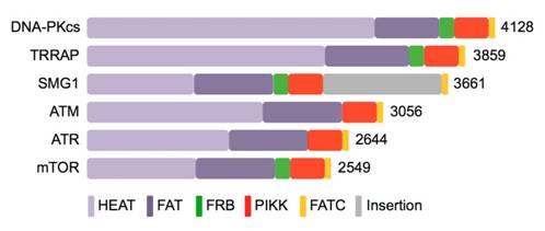
Citation: Angel Rivera-Calzada, Andrés López-Perrote, Roberto Melero, Jasminka Boskovic, Hugo Muñoz-Hernández, Fabrizio Martino, Oscar Llorca. Structure and Assembly of the PI3K-like Protein Kinases (PIKKs) Revealed by Electron Microscopy[J]. AIMS Biophysics, 2015, 2(2): 36-57. doi: 10.3934/biophy.2015.2.36
| [1] | Qing Song, Yu Mao, Mark Wilkins, Fernando Segato, Rolf Prade . Cellulase immobilization on superparamagnetic nanoparticles for reuse in cellulosic biomass conversion. AIMS Bioengineering, 2016, 3(3): 264-276. doi: 10.3934/bioeng.2016.3.264 |
| [2] | Mohamed Neifar, Rim Chatter, Habib Chouchane, Raya Genouiz, Atef Jaouani, Ahmed Slaheddine Masmoudi, Ameur Cherif . Optimization of enzymatic saccharification of Chaetomorpha linum biomass for the production of macroalgae-based third generation bioethanol. AIMS Bioengineering, 2016, 3(3): 400-411. doi: 10.3934/bioeng.2016.3.400 |
| [3] | Paula García-Fraile, Esther Menéndez, Raúl Rivas . Role of bacterial biofertilizers in agriculture and forestry. AIMS Bioengineering, 2015, 2(3): 183-205. doi: 10.3934/bioeng.2015.3.183 |
| [4] | Charlotte E. Bjorck, Paul D. Dobson, Jagroop Pandhal . Biotechnological conversion of methane to methanol: evaluation of progress and potential. AIMS Bioengineering, 2018, 5(1): 1-38. doi: 10.3934/bioeng.2018.1.1 |
| [5] | Kevyn Melo Lotas, Raissa Sayumy Kataki Fonseca, Joice Camila Martins da Costa, Ana Claudia Alves Cortez, Francisca das Chagas do Amaral Souza, Márcio Rodrigues Barreto, Lívia Melo Carneiro, João Paulo Alves Silva, Eveleise Samira Martins Canto, Flávia da Silva Fernandes, João Vicente Braga de Souza, Érica Simplício de Souza . Optimization of laccase production by Pleurotus ostreatus (Jacq.) P. Kumm. using agro-industrial residues: a comparative study on peels of tucumã (Astrocaryum aculeatum G. Mey.) and pupunha (Bactris gasipaes Kunth) fruits. AIMS Bioengineering, 2024, 11(4): 561-573. doi: 10.3934/bioeng.2024025 |
| [6] | Ronald C. Sims, Sean K. Bedingfield, Reese Thompson, Judith L. Sims . Bioenergy from wastewater-based biomass. AIMS Bioengineering, 2016, 3(1): 103-124. doi: 10.3934/bioeng.2016.1.103 |
| [7] | Dyoni M. de Oliveira, Victor Hugo Salvador, Thatiane R. Mota, Aline Finger-Teixeira, Rodrigo F. de Almeida, Douglas A. A. Paixão, Amanda P. De Souza, Marcos S. Buckeridge, Rogério Marchiosi, Osvaldo Ferrarese-Filho, Fabio M. Squina, Wanderley D. dos Santos . Feruloyl esterase from Aspergillus clavatus improves xylan hydrolysis of sugarcane bagasse. AIMS Bioengineering, 2017, 4(1): 1-11. doi: 10.3934/bioeng.2017.1.1 |
| [8] | Sezer Okay, Mehmet Sezgin . Transgenic plants for the production of immunogenic proteins. AIMS Bioengineering, 2018, 5(3): 151-161. doi: 10.3934/bioeng.2018.3.151 |
| [9] | Zongyuan Zhu, Rachael Simister, Susannah Bird, Simon J. McQueen-Mason, Leonardo D. Gomez, Duncan J. Macquarrie . Microwave assisted acid and alkali pretreatment of Miscanthus biomass for biorefineries. AIMS Bioengineering, 2015, 2(4): 449-468. doi: 10.3934/bioeng.2015.4.449 |
| [10] | Joe A. Lemire, Marc A. Demeter, Iain George, Howard Ceri, Raymond J. Turner . A novel approach for harnessing biofilm communities in moving bed biofilm reactors for industrial wastewater treatment. AIMS Bioengineering, 2015, 2(4): 387-403. doi: 10.3934/bioeng.2015.4.387 |
Cellulose is one of the most important polysaccharides and the major component of plant cell walls. Bacteria also synthesize cellulose, which has higher purity and hydrophilicity than plant cellulose, containing no lignin or hemicelluloses (different degree of crystallization). Moreover, it has greater tensile strength, versatility and moldability. Due to its particular properties, biotechnological industries require the use of bacterial cellulose for different applications (for review see Keshk, 2014 [1]) and novel studies report cellulose uses in novel applications in medicine, design of new materials or bioremediation [2,3,4].
Despite the proven importance of cellulose for the industry, the inner mechanisms of bacterial cellulose biosynthesis and its regulation are not fully characterized. Only the studies based in Gluconacetobacter xylinum, model bacterium for cellulose biosynthesis, are well described and clarified [5]. Recently, these matters are attracting the attention of the scientific community. Several studies reported cellulose-producing bacteria and alsothe characterization of cellulose biosynthesis in Pseudomonas spp., Rhizobium leguminosarum, Escherichia coli, Salmonella spp, Gluconoacetobacter hansenii and Rhodococcus sphaeroides [6,7,8,9,10,11]. Bacterial cellulose biosynthesis involves the participation of several enzyme encoding genes, including cellulose synthases, cyclic di-GMP binding domain/protein and cellulases, which must be coordinated and tightly regulated. Those genes are usually forming an operon, called bcsABZ or celABC, depending on the microorganism [8].
First studies indentifying cyclic di-GMP as allosteric regulator of cellulose biosynthesis in bacteria were published years ago [5,12] using the model bacteria G. xylinum and A. tumefaciens. Nowadays, the role of this molecule in bacterial cellulose biosynthesis is fully accepted but further characterization of the entire bacterial cellulose biosynthesis regulation is necessary [13,14,15].
The biological significance of cellulose production by bacteria is still not well addressed [8], but, for example, it is well described how Rhizobium species produce cellulose microbibrills for attachment to biotic and/or abiotic surfaces [16,17]. Moreover, cellulose is one of the main components of biofilms, constituting the inner skeleton of the extracellular matrix [18,19]. The survival of the bacteria seems to be the main reason for bacteria to produce cellulose, but cellulases are also involved in the survival strategies. About a half of the existing bacteria have genes for degrading plant biomass, such as pectinases, cellulases and hemicellulases [20].
Cellulases are enzymes that degrade the ß-1,4-glucan linkages of cellulose microfibrills, which are present in plant biomass and also in microorganism cell walls, including pathogens. Recently, the sequencing of bacterial genomes revealed different kinds of enzymes involved in polysaccharide degradation, including cellulose. The present review is focused in bacterial cellulases, which degrade ß-1,4-glucan chains (endoglucanases).
Bacterial cellulases are classified into the group EC 3.2.1.4, according to the IUBMB Enzyme Nomenclature. Cellulase (4-β-D-glucan 4-glucanohydrolase) catalyzes the endohydrolysis of ß-1,4-D-glucosidic linkages in cellulose, lichenin and cereal ß-d-glucans. These enzymes also hydrolyse 1,4-linkages in β-D-glucans, as well as containing 1,3-linkages.
Cellulases are functionally organized in categories, which are included in the CaZy database [21]. Glycosil Hydrolases comprise the functional category in which cellulases are grouped. The protein structure and domains depends on the GH family [22,23].
The number of GH families continuesto grow steadily over the years. In 1995, there were 22 families of GHs [23] and the number increased until the creation of clans of related families, which shared catalytic machinery and folding. Nowadays, there are 133 GH families and thirteen clans are defined, according to CaZy database. In Supplementary Table 1, all of the bacterial cellulases belonging to the GH families, containing EC 3.2.1.4 valid cellulases, are listed. Moreover, there are 2538 bacterial cellulases not yet assigned to any category.
Cellulase is a complex term to denominate a group of three enzymes: i) endo-1,4-β-D-glucanase (endoglucanase), exo-1,4-β-D-glucanase (exoglucanase) and β-glucosidase. These enzymes, in synergic combination, are the responsible of cellulose hydrolysis. Our interests are focused in endoglucanases, which cleave randomly linear cellulose chains. Specifically, this review deals with bacterial endoglucanases, which are susceptible of being applied to industry.
The most common cellulolytic bacteria are Acetivibrio cellulolyticus, Bacillus spp., Cellulomonas spp., Clostridium spp., Erwinia chrysanthemi, Thermobispora bispara, Ruminococcus albus, Streptomyces spp., Thermonospora spp and Thermobifida fusca [24]. Nevertheless, nowadays there is an increasing interest in the search of new cellulolytic bacterial strains.
In the last two years, at least 9 novel bacterial species were officially described directly as cellulose-degrading bacteria in the International Journal of Systematic and Evolutionary Microbiology (IJSEM), the official journal of record for novel prokaryotic taxa. Streptomyces abietis [25], Kallotenue papyrolyticum [26], Ornatilinea apprima [27], Bacteroides luti [28], Alicyclobacillus cellulosilyticus [29], Anaerobacterium chartisolvens [30], Caldicellulosiruptor changbaiensis [31], Herbinix hemicellulosilytica [32] and Pseudomonas coleopterorum [33] are the novel cellulolytic species, which were isolated from a huge variety of environments. Furthermore, species with cellulolytic activity are published in other journals. For example, Huang et al. [34] reported for the first time the cellulolytic activity of Siphonobacter aquaeclarae, Cellulosimicrobium funkei, Paracoccus sulfuroxidans, Ochrobactrum cytisi, Ochrobactrum haematophilum, Kaistia adipata, Devosia riboflavina, Labrys neptuniae, Ensifer adhaerens, Shinella zoogloeoides, Citrobacter freundii and Pseudomonas nitroreducens from the gut of Holotrichia parallela larvae. More recently, Hameed et al. [35] isolated and characterized a novel cellulose-degrading marine bacterium designated Oricola cellulosilytica, affiliated to the family Phyllobacteriaceae from surface seashore water. Whether or not those new species produce interesting cellulases, further studies will be mandatory.
Nowadays, most of the studies about production of thermostable cellulases are focused on the utilization of cellulase-producing thermo/alkalophiles and also, on the improvement of cellulase production by optimizing its nutritional and environmental necessities or by engineering new high-producer recombinants or cellulase-producing transgenic plants, such as transgenic tobacco [36,37,38].
Thermo/alkalophilic cellulose-degrading bacteria have been isolated from various environments such as gold mines [39], marine plants [40], marine depth [41,42], composting plants [43], among others. Metagenomics is recently the most used strategy to discover new cellulases [44,45,46,47,48].
Bacterial cellulases are inducible enzymes in the presence of cellulosic substrates such as pure commercial substrates (i. e., carboxymethyl-cellulase or Avicel), residual substrates (agro-wastes) or lignocellulosic plant biomass. Attri & Garg [49] used agro-waste substrates to develop a medium for the isolation of cellulolytic, pectinolytic and xylanolytic bacteria. They obtained results using this low-cost isolation method, which can be compared to the results obtained with pure commercial substrates. Also, Singh and Singh [50] obtained good results by using residual bagasses (sugarcane pulp).
Lately, there are many reseachers using classical methods for cellulose-degrading bacteria screening, such as the use of Congo Red and Gramxs Iodine [51,52]. However, new techniques are being developed, increasing importance and usefulness for new cellulolytic strains screening. For example, Taha et al. [53] developed a microplate method based in Biolog plates, revealing its effectiveness and accuracy for screening native lignocellulosic-straw-degrading bacteria.
There are several methods for measuring cellulolytic activity, most of them involved the measuring of the accumulation hydrolysis product or the reduction of the final product. Zhang et al. [54] and Sadhu & Maiti [24] reviewed some of the most useful techniques. Nevertheless, optimization of media and culture parameters are also used as mandatory for obtaining maximum yields, being relatively cheap alternatives for industrial uses [36].
To obtain maximum cellulase yields, Sadhu et al. [55] performed random mutagenesis in a cellulolytic strain belonging to the genus Bacillus by using N-methyl-N'-nitro-N-nitrosoguanidine (NTG), which leads to transitional mutations between AT and GC nucleotides. They obtained a mutant strain with enhanced CMCase activity. Similar results were obtained in Cellulomonas sp, also employing NTG [56], but the characterization of these mutants is still unclear.
Involving genetic modification, there are two major strategies for the improvement of cellulases or cellulase complex component: i) rational design and ii) directed evolution [57]. There are three kinds of cellulase systems: single cellulases, multifunctional cellulases and cellulosomes. The benefits and the modifications (classical, random mutagenesis or genetic modification) of each one are being tested and used for years to obtain maximum yield and cellulase efficiency. Above all the studies, the activity of the bifunctional cellulase system of Caldicellulosiruptor bescii is significatively superior to any known system of cellulose degradation [20].
The production of recombinant enzymes involves a well-known technology and remarkably advanced processes are directed to an efficient development and scaling-up productions for industries [47], following a combination between directed evolution and rational design [58,59]. However, the still limited knowledge of cellulase substrates, relationships and regulation of cellulosic activity compromises rational design, leading to the use of directed evolution as a tool for higher rate of success [54]. Several authors reviewed and compared recombinant cellulases expression systems [24,60,61,62,63]. In this section, we will focus in organisms that are described to homo/heterologous express bacterial cellulases.
Some studies report the use of directed evolution techniques in combination with a rational design to overexpress cellulases in their own bacterial source. Genera such as Bacillus (B. subtilis) and Clostridium (C. thermocellum) were used as a homologous cellulases production system, due to their easy genetic modification and other proper features. However, the use of these bacteria has disadvantages such as low protein yields, high production costs or need of enriched media [63].
Munjal et al. [64] reported the use of an ethanologenic E.coli strain to express a ß-1,4-endoglucanase and a ß-1,4-glucosidase from other E.coli strain under a constitutive promoter, with the potentiality for fermenting biomass hydrolysates.
Chung et al. [65] engineered Caldicellulosiruptor bescii, a bacterium able to degrade lignocellulosic biomass by itself. They cloned and performed a homologous expression of a multimodular cellulase, called CelA, which contains GH9 and GH48 domains. This report gives light in the understanding of how transglycosilation is involved in protein functionality and structure. Remarkably, this particular work shows a successful way to improve and enhance the cellulolytic activity in this important thermophilic bacterium with a potential for biotechnologic applications in industry.
Furthermore, a classical method to overproduce an interesting protein or enzyme by cloning its codifying genes in a high copy number plasmid, compatible with its host, is still commonly used. For example, Robledo et al. [66] performed this method to obtain a homologous overexpression of cellulase CelC2 from Rhizobium leguminosarum bv trifolii ANU843, which increased threefold its cellulolytic activity.
The strategies based in heterologous expression are focused in the use of non-cellulolytic micro/organisms that have high production ratio for expressing microbial cellulases [67]. Bacteria such as Escherichia coli, different species from the genus Bacillus, Pseudomonas fluorescens, Ralstonia eutropha and Zymomonas mobilis; yeasts such as Saccharomyces cerevisiae and Pichia pastoris and filamentous fungi from genera Aspergillus and Trichoderma genera are the most used in research and industry, considered as host systems for producing recombinant enzymes. Furthermore, cell cultures of mammals, plants or insects and transgenic plants and/or animals are used for protein expression [68].
Plants are considered as non-expensive alternative systems for production of recombinant enzymes. The approaches to achieve an efficient recombinant enzyme expression involve their expression from the plant nuclear genetic material, plastidic genome or plant tissues harbouring viral particles. Also, there are several strategies to enhance recombinant enzyme production yields (reviewed in [69]). The most used plants for this kind of studies are model plants such as Arabidopsis thaliana, Zea mays, Medicago sativa/truncatula, Nicotiana tabacum and Populus alba.
Several studies report the expression of cellulases in plants for research purpose, to elucidate the mode of action and effects of bacterial/fungal/plant cellulases in cellulose biosynthesis, but our interests are focused in the industrial applications. Ziegelhoffer et al. [70] expressed thermostable cellulases E2 and E3 from Thermobifida (former Thermomonospora) fusca in Medicago sativa, Solanum tuberosum and Nicotiana tabacum, obtaining low but promising expression patterns.
Brunecki et al. [71] reported the expression of a GH5 family endocellulase (E1 endoglucanase) from Acidothermus cellulolyticus, in Zea mays and Nicotiana tabacum plants, resulting in a reduction of cell walls recalcitrance to chemical pretreatments in both plants, suggesting the possibility of an application for improving plant biomass bioconversion. Petersen & Bock [72] reported the high expression of a thermostable cellulase cocktail in plastids of Nicotiana plants, also with the purpose of biotechnological application.
The most used fungal expression system is Saccharomyces cerevisiae, but Pichia pastoris and Kluyveromyces sp., especially this last one, are raising importance due to their high yields in heterologous cellulase productions. However, nowadays comparisons between those systems are difficult, because there is not enough knowledge to overcome the low cellulolytic activity of the expressed cellulases [61]. It is quite common to express fungal cellulases in Saccharomyces, such as T. reseei and other fungal sources [61], but there are some studies reporting the expression of clostridial mini-cellulosomes in this yeast [73]. Indeed, Clostridium species possess cellulosomes, cell-bound complexes of cell wall-degrading enzymes, which actsynergistically, being yeasts their used platform for expression [for review about cellulosomes and related information see 20, 74, 75, 76].
E. coli and Bacillus are vastly the most used as bacterial systems for expressing recombinant proteins. Moreover, other bacteria are also used as platforms, including Zymomonas mobilis and Streptomyces lividans.
E.coli is a commonexpression system for cellulases in industry and has several advantages, such as its well-characterized genome, its availability in commercial forms and kits and its ease for modification. However, there are some disadvantages that must be taken in account such as its limited secretion (thick outer membrane, incorrect transportation across membrane), degradation of linker sequences, reduction in cellulolytic activity and the possibility of formation of inclusion bodies. Zymomonas mobilis presented itself as an alternative to the use of yeast due to its versatility, fermenting a wide range of sugars; and also, as an alternative to E. coli due to its higher yields and the ability to express recombinant proteins intra- and extracellularly. Streptomyces lividans has proved its potentiality for large-scale biopharmaceutical production and can secrete recombinant proteins [62].
Nevertheless, the vast majority of authors selected E.coli as expression systems. Liu et al. [46] report the expression of an endoglucanase, Cel5G, indentified by metagenomics and presented the closest similarity with the myxobacterium Plesiocystis pacifica. Shinoda et al. [77] isolated a strain of Paenibacillus cookii that produced a novel endoglucanase with chitosanase activity and cloned into an E.coli strain. This study aims to understand the underlying mechanisms of degradation of ß-1,4-glucan linkages for potential applications. Shi et al. [78] reported the heterologous expression of the novel thermo-halotolerant endoglucanase Cel5H from Dyctioglomus thermophilum in E. coli. More recently, Wei et al. [79] cloned an endoglucanase from a Bacillus subtilis strain, isolated from termite gut, in E. coli.
All of these studies try to characterize and overproduce new bacterial cellulases with special features such as thermostability, halotolerance and synergy with other enzymes, among others, which will have high potential for industrial applications.
Over many decades, microbial cellulases have shown their potential applications in several fields of industry, being the most frequently used enzyme group in various industrial processes, such as biofuel production, food and wine biotechnology, pulp and paper production, bio-de-inking, textile and laundry industry, conversion of cellulosic biomass and applications in research and development and also, in medicine and agriculture [24,80,81,82]. In this review, we aim to show the latest advances in all of these fields.
Industry searches for low-cost mechanisms of cellulase production and maintenance, but more specific applications are needed depending on the biotechnological process in which they will be involved. For that reason, the study and characterization of new specific and improved cellulases is totally required. Amongst other features, their applications in industry demands the identification of proper bacterial producers and highly stable enzymes, which remain active at extreme pHs and temperatures, without forgetting the primacy for obtaining economically low-cost production cellulolytic enzymes.
Generally, cellulases are used in textile industry for biopolishing of fabrics and producing a stonewashed-look in denims, as well as they are used in household laundry detergents for improving softness and brightness of fabrics made of cotton. Features as color care, cleaning and anti-redeposition are a pursued in a proper washing powder or detergent. Moreover, compatibilitywith other ingredients present in formulations, temperature stability, andactivity under alkaline conditions are all important performance characteristics for cellulases, which are to be employed in textile and detergent applications [47,59].
Fungal cellulases from T. reseei are the most used, but some bacterial alternatives are already available. Actinomycetes from the generaStreptomyces and Thermobifida and other genera of bacteria, such as Pseudomonas and Sphingomonas, are susceptible to be used for decolourisation and degradation of textile dyes [83]
Cellulases, in combination with other enzymes, are added to detergents to catalyze the breakdown of chemical bonds in presence of water under thermophilic and/or alkalinophilic conditions, as well as in the presence of the rest of detergent components [47]. Since alkaline or neutral conditions are preferable for enzymatic processing of denim fibers, Anish et al. [84] reported the use of an alkaline-stable endoglucanase of Thermomonospora sp, in contraposition of the use of fungal commecial cellulases. Furthermore, the genus Bacillus showed a high potential to produce cellulases and/or endoglucanases directly applied in these industries. Ladeira et al. [85] reported the production of a cellulase by the thermophilic bacterium Bacillus sp. SMIA-2. This enzyme retained the major part of its activity after incubation at different temperatures and conditions and in the presence of several brands of commercial detergents. Moreover, there is a study that applies the use of a recombinant endoglucanase from a species of the genus Bacillus to soften cotton fabrics [86].
Cellulases are widely used in food processing and animal feed production, mainly in combination with hemicellulases and pectinases. Their uses are varied; for example, they are used in production of fruit and vegetable juices, carotenoids and degradation of plant cell walls for wine and beer industries, among others.
In fruit juice production industry, cellulases produced by bacteria, such as Bacillus and Paenibacillus, besides other cellulases of fungal origin, are used as accessory enzymes for clarifying fruit juices [87]. Moreover, cellulases in combination with pectinases, degrades orange, sweet potato and carrot cell walls, so indirectly, cellulases are involved in the extraction of carotenoids, which are susceptible to be used in food indrustry as colouring substances [88] or degrades grapefruit peels to produce sugars for different industrial applications, including food industries [89]. Also, cellulases participate in the extraction of phenolic compounds from grape pomace [90].
Cellulases applications in wine and beer industries are also registered. In this sense, Bamforth [91] reported that several endo-1,4-ß-D glucanases, from unknown origin, are important in plant cell wall degradation and they are involved in the malting of barley.
Other application involves the use of cellulases in animal feed to enhance digestibility of cereal-based food and to increase nutritive values for a higher quality of forages [92,93,94]. Bacillus subtilis cellulases can be applied for soya grain hull degradation to enrich its nutricional value for monogastric animals feed application [95].
Gathering all of these studies in the application of cellulases in food and animal feed processing industries, we can conclude that there are opportunities to satisfy unmet needs for developing new approaches to identify bacterial cellulases instead of fungal-origin cellulases. One of the best alternatives is focused on the use of metagenomics for searching new glycosyl hydrolases [96]. Using metagenomics, Voget et al. [44] cloned and identified an endoglucanase with halotolerance properties from soil, which belongs to the GH5 family, showing high similarity to a Cellvibrio mixtus cellulase. Other metagenomic studies characterized cellulasesfrom aquatic communities of a botanical garden and soil samples, which belonged to GH5 and GH9 families and presented high similarities with cellulases from Cellvibrio japonicas [97]
Nowadays, none of the cellulases commonly used for food processing have a bacterial origin. Nevertheless, food industry needs the discovery of new cellulases adapted to each different process. The application of metagenomic tools will probably identify new bacterial candidates for being applied in food processing, among other biotechnological applications.
The use of cellulases in paper and pulp industries are mostly based in the capacity of de-inking of papers, but is still an emerging application. The presence of cellulases in pulp and paper preparation is not desired, due to the possibility of fiber degradation and loss of viscosity; however, several patents and studies specify the use of cellulases, particularly alkaline cellulases, for paper deinking, especially in deinking of office waste paper, drainage improvement, fiber modification and/or paper recycling [98,99,100].
Cellulases applied in this kind of industry are fungal cellulases, mostly commercial formulations with enzymatic cocktails derived from Trichoderma reseei and Aspergillus niger. To the best of our knowledge, there is only one study in which a bacterial cellulase, named CelB and indentified in Paenibacillus sp. BP-23, is used for improving paper properties due to an improvement of the drainage process [101]. In conclusion, it seems that the use of bacterial cellulases is still not yet a reality in this field.
The degradation of plant biomass is an expensive process, which currently requires 3 steps: i) physicochemical pretreatments, ii) enzymatic hydrolysis and iii) fermentation. Cellulases have a potential central role in the bioconversion of renewable lignocellulosic biomass involving the hydrolysis of this biomass. Apart from the other two enzymes neccessary for hydrolyzing cellulose, endoglucanases (cellulases) randomly hydrolyzes ß-glycoside bonds of internal amorphous regions in cellulose to produce simple chains of oligosaccharides with various degrees of polymerization and generate new chain ends [60]. It is estimated a 40% of reduction in costs by using cellulolytic microbes for bioprocessing and eliminating pretreatments [102]. Currently, a significant reduction in costs is required to ensure the permanence and viability of commercial cellulase production. Therefore, the use of bacterial cellulases or direct use of cellulolytic bacteria constitutes a low-cost alternative, provided that they are compatible with bioreactor and processing environments.
Fungal strains have been used widely for bioconversion of lignocellulosic biomass into biofuels and derivatives, being Trichoderma reseei the main industrial source of cellulases and hemicellulases used to depolymerize plant biomass to simple sugars [47]. However, a report showed that an extreme thermophilic bacterium, identified as Caldicelluloseruptor bescii [103] produces a cellulase, combined in a system with a hemicellulase, which has higher activity than T. reesei cellulase in degrading different substrates [104]. Thermophilic bacteria become proper sources for identification and characterization of efficient cellulases for lignocellulosic material bioconversion to bioethanol and biofuels. Recently, Scully and Orlygsson [105] published a comprehensive review, addressing the recent advances in this field and reporting a vast number of thermophilic strains with high cellulolytic potentiality. However, there are more studies reporting the effective production of cellulases by other thermophilic isolates, such as Bacillus sp. SMIA-2 [85], Geobacillus sp. T1 [106], clostridia consortium [43], marine extremophiles (reviewed by [42]), the anaerobic thermophilic hydrogen-producer Thermosipho sp. [41], among others . Moreover, solvent-thermoestable-alkalophilic cellulases produced by strains such as Bacillus vallismortis RG-07 [107] should be taken into consideration for future applications in this field. Those examples will help to fulfill the expectations and the use of novel bacteria for a more efficient lignocellulosic biomass bioconversion, using different substrates such as straws, baggasses or other agro-waste residues.
Recently, microalgal biomass showed its potential as alternative biofuel source; however, biofuel production cannot compete with traditional fuel output. The solution remains in reducing the cost of microalgal biomass production [108]. Muñoz et al [109] presented an interesting study involving the action of bacterial cellulases isolated from marine bivalves applied as pretreatment to obtain biogas from microalgae. They isolated strains belonging to four different genera (Raoultella, Aeromonas, Pseudomonas and Chyseobacterium) with cellulolytic activity, which hydrolyze microalgae cell walls and expose them to methanogenic bacteria, responsables of biogas production. The employment of commercial cellulases will increase the economical costs, whereas the use of cellulolytic bacteria is a potential low-cost alternative.
All of these are valuable results, but nowadays the efforts are addressed to identify or develop single microorganisms with combined abilities in the hydrolysis and fermentation steps. Recent studies indicated that Caldicelluloseruptor bescii has both abilities and can be used for direct bioconversion of lignocellulosic biomass to bioethanol [65].
To the best of our knowledge, there are few evidences of the application of bacterial cellulases indirectly or directly in medicine. However, there are some applications that should be pointed out.
Several studies indicate an indirect application of cellulases, mainly of fungal origin, in degradation of chitosan, in coordination with chitinases and lysozymes, among others possible enzymes.
To obtain chitosan, a partial degradation of chitin must take place. As cellulose, chitin is an important polysaccharide that provides structural integrity, stability and protection to animals, such as marine animals and insects, as well as forms part of fungi cell walls. Chitin is a poly-ß-1,4-N-acetyl-D-glucosamine, conforming crystalline microfibrills that constitute structural components of cell walls and exoskeletons. Chitosan is the most important semicrystalline derivative form of chitin, obtained by partial deacetylation (around 50%, soluble in aqueous solution) under alkaline conditions or enzyme hydrolysis. Chitosan and its derivatives have many medical applications, such as surgical sutures, bone rebuilding, production of artificial skin, anticoagulant, antibacterial agent, hemostatic dressings, anticancer and anti-diabetic agents (in combination with metals), hypocholesterolemic effectors (LMWCs), elaboration of cosmetics, production of biopharmaceutics and encapsulation of diverse materials [110,111,112].
Thanks to the combined effect of unspecific cellulases, chitosanases and lysozymes, regular chitosan turns into low molecular weight chitosan (LMWC), which is easier to use in several biomedical applications [112,114]. There are several studies reporting how cellulases hydrolyze chitosan and their potential biomedical effects [113,114].
Qin et al. [113] reported that cellulase-treated chitosan has antitumoral activity, showing a maximum inhibitory rate between 30 and 50% (depend of administration) of tumors in mice. Lin et al. [114] showed the antibacterial activity of LMWCs obtained by Trichodermacommercial enzymes. Cellulase-treated chitosan hydrolysate loses its antibacterial capacity, perhaps due to an intensive hydrolysis. For that reason, the complete knowledge of this process is essential in order to optimize productions and to obtain economically reasonable products.
Some of the microbial cellulases used to treat chitosan have fungal origin [115,116]. In 2006, Liu & Xia [115] purified and characterized a bifunctional enzyme (chitosanase/cellulase) from Trichoderma viridae commercial formulations. Xie et al. [116] compared cellulases from different origins for depolymerizing chitosan, concluding that the application of a crude Aspergillus cellulase preparation constitutes a low-cost and effective method to produce LMWCs and chitooligosaccharides for biomedical industries, also, due to the unspecificity of the reaction and its promptness.
However, there are some reports that described chitosan treated with bacterial cellulases [77,117,118,119]. Pedraza-Reyes & Gutierrez-Corona [117] reported the characterization of a bifunctional chitosanase-cellulase from Myxobacter sp. AL-1, which had similarities with three endocellulases of Bacillus subtilis. Tanabe et al. [118] purified a novel chitosanase from Streptomyces griseus HUT 6037, showing transglycosilation activity and a high similarity to E5 endoglucanase from Thermomonospora fusca, former name of Thermobifida fusca [120], which belongs to GH5 family (see Supplementary Table1). This endoglucanase is a cellulase [121], so the enzyme characterized in this study was also a bifunctional enzyme. Also, Sinha et al [119] characterized a bifunctional chitosanase-cellulase from Streptomyces sp. Shinoda et al. [77] reported the cloning and characterization of an endoglucanase (cellulase) from Paenibacillus cookii. This enzyme showed hydrolysis activity against carboxymethyl-cellulose (amorphous cellulose), chitosan and lichenan but not against Avicel (crystalline cellulose), chitin or xylan. Moreover, the authors characterized its optimal temperature and pH.
All of these studies pointed out the necessity of further studies to characterize the existing cellulases, optimizing their conditions and to indentify novel cellulases with enhanced features. Also, industry pursues low-cost alternatives, which is another point to keep in mind.
A direct medical application of cellulases involves the treatment of phytobezoars. A phytobezoar is a concretion in the gastrointestinal tract composed by plant-origin swallowed substances, such as indigestible vegetable and/or fruit fibers, which remain stagnated. Surgical intervention is sometimes required [122]; however, some of the medical cases are treated and resolved without surgery using cellulases, Diet Coke or a combination of both [123,124,125]. Treatment with cellulases should be the principal choice [125]. There is no evidence of the use of bacterial cellulases to treat phytobezoars, but fungal cellulases are commonly used.
Human gut microbiota and human fecesare excellent sources of cellulolytic bacteria [126,127], as also are sources of novel bacterial species with this activity, such as Bacteroides cellulosilyticus [128] or Ruminococcus champanellensis [129]. Future studies are necessary to assess and compare the efficacy of cellulase treatment as unique therapy, as well as to evaluate the use of human gut and/or feces cellulolytic bacteria to improve the efficacy of cellulases treatments, not only for phytobezoars, but also for several other diseases.
Other application for bacterial cellulases involved their use to disrupt pathogenic organisms cell walls and bacterial biofilm structures. Cellulases can be applied to degrade cell walls of pathogenic protists, such as Acanthamoeba, which produces blinding keratitis as well as granulomatous amoebic encephalitis [130]. Recently, Lakhundi et al [131] proponed the use of specific cellulases produced by cellulolytic bacteria for overcome this disease, which ideally will not affect human healthy cells. Therefore, studies should focus in testing different cellulase against Acanthamoeba cells and/or other opportunistic pathogens.
Cellulases degrade cellulose, one of the major components of biofilms, representing an important part of the biofilm matrix [18,19]. Biofilms are bacterial communities imbibed in a extracellular matrix, mainly composed by polysaccharides, which constitutes a survival tool for bacteria. Pathogenic microoorganisms also produce biofilms, permiting to spread themselves. This is a matter to concern about, due to the inaccessibility of many drugs to break through the biofilm structure. Some studies and patents involve the direct application of fungal or bacterial cellulases, mainly from Bacillus species, as antibiofilm agents for medical implants [132], diverse prostethic materials [134], treatment of cystic fibrosis, such as the originated by Pseudomonas aeruginosa or Burkholderia cepacia [133,134], treatment of nosocomial infections [135], among others. In all of these cases, cellulase action is based in controlling biofilm formation on different sustrates or surfaces. Further studies should involve the combination of cellulases with other treatments or enzymes (enzymatic cocktails) to increase their effectiveness.
The use of Plant Growth-Promoting Rhizobacteria (PGPRs) in agriculture appears to be the key for reducing chemical fertilization treatments, increasing plant development and also, controlling potential plant pathogens, protecting plants from diseases. PGPRs present several mechanisms that directly benefit plants, such as nitrogen fixation, phosphate solubilization, siderophores production and plant mimic hormones biosynthesis; and also, indirect mechanisms, decreasing the potential deleteral effects of pathogens by antibiotic production, competition with pathogens or lytic enzymes production [136]. Bacteria presenting these kinds of indirect PGPR mechanisms are considered biocontrol agents. Several biocontrol bacteria secrete lytic enzymes such as chitinases, proteases and cellulases, among others, which have the ability to lyse cell walls, mainly from pathogenic fungi [137]. It has been reported that the production of bacterial cellulases in synergy with antibiotic production by other bacteria can suppress diseases caused by pathogenic fungi [138]. Nevertheless, cellulases can also work synergistically with other antifungal compounds. Paenibacillus ehimensis KWN38 produces antifungal compounds in combination with lytic enzymes, especially cellulases and ß-glucanases, which protect crops against pathogenic oomycetes, such as strains belonging to the genus Phytophthora [139]. Moreover, cellulases produced by this Paenibacillus species might help in rhizospheric soil descomposition, increasing the availability of nutrient for the plant [140].
In this sense, cellulose-degrading bacteria from agricultural soils produce cellulases, which might play a role in plant colonization, increasing the competitiveness to reach inner plant niches. Compant et al. [141] reported that Burkholderia sp strain PsJN, an endophytic PGPR with biocontrolling features, produces cell wall-degrading enzymes such as cellulases and pectinases, which increased the internal colonization of Vitis vinifera roots. This strain colonizes root surfaces, penetrating through intracellular spaces from epidermis to xylem vessels, where bacteria can be found. Colonizing niches inside root might help bacteria to perform their beneficial effects directly in contact with plants. Robledo et al. [66] suggested that cellulase CelC2 from Rhizobium leguminosarum bv trifolii, even its effects are negative for legume crops when its expression is unbalanced, may play an important role as biotechnological target for improving root colonization and penetration in cereal crops, improving their yields and nutritional contents. However, further studies should be performed in order to characterize and improve bacterial cellulases applications to this field.
In the future, the industrial cellulases market is expected to increase its volume, due to the advances in biotechnological applications. The advances in new cellulase-based strategies will be determinant for the success of a wide range of industries, as well as the optimization of each particular condition will be essential for reducing production costs. Overall, the efforts are focused in the characterization and/or engineering of thermo/alkalinostable cellulases to increase degradation yields. Future targets for genetic manipulation and optimization will include the use of the cellulolytic system of Clostridium thermocellum for engineering new strains, depending of the concrete industrial application and the fully characterization of the promising thermophilic bacterium Caldicellulosiruptor bescii.
RR would like to thank Junta de Castilla y León (Regional Government, Grant SA169U14) and MINECO (Central Government, Grant AGL2011-29227). EM acknowledges MINECO (Central Government, Grant AGL2011-29227 and AGL2013-48098-P) for funding her position. PGF is thankful to the European Social Fund and the state budget of the Czech Republic (CZ.1.07/2.3.00/30.0003), which funds her position.
All authors declare no conflicts of interest in this paper.
| [1] | Baretic D, Williams RL (2014) PIKKs - the solenoid nest where partners and kinases meet. Curr Opin Struct Biol 29C: 134-142. |
| [2] |
Lovejoy CA, Cortez D (2009) Common mechanisms of PIKK regulation. DNA Repair (Amst) 8: 1004-1008. doi: 10.1016/j.dnarep.2009.04.006

|
| [3] |
Lempiainen H, Halazonetis TD (2009) Emerging common themes in regulation of PIKKs and PI3Ks. EMBO J 28: 3067-3073. doi: 10.1038/emboj.2009.281
|
| [4] |
van der Burg M, van Dongen JJ, van Gent DC (2009) DNA-PKcs deficiency in human: long predicted, finally found. Curr Opin Allergy Clin Immunol 9: 503-509. doi: 10.1097/ACI.0b013e3283327e41
|
| [5] |
Savitsky K, Bar-Shira A, Gilad S, et al. (1995) A single ataxia telangiectasia gene with a product similar to PI-3 kinase. Science 268: 1749-1753. doi: 10.1126/science.7792600
|
| [6] | Weber AM, Ryan AJ (2014) ATM and ATR as therapeutic targets in cancer. Pharmacol Ther. |
| [7] |
Roberts TL, Ho U, Luff J, et al. (2013) Smg1 haploinsufficiency predisposes to tumor formation and inflammation. Proc Natl Acad Sci U S A 110: E285-294. doi: 10.1073/pnas.1215696110
|
| [8] |
Zoncu R, Efeyan A, Sabatini DM (2011) mTOR: from growth signal integration to cancer, diabetes and ageing. Nat Rev Mol Cell Biol 12: 21-35. doi: 10.1038/nrm3025
|
| [9] |
Kong X, Shen Y, Jiang N, et al. (2011) Emerging roles of DNA-PK besides DNA repair. Cell Signal 23: 1273-1280. doi: 10.1016/j.cellsig.2011.04.005
|
| [10] | Kruger A, Ralser M (2011) ATM is a redox sensor linking genome stability and carbon metabolism. Sci Signal 4: pe17. |
| [11] |
Oliveira V, Romanow WJ, Geisen C, et al. (2008) A protective role for the human SMG-1 kinase against tumor necrosis factor-alpha-induced apoptosis. J Biol Chem 283: 13174-13184. doi: 10.1074/jbc.M708008200
|
| [12] |
Bosotti R, Isacchi A, Sonnhammer EL (2000) FAT: a novel domain in PIK-related kinases. Trends Biochem Sci 25: 225-227. doi: 10.1016/S0968-0004(00)01563-2
|
| [13] |
Keith CT, Schreiber SL (1995) PIK-related kinases: DNA repair, recombination, and cell cycle checkpoints. Science 270: 50-51. doi: 10.1126/science.270.5233.50
|
| [14] |
Brewerton SC, Dore AS, Drake AC, et al. (2004) Structural analysis of DNA-PKcs: modelling of the repeat units and insights into the detailed molecular architecture. J Struct Biol 145: 295-306. doi: 10.1016/j.jsb.2003.11.024
|
| [15] |
Perry J, Kleckner N (2003) The ATRs, ATMs, and TORs are giant HEAT repeat proteins. Cell 112: 151-155. doi: 10.1016/S0092-8674(03)00033-3
|
| [16] |
Sommer LA, Schaad M, Dames SA (2013) NMR- and circular dichroism-monitored lipid binding studies suggest a general role for the FATC domain as membrane anchor of phosphatidylinositol 3-kinase-related kinases (PIKK). J Biol Chem 288: 20046-20063. doi: 10.1074/jbc.M113.467233
|
| [17] |
Lucero H, Gae D, Taccioli GE (2003) Novel localization of the DNA-PK complex in lipid rafts: a putative role in the signal transduction pathway of the ionizing radiation response. J Biol Chem 278: 22136-22143. doi: 10.1074/jbc.M301579200
|
| [18] |
Dames SA (2010) Structural basis for the association of the redox-sensitive target of rapamycin FATC domain with membrane-mimetic micelles. J Biol Chem 285: 7766-7775. doi: 10.1074/jbc.M109.058404
|
| [19] |
Sancak Y, Bar-Peled L, Zoncu R, et al. (2010) Ragulator-Rag complex targets mTORC1 to the lysosomal surface and is necessary for its activation by amino acids. Cell 141: 290-303. doi: 10.1016/j.cell.2010.02.024
|
| [20] |
Morita T, Yamashita A, Kashima I, et al. (2007) Distant N- and C-terminal domains are required for intrinsic kinase activity of SMG-1, a critical component of nonsense-mediated mRNA decay. J Biol Chem 282: 7799-7808. doi: 10.1074/jbc.M610159200
|
| [21] |
Yang H, Rudge DG, Koos JD, et al. (2013) mTOR kinase structure, mechanism and regulation. Nature 497: 217-223. doi: 10.1038/nature12122
|
| [22] | Sirbu BM, Cortez D (2013) DNA damage response: three levels of DNA repair regulation. Cold Spring Harb Perspect Biol 5: a012724. |
| [23] |
Ochi T, Wu Q, Blundell TL (2014) The spatial organization of non-homologous end joining: from bridging to end joining. DNA Repair (Amst) 17: 98-109. doi: 10.1016/j.dnarep.2014.02.010
|
| [24] |
Spagnolo L, Rivera-Calzada A, Pearl LH, et al. (2006) Three-dimensional structure of the human DNA-PKcs/Ku70/Ku80 complex assembled on DNA and its implications for DNA DSB repair. Mol Cell 22: 511-519. doi: 10.1016/j.molcel.2006.04.013
|
| [25] |
Hammel M, Yu Y, Mahaney BL, et al. (2010) Ku and DNA-dependent protein kinase dynamic conformations and assembly regulate DNA binding and the initial non-homologous end joining complex. J Biol Chem 285: 1414-1423. doi: 10.1074/jbc.M109.065615
|
| [26] |
Lee JH, Paull TT (2005) ATM activation by DNA double-strand breaks through the Mre11-Rad50-Nbs1 complex. Science 308: 551-554. doi: 10.1126/science.1108297
|
| [27] |
Zou L, Elledge SJ (2003) Sensing DNA damage through ATRIP recognition of RPA-ssDNA complexes. Science 300: 1542-1548. doi: 10.1126/science.1083430
|
| [28] |
Heitman J, Movva NR, Hall MN (1991) Targets for cell cycle arrest by the immunosuppressant rapamycin in yeast. Science 253: 905-909. doi: 10.1126/science.1715094
|
| [29] |
Laplante M, Sabatini DM (2012) mTOR signaling in growth control and disease. Cell 149: 274-293. doi: 10.1016/j.cell.2012.03.017
|
| [30] |
Shimobayashi M, Hall MN (2014) Making new contacts: the mTOR network in metabolism and signalling crosstalk. Nat Rev Mol Cell Biol 15: 155-162. doi: 10.1038/nrm3757
|
| [31] |
Yamashita A (2013) Role of SMG-1-mediated Upf1 phosphorylation in mammalian nonsense-mediated mRNA decay. Genes Cells 18: 161-175. doi: 10.1111/gtc.12033
|
| [32] |
Yamashita A, Izumi N, Kashima I, et al. (2009) SMG-8 and SMG-9, two novel subunits of the SMG-1 complex, regulate remodeling of the mRNA surveillance complex during nonsense-mediated mRNA decay. Genes Dev 23: 1091-1105. doi: 10.1101/gad.1767209
|
| [33] |
Ivanov PV, Gehring NH, Kunz JB, et al. (2008) Interactions between UPF1, eRFs, PABP and the exon junction complex suggest an integrated model for mammalian NMD pathways. Embo J 27: 736-747. doi: 10.1038/emboj.2008.17
|
| [34] |
Kashima I, Yamashita A, Izumi N, et al. (2006) Binding of a novel SMG-1-Upf1-eRF1-eRF3 complex (SURF) to the exon junction complex triggers Upf1 phosphorylation and nonsense-mediated mRNA decay. Genes Dev 20: 355-367. doi: 10.1101/gad.1389006
|
| [35] |
Murr R, Vaissiere T, Sawan C, et al. (2007) Orchestration of chromatin-based processes: mind the TRRAP. Oncogene 26: 5358-5372. doi: 10.1038/sj.onc.1210605
|
| [36] |
McMahon SB, Wood MA, Cole MD (2000) The essential cofactor TRRAP recruits the histone acetyltransferase hGCN5 to c-Myc. Mol Cell Biol 20: 556-562. doi: 10.1128/MCB.20.2.556-562.2000
|
| [37] |
Sibanda BL, Chirgadze DY, Blundell TL (2010) Crystal structure of DNA-PKcs reveals a large open-ring cradle comprised of HEAT repeats. Nature 463: 118-121. doi: 10.1038/nature08648
|
| [38] |
Stark H (2010) GraFix: stabilization of fragile macromolecular complexes for single particle cryo-EM. Methods Enzymol 481: 109-126. doi: 10.1016/S0076-6879(10)81005-5
|
| [39] |
Boskovic J, Rivera-Calzada A, Maman JD, et al. (2003) Visualization of DNA-induced conformational changes in the DNA repair kinase DNA-PKcs. EMBO J 22: 5875-5882. doi: 10.1093/emboj/cdg555
|
| [40] |
Melero R, Uchiyama A, Castano R, et al. (2014) Structures of SMG1-UPFs complexes: SMG1 contributes to regulate UPF2-dependent activation of UPF1 in NMD. Structure 22: 1105-1119. doi: 10.1016/j.str.2014.05.015
|
| [41] |
Dames SA, Mulet JM, Rathgeb-Szabo K, et al. (2005) The solution structure of the FATC domain of the protein kinase target of rapamycin suggests a role for redox-dependent structural and cellular stability. J Biol Chem 280: 20558-20564. doi: 10.1074/jbc.M501116200
|
| [42] | Leone M, Crowell KJ, Chen J, et al. (2006) The FRB domain of mTOR: NMR solution structure and inhibitor design. Biochemistry 45: 10294-10302. |
| [43] |
Yip CK, Murata K, Walz T, et al. (2010) Structure of the human mTOR complex I and its implications for rapamycin inhibition. Mol Cell 38: 768-774. doi: 10.1016/j.molcel.2010.05.017
|
| [44] |
Williams DR, Lee KJ, Shi J, et al. (2008) Cryo-EM structure of the DNA-dependent protein kinase catalytic subunit at subnanometer resolution reveals alpha helices and insight into DNA binding. Structure 16: 468-477. doi: 10.1016/j.str.2007.12.014
|
| [45] |
Rivera-Calzada A, Maman JD, Spagnolo L, et al. (2005) Three-dimensional structure and regulation of the DNA-dependent protein kinase catalytic subunit (DNA-PKcs). Structure 13: 243-255. doi: 10.1016/j.str.2004.12.006
|
| [46] | Kuhlbrandt W (2014) Cryo-EM enters a new era. Elife 3: e03678. |
| [47] |
Llorca O, Rivera-Calzada A, Grantham J, et al. (2003) Electron microscopy and 3D reconstructions reveal that human ATM kinase uses an arm-like domain to clamp around double-stranded DNA. Oncogene 22: 3867-3874. doi: 10.1038/sj.onc.1206649
|
| [48] |
Arias-Palomo E, Yamashita A, Fernandez IS, et al. (2011) The nonsense-mediated mRNA decay SMG-1 kinase is regulated by large-scale conformational changes controlled by SMG-8. Genes Dev 25: 153-164. doi: 10.1101/gad.606911
|
| [49] |
Adami A, Garcia-Alvarez B, Arias-Palomo E, et al. (2007) Structure of TOR and its complex with KOG1. Mol Cell 27: 509-516. doi: 10.1016/j.molcel.2007.05.040
|
| [50] |
Chiu CY, Cary RB, Chen DJ, et al. (1998) Cryo-EM imaging of the catalytic subunit of the DNA-dependent protein kinase. J Mol Biol 284: 1075-1081. doi: 10.1006/jmbi.1998.2212
|
| [51] |
Leuther KK, Hammarsten O, Kornberg RD, et al. (1999) Structure of DNA-dependent protein kinase: implications for its regulation by DNA. EMBO J 18: 1114-1123. doi: 10.1093/emboj/18.5.1114
|
| [52] |
Grinthal A, Adamovic I, Weiner B, et al. (2010) PR65, the HEAT-repeat scaffold of phosphatase PP2A, is an elastic connector that links force and catalysis. Proc Natl Acad Sci USA 107: 2467-2472. doi: 10.1073/pnas.0914073107
|
| [53] |
Forwood JK, Lange A, Zachariae U, et al. (2010) Quantitative structural analysis of importin-beta flexibility: paradigm for solenoid protein structures. Structure 18: 1171-1183. doi: 10.1016/j.str.2010.06.015
|
| [54] |
Knutson BA (2010) Insights into the domain and repeat architecture of target of rapamycin. J Struct Biol 170: 354-363. doi: 10.1016/j.jsb.2010.01.002
|
| [55] |
Spagnolo L, Barbeau J, Curtin NJ, et al. (2012) Visualization of a DNA-PK/PARP1 complex. Nucleic Acids Res 40: 4168-4177. doi: 10.1093/nar/gkr1231
|
| [56] |
Morris EP, Rivera-Calzada A, da Fonseca PC, et al. (2011) Evidence for a remodelling of DNA-PK upon autophosphorylation from electron microscopy studies. Nucleic Acids Res 39: 5757-5767. doi: 10.1093/nar/gkr146
|
| [57] |
Bakkenist CJ, Kastan MB (2003) DNA damage activates ATM through intermolecular autophosphorylation and dimer dissociation. Nature 421: 499-506. doi: 10.1038/nature01368
|
| [58] | Perry JJ, Tainer JA (2011) All stressed out without ATM kinase. Sci Signal 4: pe18. |
| [59] |
Guo Z, Kozlov S, Lavin MF, et al. (2010) ATM activation by oxidative stress. Science 330: 517-521. doi: 10.1126/science.1192912
|
| [60] |
Shiloh Y, Ziv Y (2013) The ATM protein kinase: regulating the cellular response to genotoxic stress, and more. Nat Rev Mol Cell Biol 14: 197-210. doi: 10.1038/nrm3546
|
| [61] |
Dynan WS, Yoo S (1998) Interaction of Ku protein and DNA-dependent protein kinase catalytic subunit with nucleic acids. Nucleic Acids Res 26: 1551-1559. doi: 10.1093/nar/26.7.1551
|
| [62] |
Neal JA, Sugiman-Marangos S, VanderVere-Carozza P, et al. (2014) Unraveling the complexities of DNA-dependent protein kinase autophosphorylation. Mol Cell Biol 34: 2162-2175. doi: 10.1128/MCB.01554-13
|
| [63] |
Meek K, Douglas P, Cui X, et al. (2007) trans Autophosphorylation at DNA-dependent protein kinase's two major autophosphorylation site clusters facilitates end processing but not end joining. Mol Cell Biol 27: 3881-3890. doi: 10.1128/MCB.02366-06
|
| [64] |
Dobbs TA, Tainer JA, Lees-Miller SP (2010) A structural model for regulation of NHEJ by DNA-PKcs autophosphorylation. DNA Repair (Amst) 9: 1307-1314. doi: 10.1016/j.dnarep.2010.09.019
|
| [65] |
Villarreal SA, Stewart PL (2014) CryoEM and image sorting for flexible protein/DNA complexes. J Struct Biol 187: 76-83. doi: 10.1016/j.jsb.2013.12.002
|
| [66] | Wu PY, Ruhlmann C, Winston F, et al. (2004) Molecular architecture of the S. cerevisiae SAGA complex. Mol Cell 15: 199-208. |
| [67] |
Chittuluru JR, Chaban Y, Monnet-Saksouk J, et al. (2011) Structure and nucleosome interaction of the yeast NuA4 and Piccolo-NuA4 histone acetyltransferase complexes. Nat Struct Mol Biol 18: 1196-1203. doi: 10.1038/nsmb.2128
|
| [68] |
Kozlov SV, Graham ME, Jakob B, et al. (2011) Autophosphorylation and ATM activation: additional sites add to the complexity. J Biol Chem 286: 9107-9119. doi: 10.1074/jbc.M110.204065
|
| [69] |
Zhao R, Davey M, Hsu YC, et al. (2005) Navigating the chaperone network: an integrative map of physical and genetic interactions mediated by the hsp90 chaperone. Cell 120: 715-727. doi: 10.1016/j.cell.2004.12.024
|
| [70] |
Boulon S, Bertrand E, Pradet-Balade B (2012) HSP90 and the R2TP co-chaperone complex: building multi-protein machineries essential for cell growth and gene expression. RNA Biol 9: 148-154. doi: 10.4161/rna.18494
|
| [71] |
Hurov KE, Cotta-Ramusino C, Elledge SJ (2010) A genetic screen identifies the Triple T complex required for DNA damage signaling and ATM and ATR stability. Genes Dev 24: 1939-1950. doi: 10.1101/gad.1934210
|
| [72] |
Takai H, Wang RC, Takai KK, et al. (2007) Tel2 regulates the stability of PI3K-related protein kinases. Cell 131: 1248-1259. doi: 10.1016/j.cell.2007.10.052
|
| [73] |
Horejsi Z, Takai H, Adelman CA, et al. (2010) CK2 phospho-dependent binding of R2TP complex to TEL2 is essential for mTOR and SMG1 stability. Mol Cell 39: 839-850. doi: 10.1016/j.molcel.2010.08.037
|
| [74] | Izumi N, Yamashita A, Iwamatsu A, et al. (2010) AAA+ proteins RUVBL1 and RUVBL2 coordinate PIKK activity and function in nonsense-mediated mRNA decay. Sci Signal 3: ra27. |
| [75] |
Pal M, Morgan M, Phelps SE, et al. (2014) Structural basis for phosphorylation-dependent recruitment of Tel2 to Hsp90 by Pih1. Structure 22: 805-818. doi: 10.1016/j.str.2014.04.001
|
| [76] |
Torreira E, Jha S, Lopez-Blanco JR, et al. (2008) Architecture of the pontin/reptin complex, essential in the assembly of several macromolecular complexes. Structure 16: 1511-1520. doi: 10.1016/j.str.2008.08.009
|
| [77] |
Lopez-Perrote A, Munoz-Hernandez H, Gil D, et al. (2012) Conformational transitions regulate the exposure of a DNA-binding domain in the RuvBL1-RuvBL2 complex. Nucleic Acids Res 40: 11086-11099. doi: 10.1093/nar/gks871
|
| [78] |
Matias PM, Gorynia S, Donner P, et al. (2006) Crystal structure of the human AAA+ protein RuvBL1. J Biol Chem 281: 38918-38929. doi: 10.1074/jbc.M605625200
|
| [79] |
Gorynia S, Bandeiras TM, Pinho FG, et al. (2011) Structural and functional insights into a dodecameric molecular machine - the RuvBL1/RuvBL2 complex. J Struct Biol 176: 279-291. doi: 10.1016/j.jsb.2011.09.001
|
| [80] |
Huen J, Kakihara Y, Ugwu F, et al. (2010) Rvb1-Rvb2: essential ATP-dependent helicases for critical complexes. Biochem Cell Biol 88: 29-40. doi: 10.1139/O09-122
|
| [81] |
Tosi A, Haas C, Herzog F, et al. (2013) Structure and subunit topology of the INO80 chromatin remodeler and its nucleosome complex. Cell 154: 1207-1219. doi: 10.1016/j.cell.2013.08.016
|
| [82] |
Nguyen VQ, Ranjan A, Stengel F, et al. (2013) Molecular architecture of the ATP-dependent chromatin-remodeling complex SWR1. Cell 154: 1220-1231. doi: 10.1016/j.cell.2013.08.018
|
| [83] |
Jha S, Dutta A (2009) RVB1/RVB2: running rings around molecular biology. Mol Cell 34: 521-533. doi: 10.1016/j.molcel.2009.05.016
|
| [84] |
Melero R, Buchwald G, Castano R, et al. (2012) The cryo-EM structure of the UPF-EJC complex shows UPF1 poised toward the RNA 3' end. Nat Struct Mol Biol 19: 498-505, S491-492. doi: 10.1038/nsmb.2287
|
| [85] |
Chakrabarti S, Jayachandran U, Bonneau F, et al. (2011) Molecular mechanisms for the RNA-dependent ATPase activity of Upf1 and its regulation by Upf2. Mol Cell 41: 693-703. doi: 10.1016/j.molcel.2011.02.010
|
| [86] |
Ali MM, Roe SM, Vaughan CK, et al. (2006) Crystal structure of an Hsp90-nucleotide-p23/Sba1 closed chaperone complex. Nature 440: 1013-1017. doi: 10.1038/nature04716
|
| [87] |
Takai H, Xie Y, de Lange T, et al. (2010) Tel2 structure and function in the Hsp90-dependent maturation of mTOR and ATR complexes. Genes Dev 24: 2019-2030. doi: 10.1101/gad.1956410
|
| 1. | N. Trivedi, C.R.K. Reddy, A.M. Lali, 2016, 79, 9780128047149, 27, 10.1016/bs.afnr.2016.07.002 | |
| 2. | Elisa Steiner, Rosa Margesin, Production and partial characterization of a crude cold-active cellulase (CMCase) from Bacillus mycoides AR20-61 isolated from an Alpine forest site, 2020, 70, 1590-4261, 10.1186/s13213-020-01607-3 | |
| 3. | Reetika Sharma, Gurvinder Singh Kocher, Sarvanan Satyanarayana Rao, Harinder Singh Oberoi, Improved Production of Multi-component Cellulolytic Enzymes Using Sweet Sorghum Bagasse and Thermophilic Aspergillus terreus RWY Through Statistical Process Optimization, 2020, 11, 1877-2641, 3355, 10.1007/s12649-019-00670-5 | |
| 4. | Ezequiel Peral-Aranega, Zaki Saati-Santamaría, Miroslav Kolařik, Raúl Rivas, Paula García-Fraile, Bacteria Belonging to Pseudomonas typographi sp. nov. from the Bark Beetle Ips typographus Have Genomic Potential to Aid in the Host Ecology, 2020, 11, 2075-4450, 593, 10.3390/insects11090593 | |
| 5. | Concetta De Santi, Bjørn Altermark, Donatella de Pascale, Nils-Peder Willassen, Bioprospecting around Arctic islands: Marine bacteria as rich source of biocatalysts, 2016, 56, 0233111X, 238, 10.1002/jobm.201500505 | |
| 6. | IZABELA BETLEJ, Studies on the diversity of substrate composition in the culture medium of Kombucha microorganisms and its influence on the quality of synthesized cellulose, 2019, 108, 1898-5912, 21, 10.5604/01.3001.0013.7676 | |
| 7. | Sanjeev Kumar Soni, Amita Sharma, Raman Soni, 2018, Chapter 1, 978-1-4939-7876-2, 3, 10.1007/978-1-4939-7877-9_1 | |
| 8. | IZABELA BETLEJ, KRZYSZTOF KRAJEWSKI, Bacterial cellulose synthesis by Kombucha microorganisms on a medium with a variable composition of nutrients, 2019, 108, 1898-5912, 53, 10.5604/01.3001.0013.7681 | |
| 9. | Astrid Müller, Joanna E. Kowalczyk, Miia R. Mäkelä, 2021, 9780128096338, 10.1016/B978-0-12-819990-9.00044-5 | |
| 10. | Sami Flimban, Sang-Eun Oh, Jin Ho Joo, Khalid A. Hussein, Characterization and Identification of Cellulose-degrading Bacteria Isolated from a Microbial Fuel Cell Reactor, 2019, 24, 1226-8372, 622, 10.1007/s12257-019-0089-3 | |
| 11. | Genome Insights into the Novel Species Microvirga brassicacearum, a Rapeseed Endophyte with Biotechnological Potential, 2019, 7, 2076-2607, 354, 10.3390/microorganisms7090354 | |
| 12. | Pragati Misra, Pradeep Kumar Shukla, K. Prasada Rao, P.W. Ramteke, 2019, 9780444642233, 227, 10.1016/B978-0-444-64223-3.00014-X | |
| 13. | Maria J. Ferreira, Angela Cunha, Sandro Figueiredo, Pedro Faustino, Carla Patinha, Helena Silva, Isabel N. Sierra-Garcia, The Root Microbiome of Salicornia ramosissima as a Seedbank for Plant-Growth Promoting Halotolerant Bacteria, 2021, 11, 2076-3417, 2233, 10.3390/app11052233 | |
| 14. | Muhammad Imran, Zahid Anwar, Muhammad Irshad, Muhammad Javaid Asad, Hassan Ashfaq, Cellulase Production from Species of Fungi and Bacteria from Agricultural Wastes and Its Utilization in Industry: A Review, 2016, 04, 2328-4846, 44, 10.4236/aer.2016.42005 | |
| 15. | Namita Singh, Anita Devi, Manju Bala Bishnoi, Rajneesh Jaryal, Avni Dahiya, Oleksandr Tashyrev, Vira Hovorukha, 2019, Chapter 4, 978-1-78923-861-7, 10.5772/intechopen.85036 | |
| 16. | Freny Shah, Bablesh Ranawat, Sandhya Mishra, 2019, 9780128179413, 207, 10.1016/B978-0-12-817941-3.00011-5 | |
| 17. | Ahana Sengupta, Anwesha Barik, Sujeet Kumar Verma, Angana Sarkar, 2020, 9780128195321, 331, 10.1016/B978-0-12-819532-1.00015-9 | |
| 18. | Ahmed M. Abdel-Azeem, Gihan A. Hasan, Marwa T. Mohesien, 2020, Chapter 12, 978-3-030-31611-2, 301, 10.1007/978-3-030-31612-9_12 | |
| 19. | Kalpana Sahoo, Rajesh Kumar Sahoo, Mahendra Gaur, Enketeswara Subudhi, Cellulolytic thermophilic microorganisms in white biotechnology: a review, 2020, 65, 0015-5632, 25, 10.1007/s12223-019-00710-6 | |
| 20. | Y Maryanty, D R Wulan, M D Hidayati, D A Rizal, F Aliffandri, Literature study of production dry cellulase from Trichoderma reesei, Aspergillus niger, and Bacillus subtilis , 2021, 1073, 1757-8981, 012009, 10.1088/1757-899X/1073/1/012009 | |
| 21. | Mohamad Hamizan Abdul Karim, Ming Quan Lam, Sye Jinn Chen, Adibah Yahya, Shafinaz Shahir, Mohd Shahir Shamsir, Chun Shiong Chong, Draft genome sequence of Parvularcula flava strain NH6-79 T, revealing its role as a cellulolytic enzymes producer, 2020, 202, 0302-8933, 2591, 10.1007/s00203-020-01967-z | |
| 22. | Aicha Asma Houfani, Tomáš Větrovský, Oscar U. Navarrete, Martina Štursová, Vojtěch Tláskal, Robert G. Beiko, Nawel Boucherba, Petr Baldrian, Said Benallaoua, Milko A. Jorquera, Cellulase−Hemicellulase Activities and Bacterial Community Composition of Different Soils from Algerian Ecosystems, 2019, 77, 0095-3628, 713, 10.1007/s00248-018-1251-8 | |
| 23. | Lorena Carro, Esther Menéndez, 2020, 9780128184691, 169, 10.1016/B978-0-12-818469-1.00014-6 | |
| 24. | Alejandro Jiménez-Gómez, Esther Menéndez, José D. Flores-Félix, Paula García-Fraile, Pedro F. Mateos, Raúl Rivas, 2016, Chapter 10, 978-3-319-32526-2, 109, 10.1007/978-3-319-32528-6_10 | |
| 25. | Muni Ramanna Gari Subhosh Chandra, Panyam Suresh Yadav, 2021, 9780128214060, 247, 10.1016/B978-0-12-821406-0.00023-0 | |
| 26. | Vartika Verma, Lavisha Rao, Nidhi Srivastava, 2021, 9780128218822, 19, 10.1016/B978-0-12-821882-2.00016-8 | |
| 27. | Pratima Bajpai, 2023, 9780443191978, 105, 10.1016/B978-0-443-19197-8.00016-5 | |
| 28. | Jovana Trbojević-Ivić, 2022, chapter 14, 9781668452691, 233, 10.4018/978-1-6684-5269-1.ch014 | |
| 29. | Sarmad Ahmad Qamar, Mahpara Qamar, Muhammad Bilal, Ram Naresh Bharagava, Luiz Fernando Romanholo Ferreira, Farooq Sher, Hafiz M.N. Iqbal, Cellulose-deconstruction potential of nano-biocatalytic systems: A strategic drive from designing to sustainable applications of immobilized cellulases, 2021, 185, 01418130, 1, 10.1016/j.ijbiomac.2021.06.079 | |
| 30. | Uroosa Ejaz, Muhammad Sohail, Abdelaziz Ghanemi, Cellulases: From Bioactivity to a Variety of Industrial Applications, 2021, 6, 2313-7673, 44, 10.3390/biomimetics6030044 | |
| 31. | Lirui Liu, Wen-Cong Huang, Yang Liu, Meng Li, Diversity of cellulolytic microorganisms and microbial cellulases, 2021, 163, 09648305, 105277, 10.1016/j.ibiod.2021.105277 | |
| 32. | Soumya Ghosh, Liliana Godoy, Kenneth Yongabi Anchang, Conrad C. Achilonu, Marieka Gryzenhout, 2021, Chapter 7, 978-3-030-85602-1, 263, 10.1007/978-3-030-85603-8_7 | |
| 33. | L M Simbolon, M S Ismet, P Ismiati, A F Ilham, E S Srimariana, Cellulolytic activity at the low temperature of associative bacteria from a seagrass ecosystem, 2023, 1137, 1755-1307, 012039, 10.1088/1755-1315/1137/1/012039 | |
| 34. | Fatma Ahmed Abo Nouh, Sara Atef Gezaf, Hebatallah H. Abo Nahas, Yousef H. Abo Nahas, Celia Vargas-De-La-Cruz, Richard A. Solorzano Acosta, Daniela Landa-Acuña, Bernabe Luis-Alaya, Ahmed M. Abdel-Azeem, 2021, Chapter 1, 978-3-030-85602-1, 1, 10.1007/978-3-030-85603-8_1 | |
| 35. | Gustavo Santoyo, Carlos Alberto Urtis-Flores, Pedro Damián Loeza-Lara, Ma. del Carmen Orozco-Mosqueda, Bernard R. Glick, Rhizosphere Colonization Determinants by Plant Growth-Promoting Rhizobacteria (PGPR), 2021, 10, 2079-7737, 475, 10.3390/biology10060475 | |
| 36. | Iqbal Achmad Sabilla, Evi Susanti, PEMURNIAN PARSIAL EKSTRAK KASAR SELULASE BACILLUS CIRCULANS DENGAN METODA PENGENDAPAN ASETON, 2019, 4, 2528-0422, 40, 10.20473/jkr.v4i1.13177 | |
| 37. | Azita Navvabi, Ahmad Homaei, Brett I. Pletschke, Nazila Navvabi, Se-Kwon Kim, Marine Cellulases and their Biotechnological Significance from Industrial Perspectives, 2022, 28, 13816128, 3325, 10.2174/1381612828666220406125132 | |
| 38. | Aiya Chantarasiri, Diversity and Activity of Aquatic Cellulolytic Bacteria Isolated from Sedimentary Water in the Littoral Zone of Tonle Sap Lake, Cambodia, 2021, 13, 2073-4441, 1797, 10.3390/w13131797 | |
| 39. | Shahnaz Sultana, Muhammad Manjurul Karim, 2024, Chapter 4, 978-981-97-3472-6, 101, 10.1007/978-981-97-3473-3_4 | |
| 40. | Ghulam Rasool, Muhammad Irfan, The Role of Microbial Diversity in Lignocellulosic Biomass Degradation: A Biotechnological Perspective, 2024, 11, 2196-9744, 613, 10.1002/cben.202300073 | |
| 41. | Josiani de Cassia Pereira, Daniela Alonso Bocchini, Eleni Gomes, Roberto da Silva, José Erick Galindo Gomes, Raísa Déli de Oliveira Sanches, David Spressão de Lima Junior, Waldir Eduardo Simioni Pereira, Gabriela Okamura da Silva, Carlos Eduardo de Oliveira do Nascimento, 2023, 9780323951838, 79, 10.1016/B978-0-323-95183-8.00008-1 | |
| 42. | Sougata Ghosh, Vikas Ghattargi, Komal E. Kaware, Shivani P. Kulkarni, Sirikanjana Thongmee, 2024, 9780443191503, 161, 10.1016/B978-0-443-19150-3.00005-9 | |
| 43. | Sana Azhar, Ayesha Aihetasham, Asma Chaudhary, Zawar Hussain, Rahat Abdul Rehman, Ghulam Abbas, Sulaiman Ali Alharbi, Mohammad Javed Ansari, Samina Qamer, Cellulolytic and Ethanologenic Evaluation of Heterotermes indicola’s Gut-Associated Bacterial Isolates, 2024, 9, 2470-1343, 12084, 10.1021/acsomega.3c10030 | |
| 44. | Wafa Cherif, Leila Ktari, Imen Hmani, Amel Ismail, Mehboob Ahmed, Micheline Grignon-Dubois, Monia El Bour, 2024, Chapter 62, 978-3-031-51903-1, 273, 10.1007/978-3-031-51904-8_62 | |
| 45. | Karan J. Pant, Paul D. Cotter, Martin G. Wilkinson, Jeremiah J. Sheehan, Towards sustainable Cleaning‐in‐Place (CIP) in dairy processing: Exploring enzyme‐based approaches to cleaning in the Cheese industry, 2023, 22, 1541-4337, 3602, 10.1111/1541-4337.13206 | |
| 46. | Alexandre Soares dos Santos, Lílian de Araújo Pantoja, 2023, Chapter 39-1, 978-981-19-6772-6, 1, 10.1007/978-981-19-6772-6_39-1 | |
| 47. | Janneth Escudero-Agudelo, Juan Martínez-Villalobos, Hector Arocha-Garza, Luis Jesús Galán-Wong, Hamlet Avilés-Arnaut, Susana De la Torre-Zavala, Systematic bioprospection for cellulolytic actinomycetes in the Chihuahuan Desert: isolation and enzymatic profiling, 2023, 11, 2167-8359, e16119, 10.7717/peerj.16119 | |
| 48. | Pradeep Kumar, Mukesh Chand, Sangeta Saini, Sandeep Kumar, 2024, 9781119791904, 335, 10.1002/9781119792888.ch11 | |
| 49. | Chia-Chi Yang, Chia-Hau Chou, Gia-Luen Guo, Yu-Ju Lin, Fu-Shan Wen, From agricultural biomass to D form lactic acid in ton scale via strain engineering of Lactiplantibacillus pentosus, 2024, 413, 09608524, 131553, 10.1016/j.biortech.2024.131553 | |
| 50. | Thippawan Wannawong, Wuttichai Mhuantong, Pipat Macharoen, Nantawan Niemhom, Jaruwan Sitdhipol, Neungnut Chaiyawan, Sarinna Umrung, Somboon Tanasupawat, Nakarin Suwannarach, Yukihiro Asami, Nattakorn Kuncharoen, Comparative genomics reveals insight into the phylogeny and habitat adaptation of novel Amycolatopsis species, an endophytic actinomycete associated with scab lesions on potato tubers, 2024, 15, 1664-462X, 10.3389/fpls.2024.1346574 | |
| 51. | Alexandre Soares dos Santos, Lílian de Araújo Pantoja, 2024, Chapter 39, 978-981-99-6726-1, 1015, 10.1007/978-981-99-6727-8_39 | |
| 52. | Komal Agrawal, Lakshana G. Nair, Venkatesh Chaturvedi, Pradeep Verma, Designing microbial cellulases using genetic engineering approach: A promising strategy towards zero-waste cellulosic biorefinery, 2023, 52, 18788181, 102830, 10.1016/j.bcab.2023.102830 | |
| 53. | Zeynep YILMAZ SERÇİNOĞLU, Bioprocessing of Agricultural and Agro-Industrial Wastes into Value-Added Products, 2023, 33, 1308-7576, 729, 10.29133/yyutbd.1254507 | |
| 54. | Likhindra Reang, Shraddha Bhatt, Rukam Singh Tomar, Kavita Joshi, Shital Padhiyar, Hiren Bhalani, JasminKumar Kheni, U. M. Vyas, M. V. Parakhia, Extremozymes and compatible solute production potential of halophilic and halotolerant bacteria isolated from crop rhizospheric soils of Southwest Saurashtra Gujarat, 2024, 14, 2045-2322, 10.1038/s41598-024-63581-z | |
| 55. | Uroosa Ejaz, Hassan Ghayas, Sabiha Yousuf, Ayaz Taj, Muhammad Sohail, Decoding whole genome of an industrially important thermophilic bacterium Anoxybacteroides rupiense, 2025, 24520144, 102207, 10.1016/j.genrep.2025.102207 |
Figures(7)

Angel Rivera-Calzada, Andrés López-Perrote, Roberto Melero, Jasminka Boskovic, Hugo Muñoz-Hernández, Fabrizio Martino, Oscar Llorca. Structure and Assembly of the PI3K-like Protein Kinases (PIKKs) Revealed by Electron Microscopy[J]. AIMS Biophysics, 2015, 2(2): 36-57. doi: 10.3934/biophy.2015.2.36


 DownLoad: