For the diagnosis of hypersensitivity to dental metals, patch testing has been a gold standard. The lymphocyte transformation test (LTT) has also been introduced for clinical use and established as an alternative method for detecting dental metal hypersensitivity. These tests, however, have some problems such as high false-positive/negative rates. In addition, patch testing involves the potential risk of sensitization primed by test allergens. To overcome these problems, we evaluated a genetic method using the single nucleotide polymorphism (SNP) rs2367563 that was originally identified as a nickel hypersensitivity-associated SNP.
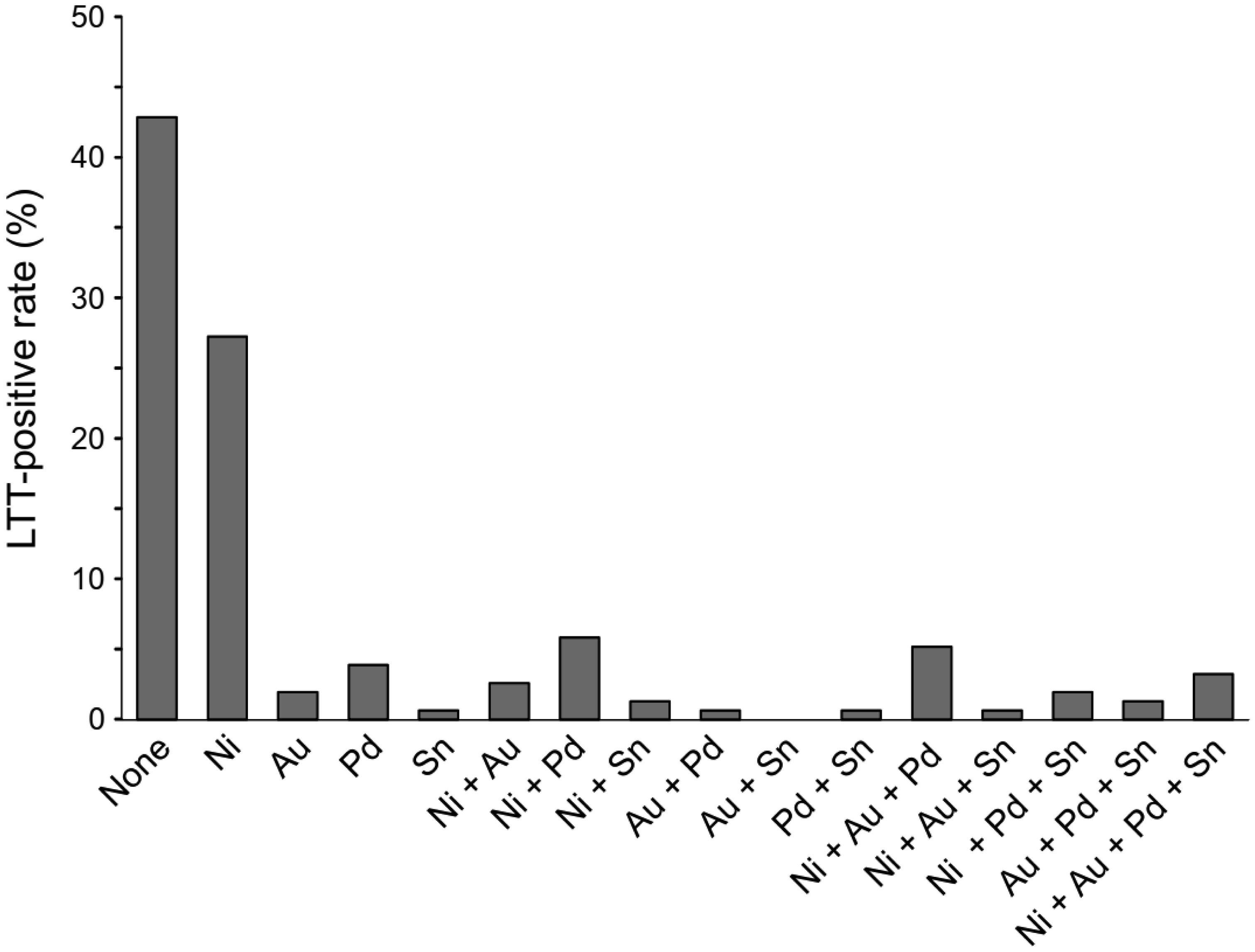
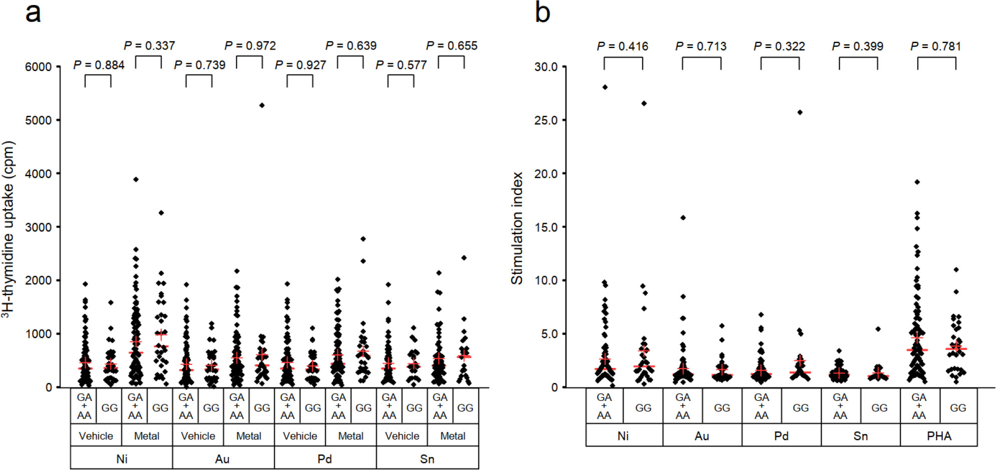
The efficacy of the rs2367563 genotyping test was evaluated with the LTT as a reference for detecting hypersensitivity to dental metals such as nickel, gold, palladium and tin in 154 Japanese participants.
The rs2367563 genotyping test yielded relatively high sensitivity (62.5–80.0%) and low specificity (21.1–26.9%). The overall sensitivity and specificity were 73.9% and 19.7%, respectively.
Genotyping of the rs2367563 involves only simple PCR-based procedures and enables the rapid screening and risk prediction of hypersensitivity to multiple dental metals without any invasive procedure and potential risk of harmful effects on the subjects. However, rs2367563 genotyping is limited to the adjunctive use due to its low specificity, and further improvement in sensitivity and specificity is needed.
Citation: Yasunari Kageyama, Yutaka Shimokawa, Kimihiko Kawauchi, Masafumi Morimoto, Koichi Aida, Tetsu Akiyama, Tsutomu Nakamura. Evaluation of the SNP rs2367563 genotyping test as an adjunctive detection tool for dental metal hypersensitivity[J]. AIMS Allergy and Immunology, 2021, 5(2): 92-101. doi: 10.3934/Allergy.2021008
For the diagnosis of hypersensitivity to dental metals, patch testing has been a gold standard. The lymphocyte transformation test (LTT) has also been introduced for clinical use and established as an alternative method for detecting dental metal hypersensitivity. These tests, however, have some problems such as high false-positive/negative rates. In addition, patch testing involves the potential risk of sensitization primed by test allergens. To overcome these problems, we evaluated a genetic method using the single nucleotide polymorphism (SNP) rs2367563 that was originally identified as a nickel hypersensitivity-associated SNP.
The efficacy of the rs2367563 genotyping test was evaluated with the LTT as a reference for detecting hypersensitivity to dental metals such as nickel, gold, palladium and tin in 154 Japanese participants.
The rs2367563 genotyping test yielded relatively high sensitivity (62.5–80.0%) and low specificity (21.1–26.9%). The overall sensitivity and specificity were 73.9% and 19.7%, respectively.
Genotyping of the rs2367563 involves only simple PCR-based procedures and enables the rapid screening and risk prediction of hypersensitivity to multiple dental metals without any invasive procedure and potential risk of harmful effects on the subjects. However, rs2367563 genotyping is limited to the adjunctive use due to its low specificity, and further improvement in sensitivity and specificity is needed.
lymphocyte transformation test
single nucleotide polymorphism
stimulation index

| [1] |
Thyssen JP, Menne T (2010) Metal allergy—a review on exposures, penetration, genetics, prevalence, and clinical implications. Chem Res Toxicol 23: 309-318. doi: 10.1021/tx9002726

|
| [2] |
Aquino M, Mucci T (2013) Systemic contact dermatitis and allergy to biomedical devices. Curr Allergy Asthma Rep 13: 518-527. doi: 10.1007/s11882-013-0365-9
|
| [3] |
Basko-Plluska JL, Thyssen JP, Schalock PC (2011) Cutaneous and systemic hypersensitivity reactions to metallic implants. Dermatitis 22: 65-79. doi: 10.2310/6620.2011.10055
|
| [4] |
Kouno M, Nishiyama A, Minabe M, et al. (2017) Retrospective analysis of the clinical response of palmoplantar pustulosis after dental infection control and dental metal removal. J Dermatol 44: 695-698. doi: 10.1111/1346-8138.13751
|
| [5] |
Martin MD, Broughton S, Drangsholt M (2003) Oral lichen planus and dental materials: A case-control study. Contact Dermatitis 48: 331-336. doi: 10.1034/j.1600-0536.2003.00146.x
|
| [6] |
Nishizawa A (2016) Dyshidrotic eczema and its relationship to metal allergy. Curr Probl Dermatol 51: 80-85. doi: 10.1159/000446785
|
| [7] |
Bjorklund G, Dadar M, Aaseth J (2018) Delayed-type hypersensitivity to metals in connective tissue diseases and fibromyalgia. Environ Res 161: 573-579. doi: 10.1016/j.envres.2017.12.004
|
| [8] |
Stejskal V, Reynolds T, Bjorklund G (2015) Increased frequency of delayed type hypersensitivity to metals in patients with connective tissue disease. J Trace Elem Med Bio 31: 230-236. doi: 10.1016/j.jtemb.2015.01.001
|
| [9] |
White JM (2012) Patch testing: What allergists should know. Clin Exp Allergy 42: 180-185. doi: 10.1111/j.1365-2222.2011.03862.x
|
| [10] |
Hindsen M, Bruze M, Christensen OB (2001) Flare-up reactions after oral challenge with nickel in relation to challenge dose and intensity and time of previous patch test reactions. J Am Acad Dermatol 44: 616-623. doi: 10.1067/mjd.2001.110873
|
| [11] |
Inerot A, Moller H (2000) Symptoms and signs reported during patch testing. Am J Contact Dermatitis 11: 49-52. doi: 10.1016/S1046-199X(00)90032-0
|
| [12] |
Theler B, Bucher C, French LE, et al. (2009) Clinical expression of nickel contact dermatitis primed by diagnostic patch test. Dermatology 219: 73-76. doi: 10.1159/000212119
|
| [13] |
Tramontana M, Hansel K, Bianchi L, et al. (2018) Flare-up of previously negative patch test and intradermal test with amoxicillin after oral provocation. Contact Dermatitis 79: 250-251. doi: 10.1111/cod.13045
|
| [14] |
Hallab NJ (2004) Lymphocyte transformation testing for quantifying metal-implant-related hypersensitivity responses. Dermatitis 15: 82-90. doi: 10.2310/6620.2004.03054
|
| [15] |
Spoerri I, Bircher AJ, Link S, et al. (2018) Delayed-type allergy to cobalt-comparison of a flow cytometric lymphocyte proliferation test with patch testing. Contact Dermatitis 79: 31-33. doi: 10.1111/cod.12990
|
| [16] |
Spoerri I, Scherer K, Michel S, et al. (2015) Detection of nickel and palladium contact hypersensitivity by a flow cytometric lymphocyte proliferation test. Allergy 70: 323-327. doi: 10.1111/all.12553
|
| [17] |
Stander S, Oppel E, Thomas P, et al. (2017) Evaluation of lymphocyte transformation tests as compared with patch tests in nickel allergy diagnosis. Contact Dermatitis 76: 228-234. doi: 10.1111/cod.12751
|
| [18] |
Cederbrant K, Hultman P, Marcusson JA, et al. (1997) In vitro lymphocyte proliferation as compared to patch test using gold, palladium and nickel. Int Arch Allergy Imm 112: 212-217. doi: 10.1159/000237456
|
| [19] |
Vamnes JS, Gjerdet NR, Morken T, et al. (1999) In vitro lymphocyte reactivity to gold compounds in the diagnosis of contact hypersensitivity. Contact Dermatitis 41: 156-160. doi: 10.1111/j.1600-0536.1999.tb06108.x
|
| [20] |
Kim DS, Kim DH, Lee H, et al. (2013) A genome-wide association study in Koreans identifies susceptibility loci for allergic nickel dermatitis. Int Arch Allergy Imm 162: 184-186. doi: 10.1159/000353235
|
| [21] |
Kitagawa M, Murakami S, Akashi Y, et al. (2019) Current status of dental metal allergy in Japan. J Prosthodont Res 63: 309-312. doi: 10.1016/j.jpor.2019.01.003
|
| [22] |
Muris J, Goossens A, Goncalo M, et al. (2015) Sensitization to palladium and nickel in europe and the relationship with oral disease and dental alloys. Contact Dermatitis 72: 286-296. doi: 10.1111/cod.12327
|
| [23] |
Raap U, Stiesch M, Reh H, et al. (2009) Investigation of contact allergy to dental metals in 206 patients. Contact Dermatitis 60: 339-343. doi: 10.1111/j.1600-0536.2009.01524.x
|
| [24] |
Yamaguchi H, Kabashima-Kubo R, Bito T, et al. (2013) High frequencies of positive nickel/cobalt patch tests and high sweat nickel concentration in patients with intrinsic atopic dermatitis. J Dermatol Sci 72: 240-245. doi: 10.1016/j.jdermsci.2013.07.009
|
| [25] |
Thyssen JP, McFadden JP, Kimber I (2014) The multiple factors affecting the association between atopic dermatitis and contact sensitization. Allergy 69: 28-36. doi: 10.1111/all.12358
|
| [26] | Clark AR, Sherertz EF (1998) The incidence of allergic contact dermatitis in patients with psoriasis vulgaris. Am J Contact Dermatitis 9: 96-99. |
| [27] |
Siddiqi A, Payne AGT, De Silva RK, et al. (2011) Titanium allergy: could it affect dental implant integration? Clin Oral Implan 22: 673-680. doi: 10.1111/j.1600-0501.2010.02081.x
|
| [28] |
Hallab N, Merritt K, Jacobs JJ (2001) Metal sensitivity in patients with orthopaedic implants. J Bone Joint Surg Am 83: 428-436. doi: 10.2106/00004623-200103000-00017
|
| [29] |
Kageyama Y, Aida K, Kawauchi K, et al. (2019) Higher incidence of zinc and nickel hypersensitivity in patients with irritable bowel syndrome. Immun Inflammation Dis 7: 304-307. doi: 10.1002/iid3.274
|
| [30] |
Kageyama Y, Shimokawa Y, Kawauchi K, et al. (2020) Higher prevalence of nickel and palladium hypersensitivity in patients with ulcerative colitis. Int Arch Allergy Imm 181: 456-461. doi: 10.1159/000506633
|
| [31] |
Kano Y, Hirahara K, Mitsuyama Y, et al. (2007) Utility of the lymphocyte transformation test in the diagnosis of drug sensitivity: Dependence on its timing and the type of drug eruption. Allergy 62: 1439-1444. doi: 10.1111/j.1398-9995.2007.01553.x
|
| [32] |
Kanda Y (2013) Investigation of the freely available easy-to-use software ‘EZR’ for medical statistics. Bone Marrow Transpl 48: 452-458. doi: 10.1038/bmt.2012.244
|
| [33] |
Larrieu-Lahargue F, Welm AL, Thomas KR, et al. (2010) Netrin-4 induces lymphangiogenesis in vivo. Blood 115: 5418-5426. doi: 10.1182/blood-2009-11-252338
|
| [34] |
Lejmi E, Leconte L, Pedron-Mazoyer S, et al. (2008) Netrin-4 inhibits angiogenesis via binding to neogenin and recruitment of Unc5B. P Natl Acad Sci USA 105: 12491-12496. doi: 10.1073/pnas.0804008105
|
| [35] |
Liu Y, Stein E, Oliver T, et al. (2004) Novel role for netrins in regulating epithelial behavior during lung branching morphogenesis. Curr Biol 14: 897-905. doi: 10.1016/j.cub.2004.05.020
|
| [36] |
Reuten R, Patel TR, McDougall M, et al. (2016) Structural decoding of netrin-4 reveals a regulatory function towards mature basement membranes. Nat Commun 7: 13515. doi: 10.1038/ncomms13515
|
| [37] |
Staquicini FI, Dias-Neto E, Li J, et al. (2009) Discovery of a functional protein complex of netrin-4, laminin γ1 chain, and integrin α6β1 in mouse neural stem cells. P Natl Acad Sci USA 106: 2903-2908. doi: 10.1073/pnas.0813286106
|
| [38] |
Villanueva AA, Falcon P, Espinoza N, et al. (2017) The netrin-4/neogenin-1 axis promotes neuroblastoma cell survival and migration. Oncotarget 8: 9767-9782. doi: 10.18632/oncotarget.14213
|
| [39] |
Xu X, Yan Q, Wang Y, et al. (2017) NTN4 is associated with breast cancer metastasis via regulation of EMT-related biomarkers. Oncol Rep 37: 449-457. doi: 10.3892/or.2016.5239
|
Figures(2) / Tables(1)

Yasunari Kageyama, Yutaka Shimokawa, Kimihiko Kawauchi, Masafumi Morimoto, Koichi Aida, Tetsu Akiyama, Tsutomu Nakamura. Evaluation of the SNP rs2367563 genotyping test as an adjunctive detection tool for dental metal hypersensitivity[J]. AIMS Allergy and Immunology, 2021, 5(2): 92-101. doi: 10.3934/Allergy.2021008


 DownLoad: