Citation: Ellie Sayers, Alex MacGregor, Simon R. Carding. Drug-microbiota interactions and treatment response: Relevance to rheumatoid arthritis[J]. AIMS Microbiology, 2018, 4(4): 642-654. doi: 10.3934/microbiol.2018.4.642
[1] | Andrew S Day . The impact of exclusive enteral nutrition on the intestinal microbiota in inflammatory bowel disease. AIMS Microbiology, 2018, 4(4): 584-593. doi: 10.3934/microbiol.2018.4.584 |
[2] | T. Amrouche, M. L. Chikindas . Probiotics for immunomodulation in prevention against respiratory viral infections with special emphasis on COVID-19. AIMS Microbiology, 2022, 8(3): 338-356. doi: 10.3934/microbiol.2022024 |
[3] | Mohammad Shamim Ahasan, Thomas B. Waltzek, Leigh Owens, Ellen Ariel . Characterisation and comparison of the mucosa-associated bacterial communities across the gastrointestinal tract of stranded green turtles, Chelonia mydas. AIMS Microbiology, 2020, 6(4): 361-378. doi: 10.3934/microbiol.2020022 |
[4] | Alexandra S McHarg, Steven Leach . The role of the gut microbiome in paediatric irritable bowel syndrome. AIMS Microbiology, 2022, 8(4): 454-469. doi: 10.3934/microbiol.2022030 |
[5] | Le Thanh Dong, Helen V. Espinoza, J. Luis Espinoza . Emerging superbugs: The threat of Carbapenem Resistant Enterobacteriaceae. AIMS Microbiology, 2020, 6(3): 176-182. doi: 10.3934/microbiol.2020012 |
[6] | Harry J. Flint, Petra Louis, Sylvia H. Duncan . Why does increased microbial fermentation in the human colon shift toward butyrate?. AIMS Microbiology, 2024, 10(2): 311-319. doi: 10.3934/microbiol.2024016 |
[7] | Souliphone Sivixay, Gaowa Bai, Takeshi Tsuruta, Naoki Nishino . Cecum microbiota in rats fed soy, milk, meat, fish, and egg proteins with prebiotic oligosaccharides. AIMS Microbiology, 2021, 7(1): 1-12. doi: 10.3934/microbiol.2021001 |
[8] | Constance J. Jeffery . Intracellular/surface moonlighting proteins that aid in the attachment of gut microbiota to the host. AIMS Microbiology, 2019, 5(1): 77-86. doi: 10.3934/microbiol.2019.1.77 |
[9] | Fatemeh Aghighi, Mahmoud Salami . What we need to know about the germ-free animal models. AIMS Microbiology, 2024, 10(1): 107-147. doi: 10.3934/microbiol.2024007 |
[10] | Chunze Zhang, Mingqian Ma, Zhenying Zhao, Zhiqiang Feng, Tianhao Chu, Yijia Wang, Jun Liu, Xuehua Wan . Gut mucosal microbiota profiles linked to development of positional-specific human colorectal cancer. AIMS Microbiology, 2024, 10(4): 812-832. doi: 10.3934/microbiol.2024035 |
With the advent of high-throughput next-generation DNA sequencing (NGS), our knowledge about the characteristics and capabilities of the human intestinal microbiota in relation to disease etiology and progression, has expanded enormously [1]. Microbial dysbiosis; an imbalance in the compositional and/or functional profiles of the intestinal microbiota, has been linked to several conditions including inflammatory bowel disease (IBD), obesity, diabetes, and rheumatoid arthritis (RA) [1,2,3,4,5]. Thus far, connections between microbial profiles and disease states have been associative. The underlying mechanisms by which resident microbes affect the etiology and progression of disease are still largely unknown. Understanding the roles that these microbes play in both the development of disease and in determining the efficacy of various treatments could facilitate the development of new treatments and interventions with the potential to improve long-term disease outcomes.
The intestinal microbiota makes up the largest and most diverse community of resident microbes in the human body [6]. It has a vast array of metabolic capabilities that is more diverse than the enzyme-coding capacity of the human genome. However, the role of the intestinal microbiota in the metabolism of xenobiotics, such as pharmaceuticals, and its subsequent effect on human physiology and treatment responses is little understood. Inter-individual microbiota variability is the largest contributor of variability in microbial community composition; although a core set of microbes and microbial functions are shared among individuals [7]. Host genetics also has a direct impact on interactions between microbes. Furthermore, microbial metabolism of xenobiotics is dependent on the specific microenvironment, including the presence of nutrients or cofactors that are either derived from exogenous sources or provided by other microorganisms in the community. Despite this complexity studies on the microbial metabolism of xenobiotics, particularly pharmaceuticals, have clearly demonstrated that the intestinal microbiota has a strong effect on clinical responses to treatment and drug toxicity [8]. Any orally-administered pharmaceuticals that are not absorbed in the upper GI-tract will eventually come into contact with the intestinal microbiota. Parenterally administered drugs may also contact the intestinal microbiota if they are excreted in bile, a process known as enterohepatic recirculation.
A good example of the importance of interactions between drugs and resident microbial populations can be seen for the vaginal microbiota where the relative abundance of particular bacteria determines the efficacy of the antiretroviral drug, tenofovir. Typically, species in the genus Lactobacillus dominate the vaginal microbiota. However, in around 39% of women taking part in a trial investigating the clinical efficacy of tenofovir, Gardnerella vaginalis replaced Lactobacillus spp. as the dominant species [9]. In those women with a G. vaginalis-dominated vaginal microbiota, concentrations of tenofovir were reduced, possibly because G. vaginalis rapidly metabolizes tenofovir before it is taken up by human cells [9]. A more detailed understanding of the mechanisms driving microbe-drug interactions may therefore be beneficial in the prevention and treatment of a number of diseases where the efficacy of drugs and topical treatments is highly variable. It is also possible that some treatment regimens could be improved by considering individual patients' microbiotas.
Several known mechanisms exist by which the intestinal microbiota can either directly, or indirectly, change the bioavailability and/or efficacy of drugs (Figure 1). Microbial and host metabolism of xenobiotics differ with respect to the type of metabolic reactions they employ. For example, microbes predominantly use reductive and hydrolytic reactions whereas their human hosts predominantly use oxidation and conjugation [8]. Orthologous enzymes for basic processes do exist between microbiota and their hosts, although the microbiota has access to a much larger library of metabolic processes than the host. This is due to the extensive diversity of species within the microbiota, which has been exploited to generate various prodrugs (e.g. sulfasalazine and metronidazole) that are activated by microbial enzymes. Many microbial processes benefit the host, for example, by converting dietary fibre to short-chain fatty acids (SCFAs) that are anti-inflammatory and provide energy to intestinal epithelial cells [10]. In contrast, microbial β-glucuronidase activity directed at the cancer drug irinotecan induces severe toxicity in the form of diarrhea which is dose-limiting and impacts on treatment efficacy [11].
Digoxin is a toxin derived from plants and is used to treat congestive heart failure and arrhythmia. In approximately 10% of patients the bioavailability and efficacy of digoxin is greatly reduced, and is replaced by an increase in cardioinactive, reduced metabolites of digoxin [12]. In the early 1980s it was shown that this lack of bioavailability could be restored by co-administration of antibiotics that depleted the intestinal microbiota [12]. Later, the same group identified the digoxin-inactivating properties of a single species of bacterium, Eggerthella lenta, that was present in the intestinal microbiota; this is the only known organism capable of reducing digoxin to dihydrodigoxin, thereby rendering it inactive [13]. E. lenta is a common member of the intestinal microbiota, but its presence alone is not sufficient to predict digoxin inactivation; only specific strains containing the cgr1 and cgr2 genes can reduce digoxin to dihydrodigoxin and the presence of these genes can be used as a marker for potential clinical non-response [14]. Interestingly, the cgr1/cgr2 operon is inhibited by the presence of the amino acid, arginine. In mice given digoxin and fed diets high in protein the concentrations of digoxin in the serum and urine were increased. This suggests that dietary supplementation with protein (or arginine) may increase digoxin bioavailability by preventing its reduction by E. lenta [14]. Supplementation with arginine may be useful to increase the bioavailability and efficacy of digoxin in people with E. lenta carrying cgr1/cgr2.
A prodrug is an inactive medication or compound that, after administration, is converted by enzymatic action into a pharmacologically active drug. Sulfasalazine (SSZ) is a prodrug used to treat inflammation in patients with IBD or RA. It is converted into an active form by the intestinal microbiota via azo reductase, an enzyme that cleaves SSZ into sulfapyridine (a potentially toxic by-product) and 5-aminosalicyclic acid (5-ASA/mesalazine) which is the main anti-inflammatory component. Azo reductases are produced by a variety of intestinal bacteria, including species of Clostridium, Eubacterium and Bacteroides [15,16]. The majority of 5-ASA is retained in the colon undergoing enterohepatic recirculation and is eventually excreted in feces, making it ideal for the treatment of IBD [17]. The abundance of azo reductase enzymes in the intestinal microbiota has not been formally measured but could potentially have an impact on treatment responses to this drug. Although one product of microbial metabolism of SSZ is active as an anti-inflammatory, the other (sulfapyridine) has an antimicrobial effect and may also be mutagenic [18]. 5-ASA/mesalazine is usually given as a standalone drug for treatment of IBD to avoid the side-effects associated with sulfapyridine. Despite this, oral mesalazine is not easily absorbed systemically, making SSZ the preferred drug for treatment of RA. However, the side-effects of SSZ may be poorly tolerated in RA patients and include nausea, headache, and gastrointestinal pain. More serious side-effects include anemia, kidney stones, and liver disease [19].
The antibiotic metronidazole is used in the treatment of various anaerobic bacterial infections and sometimes Crohn's disease, and is activated by the microbes that eventually become its target. The inactive drug is transported into bacterial cells and activated by nitro reductase under anaerobic or microaerophilic conditions, making it specific to low-oxygen environments such as the large intestine [20]. Activation leads to the generation of free radicals, mutagenicity and the formation of acetamide, a potentially carcinogenic by-product [21]. Metabolites of metronidazole lead to bacterial cell death and are expelled in feces with the possibility of contaminating the environment [22].
Immune checkpoint inhibitors (ICIs) are drugs that block the action of the particular inhibitory proteins that prevent T cells from killing cancer cells. For example, the combined effect of the protein PD-L1 (CD274) on tumor cells and the protein PD-1 (CD279) on T cells prevents the T cells from killing tumor cells [23]. Similarly, the protein CTLA4, which is expressed by activated T cells, acts as an “off switch” when bound to CD80 or CD86 on antigen-presenting cells [23]. These blocks to the tumoricidal activity of T cells can be overcome with ICIs and monoclonal antibodies that block the interactions of inhibitory proteins with T cells, enabling T cells to attack cancer cells.
The efficacy (as anti-cancer agents) and toxicity of ICIs with anti-PD-1, anti-PD-L-1 and anti-CTLA-4 activity has been associated with the presence or absence of particular microorganisms in the intestinal microbiota [24,25,26,27,28]. In germ-free or antibiotic-treated conventional mice, attempts to block CTLA-4 interactions with CD80/86 using anti-CTLA-4 antibodies are ineffective at activating T-cells and reducing tumor progression, suggesting that the intestinal microbiota is required for the beneficial effect of blocking CTLA-4 [28]. The introduction of Bacteroides fragilis to germ-free mice restored their ability to respond to anti-CTLA-4 antibodies by increasing T-cell responses close to the tumor site. The higher abundance of members from the phylum Bacteroidetes was also associated with protection from anti-CTLA-4-induced colitis, further confirming the benefits that these bacteria could have in anti-CTLA-4 antibody therapy [24]. Pre-treatment abundance of Akkermansia muciniphila in the intestinal microbiota of patients undergoing anti-PD-1 therapy, and co-administration of Bifidobacterium species with anti-PD-L-1 therapy, increased treatment efficacy via a similar mechanism to that for anti-CTLA-4 antibodies [25,26]. It is therefore likely that the presence of particular intestinal bacteria could be helpful in increasing the efficacy of drugs targeting immune system pathways, or where stimulation of immunity is important.
Aside from xenometabolism and the presence of treatment-enhancing bacteria that change drug bioavailability and efficacy, it is possible that drug-induced changes in the composition and function of the intestinal microbiota could also be an important mechanism influencing disease progression. One such example of this is metformin, a commonly prescribed drug for treatment of hyperglycemia in patients with type 2 diabetes (T2D). A central role for the GI-tract and/or the intestinal microbiota in metformin activity is supported by the reduced efficacy of intravenous metformin, and of oral metformin after the administration of antibiotics [29,30]. The mechanisms involved in the glucose-lowering effect of metformin are still unclear, although its primary action is thought to be via inhibition of gluconeogenesis in the liver via activation of AMP-activated protein kinase (AMPK), which alters the expression of gluconeogenesis-associated genes [31]. Like many other chronic diseases, T2D is associated with intestinal microbial dysbiosis [32]. The beneficial effects of metformin may also be achieved via manipulation of the microbiota, for example, by enhancing the growth of mucin-degrading and probiotic Akkermansia species [29]. Longitudinal studies of the microbiotas of mice and humans following metformin administration showed an increase in abundance of Akkermansia species and other mucin-degrading taxa such as Clostridium cocleatum and Bifidobacterium bifidum which also produce beneficial SCFA [33,34,35]. Glucose tolerance was improved in mice on a high-fat diet when they were orally administered Akkermansia species. This suggests that increased abundance of Akkermansia species following metformin treatment contributes to this drug's mode of action [29]. Additionally, Akkermansia inhibits hyperglycemia and increases expression of peptides involved in glucose homeostasis [36].
With changes in microbial diversity and composition come changes in the metabolic capabilities of the microbiota, potentiating differences in its xenobiotic capabilities. Healthy individuals may respond uniquely to drug treatments based on the community of species present in their microbiota. When the microbiota is dysbiotic, variability in responses could increase with unknown consequences for drug efficacy. In this section we will use current knowledge to discuss the possible mechanisms for drug-microbiota interactions in the treatment of early RA with methotrexate (MTX).
RA is a chronic debilitating disease of unknown etiology in which the synovial lining of the joints becomes inflamed. Changes in the intestinal microbiota of patients with RA include an increased abundance of Prevotella copri during early disease, an increase in abundance of Lactobacillus species, and a decrease in abundance of Bacteroides species. However, results from different studies are inconsistent, most likely as a consequence of differences in study design and methodology (particularly sequencing and analysis), and/or the characteristics of the subject population [2,3,4,5]. Current treatments available for RA include a range of disease-modifying anti-rheumatic drugs (DMARDs) of which MTX is usually the first-line treatment.
Clinical responses to MTX are highly variable with 50% of patients achieving a 20% reduction in their swollen-and-tender-joint count (American College of Rheumatology 20; ACR20 response) and 31% achieving a good response after 3 months according to the criteria of the European League Against Rheumatism (EULAR) [37]. Despite investigations into the predictive capabilities of age, sex, BMI, smoking status, genetics and serum biomarkers, there are currently no reliable indicators of MTX treatment response [38]. Recently, differences in the composition of the intestinal microbiota before MTX treatment were shown to be correlated with clinical responses after 3 months of treatment, suggesting that the intestinal microbiota played a role in determining clinical response [4]. Additionally, compositional changes in the microbiota were induced by MTX, raising the possibility that changes in the microbiota are involved in the drug's mode of action.
Once acquired into a cell, MTX is polyglutamated by folylpolyglutamate synthase (FPGS) to MTX-PG, which is thought to be the more potent than MTX itself (Figure 2). The concentration of various lengths of MTX-PGs in red blood cells is associated with treatment responses [39,40]. After removal of the polyglutamate tail by either folate hydrolase or glutamate carboxypeptidase II, MTX-PG may then be exported. Inside the cell, MTX and MTX-PG act as competitive inhibitors of dihydrofolic reductase (DHFR), leading to a decrease in the number of metabolites required for the generation of compounds involved in DNA synthesis; this is essentially an induced folate deficiency. MTX-PG also inhibits 5-aminoimidazole-4-carboxamide ribonucleotide (AICAR) transformylase, which causes accumulating adenosine to leave the cell and bind receptors on surrounding cells, thus delivering an anti-inflammatory effect [41]. As the folate pathway is not limited to humans, it is possible that the assimilation or metabolism of MTX by intestinal bacteria could influence MTX bioavailability and efficacy.
After internalisation via one of the various folate transporters, MTX is polyglutamated by the enzyme FPGS into a variety of MTX-PGs in humans. MTX-PG has a higher affinity for its target proteins than MTX, but also a lower affinity for folate transporters than MTX. This means that MTX-PGs are poorly transported in and out of cells. Removal of glutamate entities from MTX-PGs by carboxypeptidase reduces its efficacy as an inhibitor of DHFR. A carboxypeptidase expressed by Pseudomonas species, which cleaves glutamate entities from MTX (CPG2), was identified in 1967 [42]. Previous studies had demonstrated assimilation and metabolism of folic acid and its analogues by various bacteria and yeasts, including Streptococcus faecalis, Enterobacter aerogenes and Candida tropicalis [43]. CPG2 is now used as a rescue agent for high-dose MTX toxicity as it rapidly reduces the number of MTX-PGs in the blood, reversing the induced folate deficiency. CPG2 has orthologous enzymes in other bacterial species, such as p-aminobenzoyl-glutamate hydrolase (PGH) in E. coli, which can also act on MTX [44]. These enzymes have essential functions in folate-requiring bacteria which could potentially assimilate and metabolize MTX, thereby altering its efficacy. In addition to removal of glutamate via carboxypeptidase, some intestinal bacteria have the capacity to add glutamate to MTX via FPGS-like enzymes. In a study investigating the impact of MTX on the metabolism of a panel of 25 intestinal microbes, at least two species could metabolize MTX to MTX-PG [45].
As MTX-PG is not transported as easily as MTX, is it unlikely that intestinal bacteria-generated MTX-PG would affect treatment outcomes because the MTX-PG would likely be retained by the cell or converted back to MTX prior to export. Additionally, removal of glutamate entities from MTX via, for instance, Pseudomonas' CPG2, would be unlikely to decrease efficacy of MTX as it would not affect post-absorption formation of MTX-PGs. However, switching from oral to parenteral administration of MTX leads to a significant increase in long and very long-chain MTX-PGs, suggesting that the oral route may inhibit formation of MTX-PGs, possibly via assimilation of MTX by the microbiota [46].
To date only one study has investigated the influence of the microbiota on determining treatment outcomes in response to MTX [4]. Shotgun metagenomics was used to characterize the salivary, dental, and fecal microbiotas of treatment-naïve RA patients before and after the use of MTX in combination with other DMARDs [4]. Differentially-abundant species were identified in baseline samples from patients that were responding well, moderately or not at all (based on DAS28 reduction) and it was found that MTX changed the composition of the fecal microbiota. In particular, four RA-enriched unclassifiable taxa and one species that was most closely related to Enterococcus faecium, decreased in abundance following treatment, suggesting that MTX may act either by decreasing the abundance of disease-associated bacteria or by restoring a “healthy” microbiota. Notably, there were more changes to the composition of the dental and salivary microbiota than to the composition of the fecal microbiota; there were increases in several healthy control-associated taxa in the dental and salivary microbiota following treatment, suggesting that these microenvironments are more susceptible to MTX-induced changes and may also be involved in treatment responses. This study also generated predictive models using the microbiota data collected, which were able to differentiate between good and poor responders. These observations provide evidence that microbiota-based variables may be important in determining whether an RA patient responds well or poorly to treatment with MTX, although the underlying mechanisms remain unknown.
MTX treatment failure in RA patients may lead to the use of other DMARDs or biological treatments such as TNF inhibitors, which also carry a variable treatment response and lack reliable predictive factors [38]. No studies have specifically linked microbiota profiles with treatment responses to TNF inhibitors in RA patients. However, investigations with IBD patients suggest partial recovery of microbial dysbiosis is induced by TNF inhibitors, and response-associated baseline microbiota profiles have been identified in patients with spondyloarthritis [47,48]. The possibility of using microbiota signatures to guide treatment (TNF inhibitors) decisions, and the effect of various lifestyle factors on treatment outcomes, is currently being investigated in a prospective cohort study in Denmark [49]. In this study the collection of several lifestyle and dietary variables alongside microbiota data will allow both identification of predictive markers in the microbiota, and also the associated lifestyle factors that can be modified to improve treatment efficacy. This is an important step forward in translating microbiota studies into the clinical management of chronic inflammatory diseases such as RA.
Our understanding of the various mechanisms by which the human intestinal microbiota may contribute to treatment responses is expanding. However, more mechanistic studies are needed to unravel the ways in which bacteria and other microbes interact with drugs to influence their bioavailability and efficacy. Associations between the microbiota and treatment responses (of which several are known) need to be followed up with mechanistic in vitro and in vivo studies to identify the specific taxa involved and define their interactions with particular drugs, the environment, and the host. From a clinical perspective, elucidation of these mechanisms could greatly improve personalized medicine because it would reduce the costs and harm to patients that is caused by “trial and error” use of drugs (such as MTX for RA patients) with highly variable treatment responses. Using fecal samples as a surrogate for the intestinal microbiota, it is possible to profile and identify the composition of the microbiota prior to treatment which could inform clinical decisions on which treatment and/or adjuvants to use, thus improving clinical response and patient quality of life. Known organisms associated with good or poor responses could be targeted. For example, prebiotics, probiotics, fecal microbiota transplant, or personalized nutrition could introduce and maintain beneficial microbes, or antibiotics could be used selectively to remove unwanted microbial populations.
Funding in support of this work was provided by the UK Biotechnology and Biological Sciences Research Council (BBSRC) under grant number BB/J004529/1 (SRC) and by a postgraduate studentship awarded by the Faculty of Medicine and Health Sciences, University of East Anglia (ES).
The authors declare no conflicts of interest in this paper.
[1] | Carding S, Verbeke K, Vipond DT, et al. (2015) Dysbiosis of the gut microbiota in disease. Microb Ecol Heal Dis 26: 26191–26199. |
[2] |
Scher JU, Sczesnak A, Longman RS, et al. (2013) Expansion of intestinal Prevotella copri correlates with enhanced susceptibility to arthritis. Elife 2: e01202. doi: 10.7554/eLife.01202
![]() |
[3] |
Liu X, Zou Q, Zeng B, et al. (2013) Analysis of fecal Lactobacillus community structure in patients with early rheumatoid arthritis. Curr Microbiol 67: 170–176. doi: 10.1007/s00284-013-0338-1
![]() |
[4] |
Zhang X, Zhang D, Jia H, et al. (2015) The oral and gut microbiotas are perturbed in rheumatoid arthritis and partly normalized after treatment. Nat Med 21: 895–905. doi: 10.1038/nm.3914
![]() |
[5] |
Chen J, Wright K, Davis JM, et al. (2016) An expansion of rare lineage intestinal microbes characterizes rheumatoid arthritis. Genome Med 8: 43–51. doi: 10.1186/s13073-016-0299-7
![]() |
[6] |
Sender R, Fuchs S, Milo R (2016) Revised estimates for the number of human and bacteria cells in the body. PLoS Biol 14: e1002533. doi: 10.1371/journal.pbio.1002533
![]() |
[7] |
Falony G, Joossens M, Vieira-Silva S, et al. (2016) Population-level analysis of gut microbiota variation. Science 352: 560–564. doi: 10.1126/science.aad3503
![]() |
[8] | Koppel N, Rekdal VM, Balskus EP (2017) Chemical transformation of xenobiotics by the human gut microbiota. Science 356: 1246–1257. |
[9] |
Klatt NR, Cheu R, Birse K, et al. (2017) Vaginal bacteria modify HIV tenofovir microbicide efficacy in African women. Science 356: 938–945. doi: 10.1126/science.aai9383
![]() |
[10] |
Koh A, De Vadder F, Kovatcheva-Datchary P, et al. (2016) From dietary fiber to host physiology: Short-chain fatty acids as key bacterial metabolites. Cell 165: 1332–1345. doi: 10.1016/j.cell.2016.05.041
![]() |
[11] | Takasuna K, Hagiwara T, Hirohashi M, et al. (1996) Involvement of b-glucuronidase in intestinal microflora in the intestinal toxicity of the antitumor camptothecin derivative irinotecan hydrochloride (CPT-11 ) in rats. Cancer Res 56: 3752–3757. |
[12] |
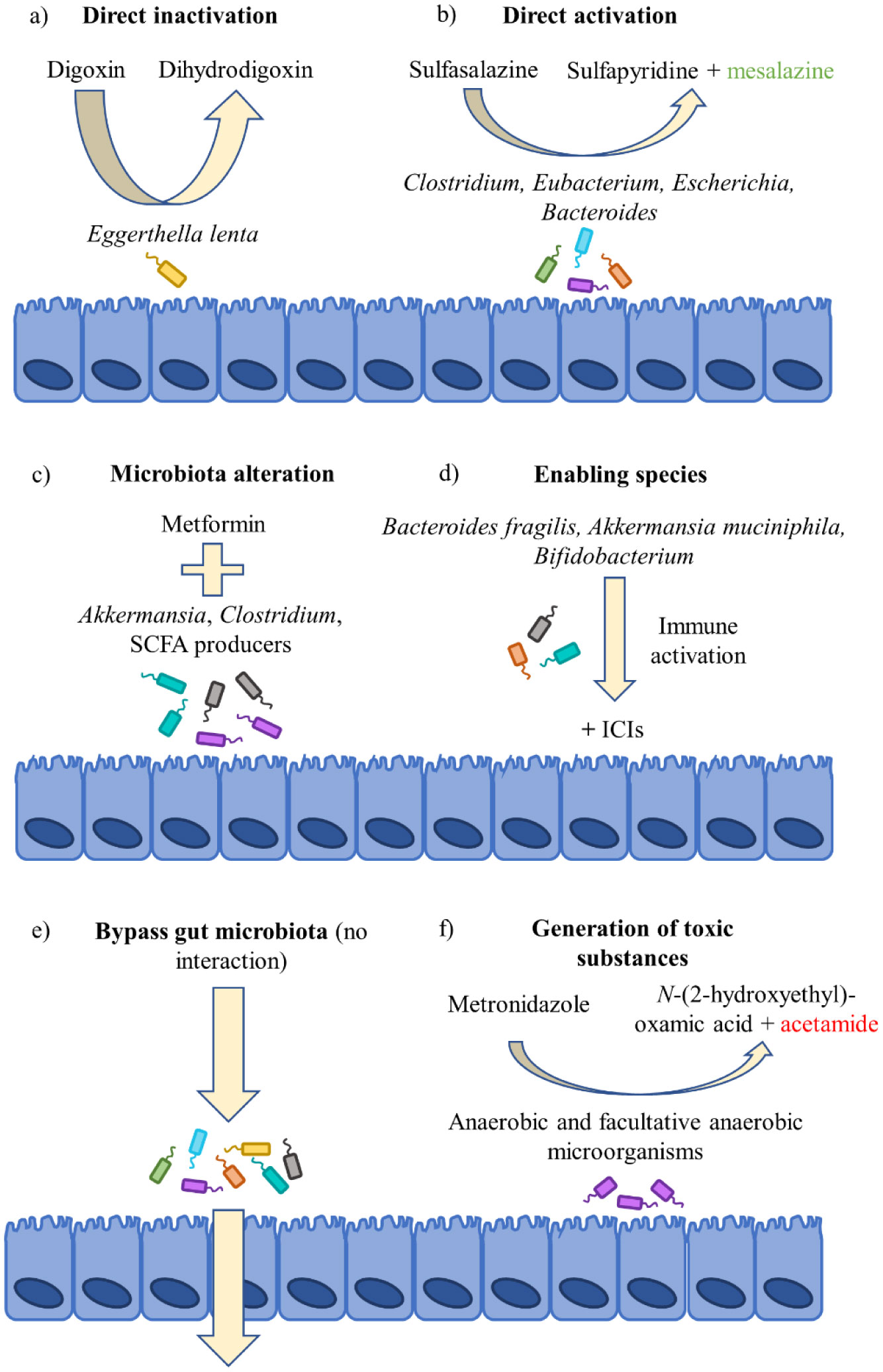
Lindenbaum J, Rund DG, Butler VP, et al. (1981) Inactivation of digoxin by the gut flora: Reversal by antibiotic therapy. N Engl J Med 305: 789–794. doi: 10.1056/NEJM198110013051403
![]() |
[13] | Dobkin J, Saha J, Butler V, et al. (1982) Digoxin-inactivating bacteria: Identification in human gut flora. Science 220: 325–327. |
[14] |
Haiser HJ, Gootenberg DB, Chatman K, et al. (2013) Predicting and manipulating cardiac drug inactivation by the human gut bacterium Eggerthella lenta. Science 341: 295–298. doi: 10.1126/science.1235872
![]() |
[15] | Rafii F, Franklin W, Cerniglia CE (1990) Azoreductase activity of anaerobic bacteria isolated from human intestinal microflora. Appl Environ Microbiol 7: 2146–2151. |
[16] | Rafii F, Cerniglia CE (1995) Reduction of azo dyes and nitroaromatic compounds by bacterial enzymes from the human intestinal tract. Environ Health Persp 103: 17–19. |
[17] |
Azadkhan A, Truelove S, Aronson J (1982) The disposition and metabolism of sulphasalazine (salicylazosulphapyridine) in man. Brit J Clin Pharmaco 13: 523–528. doi: 10.1111/j.1365-2125.1982.tb01415.x
![]() |
[18] |
Bishop JB, Witt KL, Gulati DK, et al. (1990) Evaluation of the mutagenicity of the anti-inflammatory drug salicylazosulfapyridine (SASP). Mutagenesis 5: 549–554. doi: 10.1093/mutage/5.6.549
![]() |
[19] |
Das KM, Eastwood MA, McManus JPA, et al. (1973) Adverse reactions during salicylazosulfapyridine therapy and the relation with drug metabolism and acetylator phenotype. N Engl J Med 289: 491–495. doi: 10.1056/NEJM197309062891001
![]() |
[20] | Dingsdag SA, Hunter N (2017) Metronidazole: an update on metabolism, structure-cytotoxicity and resistance mechanisms. J Antimicrob Chemoth 73: 265–279. |
[21] |
Bendesky A, Menéndez D, Ostrosky-Wegman P (2002) Is metronidazole carcinogenic? Mutat Res-Rev Mutat 511: 133–144. doi: 10.1016/S1383-5742(02)00007-8
![]() |
[22] |
Chrystal EJT, Koch RL, McLafferty MA, et al. (1980) Relationship between metronidazole metabolism and bactericidal activity. Antimicrob Agents Ch 18: 566–573. doi: 10.1128/AAC.18.4.566
![]() |
[23] |
Pardoll DM (2012) The blockade of immune checkpoints in cancer immunotherapy. Nat Rev Cancer 12: 252–264. doi: 10.1038/nrc3239
![]() |
[24] |
Dubin K, Callahan MK, Ren B, et al. (2016) Intestinal microbiota analyses identify melanoma patients at risk for checkpoint-blockade-induced colitis. Nat Commun 7: 10391–10397. doi: 10.1038/ncomms10391
![]() |
[25] |
Sivan A, Corrales L, Hubert N, et al. (2015) Commensal Bifidobacterium promotes antitumor immunity and facilitates anti-PD-L1 efficacy. Science 350: 1084–1089. doi: 10.1126/science.aac4255
![]() |
[26] |
Routy B, Le Chatelier E, Derosa L, et al. (2018) Gut microbiota influences efficacy of PD-1-based immunotherapy against epithelial tumors. Science 359: 91–97. doi: 10.1126/science.aan3706
![]() |
[27] |
Matson V, Fessler J, Bao R, et al. (2018) The commensal microbiota is associated with anti- PD-1 efficacy in metastatic melanoma patients. Science 359: 104–108. doi: 10.1126/science.aao3290
![]() |
[28] |
Vétizou M, Pitt JM, Daillère R, et al. (2015) Anticancer immunotherapy by CTLA-4 blockade relies on the gutmicrobiota. Science 350: 1079–1084. doi: 10.1126/science.aad1329
![]() |
[29] | Shin NR, Lee JC, Lee HY, et al. (2014) An increase in the Akkermansia spp. population induced by metformin treatment improves glucose homeostasis in diet-induced obese mice. Gut 63: 727–735. |
[30] |
Bonora E, Cigolini M, Bosello O, et al. (1984) Lack of effect of intravenous metformin on plasma concentrations of glucose, insulin, C-peptide, glucagon and growth hormone in non-diabetic subjects. Curr Med Res Opin 9: 47–51. doi: 10.1185/03007998409109558
![]() |
[31] |
Foretz M, Guigas B, Bertrand L, et al. (2014) Metformin: From mechanisms of action to therapies. Cell Metab 20: 953–966. doi: 10.1016/j.cmet.2014.09.018
![]() |
[32] |
Qin J, Li Y, Cai K, et al. (2012) A metagenome-wide association study of gut microbiota in type 2 diabetes. Nature 490: 55–60. doi: 10.1038/nature11450
![]() |
[33] |
Forslund K, Hildebrand F, Nielsen T, et al. (2015) Disentangling type 2 diabetes and metformin treatment signatures in the human gut microbiota. Nature 528: 262–266. doi: 10.1038/nature15766
![]() |
[34] |
De La Cuesta-Zuluaga J, Mueller NT, Corrales-Agudelo V, et al. (2017) Metformin is associated with higher relative abundance of mucin-degrading akkermansia muciniphila and several short-chain fatty acid-producing microbiota in the gut. Diabetes Care 40: 54–62. doi: 10.2337/dc16-1324
![]() |
[35] |
Lee H (2014) Effect of metformin on metabolic improvement and gut microbiota. Appl Environ Microbiol 80: 5935–5943. doi: 10.1128/AEM.01357-14
![]() |
[36] |
Everard A, Belzer C, Geurts L, et al. (2013) Cross-talk between Akkermansia muciniphila and intestinal epithelium controls diet-induced obesity. P Natl Acad Sci USA 110: 9066–9071. doi: 10.1073/pnas.1219451110
![]() |
[37] | Lie E, Uhlig T, van der Heijde D, et al. (201) Effectiveness of sulfasalazine and methotrexate in 1102 DMARD-nave patients with early RA. Rheumatology 51: 670–678. |
[38] |
Wijbrandts CA, Tak PP (2017) Prediction of response to targeted treatment in rheumatoid arthritis. Mayo Clin Proc 92: 1129–1143. doi: 10.1016/j.mayocp.2017.05.009
![]() |
[39] |
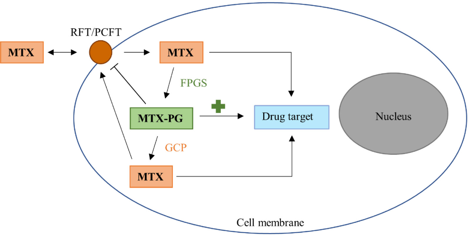
Cutulo M, Sulli A, Pizzorni C, et al. (2001) Anti-inflammatory mechanisms of methotrexate in rheumatoid arthritis. Ann Rheum Dis 60: 729–735. doi: 10.1136/ard.60.8.729
![]() |
[40] |
Takahashi C, Kaneko Y, Okano Y, et al. (2017) Association of erythrocyte methotrexate- polyglutamate levels with the efficacy and hepatotoxicity of methotrexate in patients with rheumatoid arthritis : a 76-week prospective study. RMD Open 3: e000363. doi: 10.1136/rmdopen-2016-000363
![]() |
[41] | Tian H, Cronstein BN (2007) Understanding the mechanisms of action of methotrexate-Implications for the treatment of rheumatoid arthritis. Bull NYU Hosp Jt Dis 65: 168–173. |
[42] | Levy CC, Goldman P (1967) The enzymatic hydrolysis of methotrexate. J Biol Chem 242: 2933–2938. |
[43] |
Webb M (1955) Inactivation of analogues of folic acid by certain non-exacting bacteria. Biochem Biophys Acta 17: 212–225. doi: 10.1016/0006-3002(55)90352-8
![]() |
[44] |
Larimer CM, Slavnic D, Pitstick LD, et al. (2014) Comparison of substrate specificity of Escherichia coli p-Aminobenzoyl-glutamate hydrolase with Pseudomonas carboxypeptidase G. Adv Enzym Res 2: 39–48. doi: 10.4236/aer.2014.21004
![]() |
[45] | Nayak R, O'Loughlin C, Fischbach M, et al. (2016) Methotrexate is an antibacterial drug metabolized by human gut bacteria-ACR Meeting Abstracts. Arthritis Rheumatol 68: 10. |
[46] |
Dervieux T, Zablocki R, Kremer J (2010) Red blood cell methotrexate polyglutamates emerge as a function of dosage intensity and route of administration during pulse methotrexate therapy in rheumatoid arthritis. Rheumatology 49: 2337–2345. doi: 10.1093/rheumatology/keq216
![]() |
[47] |
Busquets D, Mas-de-Xaxars T, López-Siles M, et al. (2015) Anti-tumour necrosis factor treatment with adalimumab induces changes in the microbiota of Crohn's Disease. J Crohns Colitis 9: 899–906. doi: 10.1093/ecco-jcc/jjv119
![]() |
[48] |
Bazin T, Hooks KB, Barnetche T, et al. (2018) Microbiota composition may predict anti-TNF alpha response in spondyloarthritis patients: An exploratory study. Sci Rep 8: 5446–5449. doi: 10.1038/s41598-018-23571-4
![]() |
[49] |
Andersen V, Holmskov U, Sørensen SB, et al. (2017) Proposal for a study on treatment selection and lifestyle recommendations in chronic inflammatory diseases: A Danish multidisciplinary collaboration on prognostic factors and personalised medicine. Nutrients 9: 499–508. doi: 10.3390/nu9050499
![]() |
1. | Vinod Chandran, Proton Rahman, Predicting therapeutic response through biomarker analysis in psoriatic arthritis, an example of precision medicine, 2020, 5, 2380-8993, 35, 10.1080/23808993.2020.1724509 | |
2. | Malavikalakshmi M. Attur, Jose U. Scher, 2019, Chapter 2, 978-3-030-23310-5, 11, 10.1007/978-3-030-23311-2_2 | |
3. | Martina Franzin, Katja Stefančič, Marianna Lucafò, Giuliana Decorti, Gabriele Stocco, Microbiota and Drug Response in Inflammatory Bowel Disease, 2021, 10, 2076-0817, 211, 10.3390/pathogens10020211 | |
4. | Ignacio Dei-Cas, Florencia Giliberto, Leonela Luce, Hernán Dopazo, Alberto Penas-Steinhardt, Metagenomic analysis of gut microbiota in non-treated plaque psoriasis patients stratified by disease severity: development of a new Psoriasis-Microbiome Index, 2020, 10, 2045-2322, 10.1038/s41598-020-69537-3 | |
5. | Xiao-Yu Cai, Yue Zhu, Chen Wang, Xiao-Yu Tang, Le Han, Jin-Ling Shu, Xian-Zheng Zhang, Fa-Qin Liang, Jing-Ru Ge, Li Xu, Dan Mei, Ling-Ling Zhang, Wei Wei, Etanercept Inhibits B Cell Differentiation by Regulating TNFRII/TRAF2/NF-κB Signaling Pathway in Rheumatoid Arthritis, 2020, 11, 1663-9812, 10.3389/fphar.2020.00676 | |
6. | Anny Camelo-Castillo, José Miguel Rivera-Caravaca, Esteban Orenes-Piñero, Inmaculada Ramírez-Macías, Vanessa Roldán, Gregory Y. H. Lip, Francisco Marín, Gut Microbiota and the Quality of Oral Anticoagulation in Vitamin K Antagonists Users: A Review of Potential Implications, 2021, 10, 2077-0383, 715, 10.3390/jcm10040715 | |
7. | Ryoichi Hashida, Yasunori Shimozuru, Jessica Chang, Ibis Agosto-Marlin, Takaki Waritani, Kuniaki Terato, Veena Taneja, New Studies of Pathogenesis of Rheumatoid Arthritis with Collagen-Induced and Collagen Antibody-Induced Arthritis Models: New Insight Involving Bacteria Flora, 2021, 2021, 2090-0430, 1, 10.1155/2021/7385106 | |
8. | Ana L. Carvalho, Christian M. Hedrich, The Molecular Pathophysiology of Psoriatic Arthritis—The Complex Interplay Between Genetic Predisposition, Epigenetics Factors, and the Microbiome, 2021, 8, 2296-889X, 10.3389/fmolb.2021.662047 | |
9. | Nadji Hannachi, Laurence Camoin-Jau, Drug Response Diversity: A Hidden Bacterium?, 2021, 11, 2075-4426, 345, 10.3390/jpm11050345 | |
10. | Maja Skov Kragsnaes, Jens Kjeldsen, Hans Christian Horn, Heidi Lausten Munk, Jens Kristian Pedersen, Søren Andreas Just, Palle Ahlquist, Jesper Rømhild Davidsen, Anna Christine Nilsson, Richard Röttger, Mogens Kruhøffer, Julian R Marchesi, Karsten Kristiansen, Robin Christensen, Torkell Ellingsen, Response to: ‘Correspondence on ‘Safety and efficacy of faecal microbiota transplantation for active peripheral psoriatic arthritis: an exploratory randomised placebo-controlled trial’’ by McGonagleet al, 2021, 0003-4967, annrheumdis-2021-220910, 10.1136/annrheumdis-2021-220910 | |
11. | Chiara Montanari, Sara Parolisi, Elisa Borghi, Lorenza Putignani, Giulia Bassanini, Juri Zuvadelli, Cristina Bonfanti, Albina Tummolo, Carlo Dionisi Vici, Giacomo Biasucci, Alberto Burlina, Maria Teresa Carbone, Elvira Verduci, Dysbiosis, Host Metabolism, and Non-communicable Diseases: Trialogue in the Inborn Errors of Metabolism, 2021, 12, 1664-042X, 10.3389/fphys.2021.716520 | |
12. | Min Wen, Taohua Liu, Mingyi Zhao, Xiqiang Dang, Shipin Feng, Xuewei Ding, Zhiquan Xu, Xiaoyan Huang, Qiuyu Lin, Wei Xiang, Xiaoyan Li, Xiaojie He, Qingnan He, Roberta Antonia Diotti, Correlation Analysis between Gut Microbiota and Metabolites in Children with Systemic Lupus Erythematosus, 2021, 2021, 2314-7156, 1, 10.1155/2021/5579608 | |
13. | Jung Hee Koh, Eun Ha Lee, Kwang Hyun Cha, Cheol-Ho Pan, Donghyun Kim, Wan-Uk Kim, Factors associated with the composition of the gut microbiome in patients with established rheumatoid arthritis and its value for predicting treatment responses, 2023, 25, 1478-6362, 10.1186/s13075-023-03013-x | |
14. | Zhexin Fan, Bo Yang, R. Paul Ross, Catherine Stanton, Feng Zhang, Jian Sun, Jianxin Zhao, Hao Zhang, Wei Chen, Propionate restores disturbed gut microbiota induced by methotrexate in Rheumatoid Arthritis: From clinic to experiments, 2021, 33, 10183647, 101545, 10.1016/j.jksus.2021.101545 | |
15. | Victor Llorenç, Yukiko Nakamura, Christina Metea, Lisa Karstens, Blanca Molins, Phoebe Lin, Antimetabolite Drugs Exhibit Distinctive Immunomodulatory Mechanisms and Effects on the Intestinal Microbiota in Experimental Autoimmune Uveitis, 2022, 63, 1552-5783, 30, 10.1167/iovs.63.3.30 | |
16. | Heike E. F. Becker, Karlijn Demers, Luc J. J. Derijks, Daisy M. A. E. Jonkers, John Penders, Current evidence and clinical relevance of drug-microbiota interactions in inflammatory bowel disease, 2023, 14, 1664-302X, 10.3389/fmicb.2023.1107976 | |
17. | Daniel A. Schupack, Ruben A. T. Mars, Dayne H. Voelker, Jithma P. Abeykoon, Purna C. Kashyap, The promise of the gut microbiome as part of individualized treatment strategies, 2022, 19, 1759-5045, 7, 10.1038/s41575-021-00499-1 | |
18. | Baskar Balakrishnan, Veena Taneja, 2022, Chapter 16, 978-981-19-1945-9, 387, 10.1007/978-981-19-1946-6_16 | |
19. | Manuel Kussmann, Markus Obermueller, Kathrin Spettel, Stefan Winkler, Daniel Aletaha, In vitro evaluation of disease-modifying antirheumatic drugs against rheumatoid arthritis associated pathogens of the oral microflora, 2021, 7, 2056-5933, e001737, 10.1136/rmdopen-2021-001737 | |
20. | S. S. Spitsina, R. Sh. Tinaeva, Role of microbioma in rheumatoid arthritis and possible methods of its correction, 2023, 2949-2807, 51, 10.33667/2078-5631-2023-9-51-56 | |
21. | Catherine O’Reilly, Susan Mills, Mary C. Rea, Aonghus Lavelle, Subrata Ghosh, Colin Hill, R. Paul Ross, Interplay between inflammatory bowel disease therapeutics and the gut microbiome reveals opportunities for novel treatment approaches, 2023, 2, 2771-5965, 10.20517/mrr.2023.41 | |
22. | Qing Zhao, Yao Chen, Weihua Huang, Honghao Zhou, Wei Zhang, Drug-microbiota interactions: an emerging priority for precision medicine, 2023, 8, 2059-3635, 10.1038/s41392-023-01619-w | |
23. | Abhimanyu Amarnani, Gregg J. Silverman, Understanding the roles of the microbiome in autoimmune rheumatic diseases, 2023, 4, 2719-4523, 177, 10.2478/rir-2023-0027 | |
24. | Shuaiqi Wang, Dianwen Ju, Xian Zeng, Mechanisms and Clinical Implications of Human Gut Microbiota-Drug Interactions in the Precision Medicine Era, 2024, 12, 2227-9059, 194, 10.3390/biomedicines12010194 | |
25. | Qin-Yi Su, Yan Zhang, Dan Qiao, Xia Song, Yang Shi, Rong-Qi Li, Ying-Fang Zhang, Sheng-Xiao Zhang, Gut microbiota dysbiosis in rheumatoid arthritis: a systematic review and meta-analysis, 2024, 2692-3106, 709, 10.37349/emed.2024.00251 | |
26. | Ting-Jia Gu, Jingwei Cai, Alexis Auster, Elizabeth Torres, Donglu Zhang, Cyrus Khojasteh, Shuai Wang, In Vitro Metabolism of Targeted Covalent Inhibitors and Their Thiol Conjugates by Gut Microbiome from Rats, Mice, and Humans, 2024, 00909556, 100027, 10.1016/j.dmd.2024.100027 | |
27. | Stefano Rodolfi, Carlo Selmi, Environmental factors and rheumatic diseases, 2025, 15216942, 102053, 10.1016/j.berh.2025.102053 |