Citation: J.R. Calvo, M.D. Maldonado. The role of melatonin in autoimmune and atopic diseases[J]. AIMS Molecular Science, 2016, 3(2): 158-186. doi: 10.3934/molsci.2016.2.158
[1] | Kim Lawson . Is there a role for melatonin in fibromyalgia?. AIMS Molecular Science, 2019, 6(4): 73-86. doi: 10.3934/molsci.2019.4.73 |
[2] | Uday Chintapula, Samir M Iqbal, Young-Tae Kim . A compendium of single cell analysis in aging and disease. AIMS Molecular Science, 2020, 7(1): 49-69. doi: 10.3934/molsci.2020004 |
[3] | Amena W. Smith, Swapan K. Ray, Arabinda Das, Kenkichi Nozaki, Baerbel Rohrer, Naren L. Banik . Calpain inhibition as a possible new therapeutic target in multiple sclerosis. AIMS Molecular Science, 2017, 4(4): 446-462. doi: 10.3934/molsci.2017.4.446 |
[4] | Kenji Chiba, Yasuhiro Maeda, Noriyasu Seki, Hirotoshi Kataoka, Kunio Sugahara . Role of sphingosine 1-phosphate (S1P) and effects of fingolimod, an S1P receptor 1 functional antagonist in lymphocyte circulation and autoimmune diseases. AIMS Molecular Science, 2014, 1(4): 162-182. doi: 10.3934/molsci.2014.4.162 |
[5] | Nicola Gaetano Gatta, Rosaria Romano, Elenamaria Fioretti, Vittorio Gentile . Transglutaminase inhibition: possible therapeutic mechanisms to protect cells from death in neurological disorders. AIMS Molecular Science, 2017, 4(4): 399-414. doi: 10.3934/molsci.2017.4.399 |
[6] | Sarmed H. Kathem, Ashraf M. Mohieldin, Surya M. Nauli . The Roles of Primary cilia in Polycystic Kidney Disease. AIMS Molecular Science, 2014, 1(1): 27-46. doi: 10.3934/molsci.2013.1.27 |
[7] | Hannah P. Priyanka, Rahul S. Nair, Sanjana Kumaraguru, Kirtikesav Saravanaraj, Vasantharekha Ramasamy . Insights on neuroendocrine regulation of immune mediators in female reproductive aging and cancer. AIMS Molecular Science, 2021, 8(2): 127-148. doi: 10.3934/molsci.2021010 |
[8] | Amal Feiroze Farouk, Areez Shafqat, Shameel Shafqat, Junaid Kashir, Khaled Alkattan, Ahmed Yaqinuddin . COVID-19 associated cardiac disease: Is there a role of neutrophil extracellular traps in pathogenesis?. AIMS Molecular Science, 2021, 8(4): 275-290. doi: 10.3934/molsci.2021021 |
[9] | Joanne L. Hopper, Natasha Begum, Laura Smith, Thomas A. Hughes . The role of PSMD9 in human disease: future clinical and therapeutic implications. AIMS Molecular Science, 2015, 2(4): 476-484. doi: 10.3934/molsci.2015.4.476 |
[10] | Rajkumar Bavaharini, Chaitanya Sree Somala, Konda Mani Saravanan, Thirunavukarasou Anand . Neurogranin in Alzheimer's Disease: Roles in synaptic function, pathology, and potential as a diagnostic biomarker. AIMS Molecular Science, 2024, 11(4): 330-350. doi: 10.3934/molsci.2024020 |
Melatonin (N-acetyl-5-methoxytryptamine) was discovered in 1958 in the bovine pineal gland by Lerner and co-workers [1]. Melatonin is the major secretory product synthesized by this the pineal gland during the night and it is the main chronobiotic hormone that regulates the circadian rhythms and seasonal changes in vertebrate physiology via its daily nocturnal increase in the blood [2,3]. Melatonin is converted in two steps from the amino acid tryptophan into serotonin and is then acetylated by arylalkylamine N-acetyltransferase (EC 2.3.1.87, AANAT), after which it is converted into melatonin by hydroxyndole-O-methyltransferase (EC 2.1.1.4, HIOMT) [4]. Although melatonin was originally recognized as a pineal hormone, subsequent studies showed that this indoleamine appeared very early during evolution. Thus, melatonin is present in bacteria, unicellular eukaryotic organisms, invertebrates and vertebrates, algae, plants and fungi, and is also found in various edibles, such as vegetables, fruit, herbs, and seeds [5,6]. In mammals, melatonin is synthesized in many tissues and organs, such as gastrointestinal, respiratory, genitourinary, immune systems, and skin [7,8,9,10,11,12,13]. Additionally, melatonin shows a remarkable functional versatility exhibiting antioxidant [14,15,16,17,18], oncostatic [19,20], antiaging [21,22], and immunomodulatory [11,12,23]effects. The molecular mechanisms responsible for the pleiotropic effects of melatonin involve mechanisms of action receptor-dependents [24] as well as receptor-independents [25,26,27].
A large body of evidence has shown a relationships between nervous, endocrine and immune systems and now it is very clear that these systems use a common chemical language for intra- and inter-system communication [28]. In this framework, currently pineal-synthesized melatonin is considered one of members of the complex neuro-endocrine-immunological network and the existence of a bidirectional communication between the pineal gland and the immune system is completely accepted [29,30]. Thus, a number of in vivo and in vitro studies have clearly documented that melatonin plays a fundamental role in the function of both innate and immune systems [11,12] and a direct correlation between melatonin production and the circadian and seasonal variations in the immune system has also been documented [31,32]. Reciprocally, immunological signals produced by the immunocompetent cells are perceived by the pineal gland and provides a feedback for the regulation of pineal function [33,34,35].
On the other hand, it is important to note that currently the skin is considered a very important organ within of the neuro-endocrine-immune network [36,37]. The skin is strategically located at the interface between the external and internal environment where detects, integrates, and responds to diverse stressors and stimuli through regulated production of different chemical messengers [37]. Thus, all of the elements controlling the hypothalamus-pituitary-adrenal axis, such as corticotropin releasing hormone (CRH), urocortin, and proopiomelanocortin (POMC)-derived neuropeptides, are expressed in the skin [38]. Moreover, the skin produces many other products biologically active, including a serotoninergic/melatoninergic system [39]. In fact, melatonin is synthesized from serotonin and the enzymatic machinery necessary for the biosynthesis of melatonin is expressed in the skin [39,40]. Moreover, both specific receptors for serotonin and melatonin are expressed in keratinocytes, melanocytes, and fibroblasts [41]. In these cells, melatonin has effects on cellular proliferation and apoptosis [42]. Moreover, melatonin exerts receptor-independent effects, such as oxidative stress protection and cellular metabolism modifications [41,43]. Finally, it is important to note that melatonin is metabolized in the skin through kynuric and indolic pathways andthe metabolites produced are 6-hydroxymelatonin, N(1)-acetyl-N(2)-formyl-5-methoxykynuramine and 5-methoxytryptamine [40,44,45,46,47]. In this context, it has been shown that melatonin and its metabolites have antiproliferative effects on human primary epidermal keratinocytes and stimulate differentiation in human epidermis, indicating to have an important function in maintaining the skin barrier [45,46,47,48].
The molecular basis for the neuroimmunomodulatory effect of melatonin on the immune system is supported by the existence of specific melatonin receptors in immune organs as well as in immunocompetent cells [11,12,49,50]. Thus, these melatonin receptors are located in plasma membrane of the immunocompetent cells. Using radioligands, specific binding sites for melatonin have been located and characterized in plasma membranes of the several types of immunocompetent cells of different species including birds [51,52,53], rodents [49,54,55], and human lymphocytes [49,56,57]. Moreover and in human lymphocytes, several second messengers such as cyclic AMP, cyclic GMP, and diacylglicerol, have been involved in the mechanism of action of melatonin in these cells [58,59]. Finally, it is important to note that the classification and denomination of plasma membrane melatonin receptors have been realized by using the official nomenclature suggested by the IUPHAR committee [60]. Thus, the expression of two types of plasma membranes melatonin receptors (called MT1 and MT2 receptors) have been reported in both organs and immune cells of different species including human [61,62,63,64,65].
In the context of mechanism of action of melatonin, it is important to note that in the past the nuclear transcription factor retinoic acid-related orphan receptor α (RORα), a member of the orphan nuclear receptor family, was proposed as the putative nuclear receptor for melatonin, but recent both crystallography data and functional data clearly show that RORα is a receptor for cholesterol, sterols and secosteroids [66,67,68]. Therefore, a putative nuclear melatonin receptor remains to be identified [50].
The immune system is functionally organized in innate immunity and adaptive immunity. Both immune responses types serve the important function of host defense against microbial infections. Normally, the immune response eradicates infecting organisms without serious injury to host tissues. However, sometimes these responses are inadequately controlled, giving rise to the called hypersensitivity diseases, or inappropriately targeted to host tissues, causing the autoimmune diseases [69]. The hypersensitivity diseases involve a particular subtype of T lymphocyte called TH2 cells, immunoglobulin E (IgE), mast cells, eosinophils, and a variety of biochemical mediators. These mediators collectively cause increased vascular permeability, vasodilation, and bronchial and visceral smooth muscle contraction. This reaction is called immediate hypersensitivity because it begins rapidly, within minutes of antigen challenge, and has major pathologic consequences. Following the immediate response, there is a more slowly developing inflammatory component called the late-phase reaction characterized by the accumulation of neutrophils, eosinophils, macrophages , and CD4+ TH2 cells. This later reaction is triggered by cytokines produced by the TH2 cells and by mast cells, as well as by lipid mediators secreted by mast cells and the term immediate hypersensitivity is commonly used to describe the combined immediate and late-phase reactions. In clinical medicine, these reactions are called allergy or atopy, and the associated diseases are called allergic, atopic, or immediate hypersensitivity diseases [70,71].
The hallmarks of allergic or atopic diseases are the activation of TH2 cells and the production of IgE antibody. Repeated bouts of these reactions can lead to chronic allergic diseases, with tissue damage and remodeling. Allergic diseases are the most common disorder of immunity, affecting 20% of all individuals in the United States. The clinical and pathologic manifestations of immediate hypersensitivity consist of the vascular and smooth muscle reaction that develops rapidly after repeated exposure to the allergen and a delayed inflammatory reaction. All these reactions may be triggered by IgE-mediated mast cell activation, but different mediators are responsible for different components of the immediate and late-phase reactions. Immediate hypersensitivity reactions are manifested in different ways, depending on the tissues affected, including rashes, sinus congestion, bronchial constriction, abdominal pain, diarrhea, and systemic shock [72,73,74,75]. In this review, we will summarize the role of melatonin in several hypersensitivity diseases, such as atopic dermatitis and asthma.
Regardingautoimmunity, we now know that the key events in the development of autoimmunity are a failure or breakdown of the mechanisms normally responsible for maintaining self-tolerance in B lymphocytes, T lymphocytes, or both, the recognition of self-antigens by autoreactive lymphocytes, the activation of these cells to proliferate and differentiate into effector cells, and the tissue injury caused by the effector cells and their products [76,77]. Autoimmunity is an important cause of disease in humans and is estimated to affect 2-5% of the U. S. A. population. Our understanding of autoimmunity has improved greatly during the past two decades, mainly because of the development of a variety of animal models of these diseases and the identification of genes that may predispose to autoimmunity [78,79]. Nevertheless, the etiology of most human autoimmune diseases remains obscure. However, at the present we know that the major factors that contribute to the development of autoimmunity are genetic susceptibility and environmental triggers, such as infections. Autoimmune diseases may be either systemic or organ specific and various effector mechanisms are responsible for tissue injury in different autoimmune diseases [76,80]. This review will summarize the role of melatonin in several autoimmune diseases, such as arthritis rheumatoid, multiple sclerosis, systemic lupus erythematosus, type 1 diabetes mellitus, and inflammatory bowel diseases.
Atopic dermatitis, also called in Clinical Medicine atopic eczema, is an increasing common childhood multifactorial and chronic inflammatory skin disease that also affects adults [81]. The immunopathological basis of atopic dermatitis is complex, but at the present time we know that it is mediated by a TH1/TH2 biphasic inflammatory response that involves several cytokines, such as IL-4, IL-5, IL-13 and IFN-γ [82,83,84]. Regarding to melatonin and atopic dermatitis, a first report shown evidences of a dysfunction of the melatonin secretion in patients with atopic eczema, possibly due to a partially reduced activity of the sympathetic nervous system, which is involved in the control of melatonin secretion [85]. Later, it was shown an elevation of salivary melatonin in patients with atopic eczema [86]. By contrary, in the phases of disease outbreaks it has been documented a reduction in the serum levels of both melatonin and β-endorphin. In the case of melatonin, the difference is statistically significant only during the day, although nocturnal levels are greater for both hormones [87]. On the other hand, it has been reported that melatonin suppresses the development of atopic dermatitis-like skin lesions in 2, 4-dinitrofluorobenzene-treated NC/Nga mice by reducing total IgE in serum and IL-4 and IFN-γ production by activated CD4+ T cells [88]. Moreover, it has been shown that melatonin inhibited the inflammatory response associated with contact hypersensitivity [89] and that melatonin treatment attenuated the delayed-type hypersensitivity (DTH) caused by sub-chronic treatment of propoxur in rats [90]. Finally, it has been recently reported that the combined use of deltaran and melatonin are available to correct the alteration in both humoral and cellular immunity observed in an experimental rat contact dermatitis [91].
The potential use of melatonin in the treatment of atopic dermatitis has been postulated. In fact, melatonin might protect skin integrity thorough both antioxidant and antiapoptotic effects [92,93,94]. Moreover, melatonin might be involved in the pathogenesis of atopic dermatitis through is regulatory effects on the production of several cytokines, such as IFN-γ and IL-4. Thus, a relationship between IFN-γ, melatonin and atopic dermatitis has been suggested [86,95,96] and it has been also postulated that melatonin might reduce IgE and IL-4 production thereby preventing the development of atopic dermatitis [88]. On the other hand, it is known that sleep disturbance is very frequent in patients with atopic dermatitis and it has been reported that melatonin supplementation is both safe and effective to improve the sleep-onset latency [97]. Despite all above mentioned, no clinical trial has investigated the administration of melatonin in human atopic dermatitis patients.
Asthma is a clinical syndrome characterized by chronic airway inflammation, airway responsiveness, and expiratory airflow limitation. Moreover, patients with asthma have circadian variations in the airway inflammation and lung function [98]. Both nocturnal symptoms and overnight decreases in lung function are a common part of the asthma clinical syndrome and also appear to be associated with asthma-related mortality [99,100]. At the present time, it is considered that this disease is developed through complex interactions between genetic and environmental factors and an imbalance between oxidative stress and antioxidant defenses may play a critical role in both the development and progression of disease [101,102]. Current treatments reduce asthma related mortality, but do not modifies the natural history of the disease [103].
An accumulating body of evidence suggests that melatonin could be involved in the pathogenesis of asthma. Thus, in bronchial asthma patients has been found alterations in circadian rhythms of melatonin and cortisol [104,105], disorders in circadian rhythm of urine 6-sulphatoxymelatonin (a melatonin metabolite) excretion [106], and asthmatic postmenopausal women treated with glucocorticosteroids showed lowered circadian secretion of melatonin [107]. Moreover, in an asthma clinical phenotype called aspirin-sensitive asthma (ASA), it has been shown a lower level of 6-sulphatoxymelatonin excretion than in aspirin-tolerant asthma (ATA) patients [106].
At the present time, it is known that platelets are involved in the pathogenesis of bronchial asthma. Preincubation of platelet-rich plasma with melatonin resulted in an increase in both the intensity and the rate of the first platelet aggregation phase in the ASA patients compared with the ATA patients and control subjects [108]. In addition, in ASA patients has been shown a reduced melatonin synthesis in platelets [109] and a melatonin expression in nasal polyps [110]. Available clinical data on ASA indicate that ASA patients have certain disturbances in the nervous, endocrine, immune, and other body systems. Thus, it has been found that such patients have a lower melatonin production in daytime, a pathology of the platelet membrane-receptor complex, and a pathological response to exogenic melatonin and acetylsalicilic acid [111].
Another clinical classification of asthma is nocturnal asthma and non-nocturnal asthma and patients with nocturnal asthma demonstrate circadian variations in airway inflammation [112]. In this context, it has been reported that melatonin showed differential immunomodulatory effects based on asthma clinical phenotype (nocturnal and non-nocturnal asthma) [112]. Moreover, it has been shown that elevated serum melatonin is associated with the nocturnal worsening of asthma [113]. These results suggest that melatonin might play a role in the pathogenesis of nocturnal asthma and also may indicate an adverse effect of exogenous melatonin in asthma [112]. Therefore, clinicians should be very aware of the importance of melatonin to nocturnal exacerbation of asthma symptoms and alert asthmatic patients that use exogenous melatonin supplementation could have potential negative effects [100].
On the other hand, it is interesting to note that it has been found an increased oxidative stress in the exacerbation period of patients with bronchial asthma and chronic obstructive pulmonary disease, whereas the antioxidant enzymes and melatonin were reduced [114,115]. Moreover, disturbed sleep is common in asthma and it has been reported that melatonin can improve sleep in these patients [116].
Asthma is an inflammatory lung disease characterized by cell migration, bronchoconstriction and hyperresponsiveness, and can be induced in experimental animals by ovalbumin (OVA) sensitization followed by a challenge. In this experimental model, pinealectomy reduced the total cell number present in the lung and bone marrow cell proliferation, without changing the number cells in the bone marrow or in the peripheral blood [117]. This fact suggests that melatonin is important in the control of cell recruitment from the bone marrow and the migration to those cells to the lung. Thus, melatonin administration to pinealectomized rats seem to restore the ability of cells to migrate from the bone marrow to the bronchoalveolar fluid [117]. Moreover, pinealectomy reduced the total inflammatory cell number in the asthmatic rat lung [118]. As consequence, it has been hypothesized that melatonin may modulate the circadian inflammatory variations in asthma by stimulating the expression of chemotactic agents in the lung epithelial cells. In fact, studies with culture lung epithelial cells suggest that melatonin might synergize with pro-inflammatory cytokines to modulate the asthma airway inflammation through promoting the expression of chemotaxins [118].
Nuclear factor-kappa B (NF-κB) is a critical transcription factor governing the expression of many cytokines that are involved in the pathogenesis of inflammatory diseases, such as asthma. In Sprague-Dawley rats sensitized with OVA, melatonin inhibited the expression of NF-κB, down-regulated the activity of inducible nitric oxide synthase (iNOS) in lung tissue and decreased the production of nitric oxide (NO) in bronchoalveolar lavage fluid (BALF). These data suggest that the inhibitory effect of melatonin probably play a role in decreasing airway hyperresponsiveness and airway inflammation of asthmatic rats models [119]. In addition, in a murine model of chronic asthma it has been described that melatonin inhibited airway collagen accumulation, probably by the inhibition of metalloproteinase-9 [120]. Finally, it is interesting to note that another mechanism which melatonin may be beneficial in asthma is by regulating the mucus production. Thus, recently it has been shown that melatonin inhibited mucus production in an asthma murine model [121].
In summary and at to the current date, the role of melatonin in asthma is controversial and it is not clear. Thus, the beneficial effects of melatonin would a consequence of its antioxidants properties, while the negative effects would a consequence of its pro-inflammatory activity [94]. Thus, further studies are required to investigate the long-term effects of melatonin on airway inflammation and bronchial hyperresponsiveness in humans and so to have a clear criterion to recommend or not the use of melatonin in these patients.
The effects of melatonin on the immune response may not always be beneficial. Thus, the effects of melatonin in different autoimmune diseases are not clear, even some studies have implicated melatonin in the development of different autoimmune diseases. Autoimmune diseases affect approximately 5% of the population in Western countries. These diseases can be systemic, such as lupus erythematosus (SLE), or organ-specific, such as type 1 diabetes mellitus (T1D) [76]. Our understanding of autoimmunity has improved greatly during the past two decades, mainly because of the development of a variety of animal models of these diseases and the identification of genes that may predispose to autoimmunity [79,122]. Nevertheless, the etiology of most human autoimmune diseases remains obscure. We now know that the autoimmunity results from a failure or breakdown of the mechanisms normally responsible for maintaining self-tolerance in B cells, T cells, or both and, consequently, the recognition of self-antigens by autoreactive lymphocytes, the activation of these cells to proliferate and differentiate into effector cells, and the tissue injury caused by the effector cells and their products [76,77,80].
In this review we will focus the effects of melatonin in several autoimmune diseases, such as rheumatoid arthritis (RA), multiple sclerosis (MS), systemic lupus erythematosus (SLE), type 1 diabetes (T1D), and inflammatory bowel disease (IBD).
Rheumatoid arthritis (RA) is a chronic inflammation of the joints characterized by progressive erosion of both cartilage and bone that is associated with the formation of proliferated pannus. The pathogenesis of RA is associated with hyperplasia, increased vascularity, and infiltration of inflammatory immune cells to the synovial membrane of the joints. Activated CD4+ T cells stimulate monocytes, macrophages, and synovial fibroblasts to produce different inflammatory cytokines, such as IL-1β, IL-6, and TNF-α. CD4+ T lymphocytes also stimulate B lymphocytes to produce immunoglobulins [123,124]. The role of melatonin on RA, a common autoimmune disease suffered by approximately 1% of the world’s population [125], is not clear [126,127].
Different studies using experimental animal models of arthritis have suggested deleterious actions for both endogenous and exogenous melatonin. Thus, an earlier study reported that constant darkness increases the development of collagen-induced arthritis and exhibit higher titers of serum anti-collagen antibodies than those kept under constant light or a normal photoperiod [128]. The effects of constant darkness were not observed in pinealectomized animals [129]. Furthermore, administration of melatonin to DBA/1 mice immunized with rat collagen II injected subcutaneously and kept under constant light, showed increased development of collagen-induced arthritis (CIA), via enhancement of T lymphocytes priming, when it was injected at the beginning of the immunization, whereas melatonin injection at the onset of the disease (day 30-39), did not affect the clinical signs of the disease [130,131]. Another study showed that melatonin increased serum anti-collagen antibody titer and IL-1β and IL-6 levels both in the serum and joints of arthritic rats, while it decreased oxidative markers in serum but not in joints. Once more, pinealectomy reduced antibodies, cytokine levels and oxidative stress in joints, but also elevated oxidative markers in serum [132]. Moreover, it has been documented increased nocturnal pineal production of melatonin induced by Freund’s adjuvant in an experimental model of arthritis [133] and a recent study found that melatonin increased the severity of CIA, probably via attenuation of the expression of cryptochrome 1 [134]. Thus, factors that enhance endogenous melatonin production might play a role in the etiology of RA. However and conversely, in an adjuvant-induced arthritis in rats, prophylactic and/or therapeutic treatment with melatonin reduced hind paw swelling similar to indomethacin [135]. These contradictory observations may result from the different dosages utilized or different experimental models used in these studies. It is interesting to note that androgens have been found to exert a protective effect against the development of RA [136]. Thus, it has been shown that rat Leydig cells express melatonin receptors and the secretion of testosterone in these cells was reduced in the presence of melatonin [137]. This effect has been suggested to be the cause of the lower incidence of RA in men.
It has been shown that the geographical distribution of RA shows a north-south gradient, with higher latitudes being associated with an increased incidence and severity of RA, suggesting that augmented melatonin production during long winter nights could be related to RA [138]. Moreover, the risk of arthritis is inversely associated with UVB exposition [139], which is radiation known to reduce pineal synthesis of melatonin [140]. In this context, it is important to note that and additional explanation for incidence of autoimmune diseases in the North is lower production of vitamin D due to shortage of UVB [141]. However, there are UVB immunosuppressive effects independent on the vitamin D production [142,143]. On the other hand, it has been reported that the clinical symptoms of RA show a circadian variation with joint stiffness and pain being more prominent in the early morning [144,145], coinciding with high levels of pro-inflammatory cytokines (especially IL-6 and TNF-α) and low serum concentrations of cortisol [145,146,147]. Interestingly, some authors have reported a rise in blood melatonin levels during the early morning in RA patients compared with healthy controls, a positive correlation between melatonin levels and disease activity scores, and an advance in the nocturnal melatonin peak compared to control subjects [138,145,148]. However, a recent study denoted that, although morning melatonin serum levels were higher in RA patients than in healthy volunteers, melatonin and RA disease activity do not correlate [149]. In this context it is possible to speculate that the higher levels of melatonin in RA patients might not be the cause of the symptoms but as a consequence of the disease, because the RA is a stressor and the stress would stimulatethe synthesis of melatonin to protect the further injury [126,127]. On the other hand, other authors have reported significantly lower levels of morning serum melatonin [150] and even other authors have been observed in RA patients that serum melatonin levels exhibit a wider plateau than in healthy people [151]. It is very interesting to note that a clinical trial of melatonin treatment in RA patients has been conducted. In this study, patients received 10 mg melatonin at night over six months. There was an increase in inflammatory indicators, such as neopterin and erythrocyte sedimentation rates, and low antioxidant profiles. However, there were not significant effects on clinical symptoms or on the levels of TNF-α, IL-1β and IL-6 [152].
Regarding in vitro studies, it is interesting to indicate that macrophages infiltrating the synovial fluid of RA show specific melatonin receptors [153] and produce high levels of IL-12 and NO after melatonin administration [154]. In addition, it has been shown that synovial fluid from RA patients has relatively high levels of melatonin [153] and that melatonin inhibits the excessive proliferation of RA fibroblast-like synoviocytes through activation of the cyclin-dependent kinase inhibitors P21 (CIP1) and P27 (KIP1) mediated by ERK [155]. On the other hand, synovial fibroblasts from RA patients show impaired circadian expression of timekeeping genes and pro-inflammatory cytokines, such as TNF-α, IL-1β, and IL-6 [156]. More recently, in a study with human mesenchymal stem cells, it has been shown that melatonin significantly reduced reactive oxygen species (ROS) and increased superoxide dismutase (SOD) expression, restored the expression of cartilage matrix and chondrogenic genes, and prevented the cartilage degradation by downregulating matrix metalloproteinases (MMPs) [157].
In summary, the immunoregulatory and antioxidant properties of melatonin have led scientist to address its involvement in RA. Nevertheless, preliminary data suggests that the utility of melatonin in the treatment of this disease is ambiguous or negative [127,158]. However, it is interesting to note that a recent paper has shown in an experimental rat model of rheumatoid arthritis that the effect of melatonin on several hematologic indices of inflammation and immunologic reactivity was more potent than that of diclofenac [159].
Another serious autoimmune disease that might be related to melatonin is multiple sclerosis (MS). MS is the most common inflammatory demyelinating disease of the central nervous system (CNS) in young adults [160], with a worldwide prevalence of 1.1-2.5 million cases [161]. This disease results from the loss of the neuronal myelin sheath because of attack by autoantigen-specific immune cells. Both adaptive and innate immune cells, as well as proinflammatory cytokines, are associated with the pathogenesis of MS [162,163]. These cytokines also lead to the generation of ROS in the affected sites and markers of oxidative stress have been reported in the sera of MS patients [164,165] and in the CNS of experimental animals [166].
An association between melatonin and MS has been suggested by several observations. Thus, although the etiology of MS is not currently fully understood, one environmental factor that appears to be implicated is latitude, so that shorter winter days could be involved in its etiology [167,168,169,170,171]. In addition, the prevalence of the disease increases in northern countries [172] and a diminished prevalence has been described in mountainous areas with respect to neighboring lower areas [173]. Furthermore, a recent study investigated the relationship between melatonin pathway and MS in a high-risk Finnish population by studying the single nucleotide polymorphisms in the genes coding for enzymes and receptors involved in the melatonin pathway. The results of this investigation showed the association of polymorphisms in the tryptophan hydroxylases 2 and melatonin receptor 1B genes with the progressive subtypes of MS and disability and suggest a dysregulation in melatonin pathway [174]. Finally and in relation to the role of melatonin in the pathogenesis of MS, epidemiological studies have hypothesized a role of the changes in pineal melatonin secretion during puberty in the onset of MS [175].
It has been suggested a relationship between an altered melatonin circadian rhythm and MS. Thus, shiftwork at a young age has been associated with increased incidence of MS, with a positive correlation between the risk of MS and the duration of shift work [176]. Moreover, it is interesting to note that sleep disruption is a frequent complaint in MS patients [177,178]. On the other hand, MS patients exhibit impaired circadian rhythms for both melatonin and its catabolic product, the 6-sulfatoxymelatonin. Furthermore, a high percentage of patients with exacerbated MS were shown to display an inverted melatonin circadian rhythm [179] and a lower total urine excretion of 6-sulfatoxymelatonin than healthy controls [180]. Additionally, patients exhibited significantly reduced night-time excretion of 6-sulfatoxymelatonin when compared with controls, which was normalized by IFN-β treatment [181]. These observations may suggest that a dysregulation of physiological level of melatonin may be involved in both the pathogenesis and severity of MS [158,182,183].
In addition to studies of melatonin action over the course of the MS, endogenous melatonin has been related to the clinical complications of disease. Thus, serum melatonin levels inversely correlate with depression in MS patients [184]. Additionally, diurnal vision impairment related to MS was shown to be linked to the melatonin circadian rhythm and, more importantly, it was improved by oral treatment with melatonin [185]. On the other hand, it has been suggested a relationship between melatonin and the prevalence of seizures associated with MS [186]. Despite everything mentioned above suggest a detrimental effect for melatonin in MS, other authors have found that neither winter-type short days nor melatonin supplementation influence the development or severity of the disease [187]. Moreover, other recent research suggests a beneficial role of melatonin in multiple sclerosis. Thus, melatonin supplementation in MS patients was a positive effect on serum antioxidant capacities and improved the quality of life of the patients [188]. More recently, it has been reported a possible role of melatonin in reducing oxidative stress in MS through its effect on sirtuin 1 (SIRT1) an antioxidant enzymes manganese superoxide dismutase (mnSOD) and catalase in peripheral blood mononuclear cells (PBMCs) from MS patients [189]. Moreover, a recent report has shown that melatonin administration improves primary progressive MS [190]. Finally, it is very important to note that a recent publication shows that natalizumab, the most currently successful treatment for MS, causes significant increase in serum melatonin levels and also reduces several oxidative stress biomarkers [191].
The role of melatonin in the pathogenesis of MS it has also been studied using experimental models. Experimental autoimmune encephalomyelitis (EAE), the most frequently used animal model of MS, has also reported contradictory results. Thus, one study showed that exogenous melatonin reduced both the severity and the duration of EAE-induced paralysis in Lewis rats. Moreover, melatonin administration also diminished macrophage and CD4+ and CD8+ T cell infiltration in the spinal cord and ICAM-1 expression in the blood vessels close to EAE lesions [192] and a recent report has shown that melatonin had a protective effect on mitochondrial injury in a murine model of MS [193]. Recently, it has been reported that melatonin protected against EAE by controlling peripheral and central T effector/regulatory balance [194]. Moreover, it has been recently shown that melatonin exhibited a therapeutic role by ameliorating the clinical severity and restricting the infiltration of inflammatory Th17 cells into the central nervous system of mice with myelin oligodendrocyte glycoprotein (MOG)-induced EAE [195]. In this same experimental model, melatonin also enhanced IL-10 expression in regulatory T cells [195]. In this context, it is important to indicate that these cellular and molecular effects of melatonin have been also demonstrated by other authors [196,197]. However, another study demonstrated that a melatonin receptor antagonist, luzindole, suppresses EAE [198]. Luzindole acts as a selective melatonin receptor antagonist, with a higher affinity for the MT2 receptor than for the MT1 receptor [199] and both receptors are expressed in lymphocytes, so that this antagonist may trigger a complex effect in lymphocytes that could explain these contradictory results. Finally, it has been reported that when new-born rats were pinealectomyzed, they suffered extensive pathological damage and severe neurological deficits after EAE induction, but adult pinealectomized rats were protected against thedevelopment of EAE [200].
Systemic lupus erythematosus (SLE) is a multifactorial autoimmune disease resulting from damage to tissues by immune cells. This disease may manifest at any age and in either sex, but women are more frequently affected than men. SLE has a yearly incidence of 1 to 10 and an estimated prevalence of approximately 20-150 cases per 100,000 people [201]. A hallmark of SLE is the formation of immune complexes in blood and tissues that cause extensive tissular damage [202]. More specifically, the pathogenesis of SLE involves activation of autoreactive T lymphocytes that subsequently initiate the hyperactivity of B lymphocytes. B cells activated leads to polyclonal hypergammaglobulinemia and immune complex deposition [202]. Either TH1 or TH2 immune responses can be involved in this disease. The levels of TH1 cytokines, such as IL-2 and IFN-γ, and of TH2 cytokines, mainly IL-4, are elevated in SLE patients [203]. However, SLE is still considered a TH1-dominat disease [204]. In addition, TNF-α and IL-6 also are involved in the disease and the importance of the IL-23-TH17 axis has also been documented [205]. In summary, the sustained immune response causes SLE patients to develop localized inflammatory episodes that give rise to a vicious circle in which the autoimmune response leads to a number of immunological abnormalities and tissue destruction [206,207].
The role of melatonin in SLE is unclear [158]. The first published paper reported a significantly enhancement in the survival of female NZB/W lupus mice when melatonin were given in the morning versus afternoon [183,208]. On the other hand, an uncoupled melatonin circadian rhythm has been described in MRL-Faslpr mice, a mouse model of SLE [209]. In MRL-Faslpr animals, IL-10 deficiency exacerbates the development of disease, including a reduction in the survival rate, increased tissues lesions, enhanced production of autoantibodies, and increased number of IFN-γ-producing T cells, suggesting that IL-10 has a protective role in SLE [210]. In this context, one study found that melatonin administration reduced serum levels of autoantibodies, decreased the production of inflammatory cytokines, and increased the production of IL-10 in female MRL-Faslpr mice [211]. By contrast, in male mice, melatonin treatment shifted the TH2 response to a TH1 profile that displayed higher levels of both inflammatory cytokines and autoantibodies [211]. Their data show a gender-dependent effect of melatonin. In fact, a later study shown that this gender-dependent effect of melatonin is through modulation of sex hormones [212]. On the other hand, another study in mice with pristine-induced lupus shown that melatonin treatment had a beneficial effect, reducing the serum titers of both anti-single-stranded DNA and anti-histone antibodies and the production of IL-6 by splenic lymphocytes [213]. Moreover, it has been shown that melatonin treatment ameliorates murine membranous nephritis through its antioxidant, antiapoptotic, and immunomodulatory effects [214].
Regarding the role of melatonin in SLE patients, very few reports have been published. Thus, a study shown no association of melatonin levels and seasonal light variations with disease activity in SLE patients from a subarctic region [215]. Moreover and recently, it has been reported a decreased daily serum melatonin levels in women with SLE and this parameter was inversely correlated with the activity of the disease [216]. In summary and based on all the above mentioned studies, it has been suggested that melatonin treatment could be beneficial for therapy in SLE.
The insulin-dependent diabetes mellitus (IDDM), also known as type 1 diabetes (T1D), is a T cell-mediated autoimmune disease in which the immune response against pancreatic β-cells causes an insulin deficit [217]. Although T1D only accounts for 5-10% of all cases of diabetes, its incidence is rising worldwide [218]. As for most autoimmune pathologies, a latitudinal gradient of T1D incidence has been demonstrated, with cases increasing with latitude. It has been proposed that this relationship is associated with UV exposure and vitamin D levels [219], although melatonin involvement cannot be ruled out.
The animal model more used to study the T1D is the newborn non-obese diabetic (NOD) mice. The NOD mouse strain spontaneously develops T cell-dependent β cell destruction resembling human T1D [220]. This mouse strain exhibits a sexual dimorphism in the development of the diabetes. Thus, 80-90% of female mice develop diabetes, while only 40-50% of male mice develop the disease [221]. It has been well documented that an imbalance between TH1 and TH2 responses predisposes NOD mice to developing the diabetes [222,223]. Thus, it has been shown in NOD mice that the CD4+ T cells show a TH1-dominant phenotype, which manifests as increased IFN-γ and decreased IL-4 production by activated CD4+ T cells [224].
Regarding melatonin and T1D, it has been shown that pinealectomy of newborn NOD mice significantly diminished survival and induced glycosuria, whereas chronic administration of melatonin increased survival and delayed the onset of the disease, facilitating the maintenance of glucose homeostasis [225]. Moreover, melatonin reduced the proliferation of splenocytes and TH1 cells, prolonging the survival of pancreatic islet grafts in NOD mice [158,226]. On the other hand and interestingly, in streptozotocin (STZ)-treated rats and in LEW.1AR1-iddm (insulin-dependent diabetes mellitus) rats, two animal models of T1D, it has been shown that low levels of insulin correlated with increased production of melatonin [227,228]. Moreover, in one of these experimental models the insulin administration normalized the pineal changes [228]. To explain the above mentioned antagonism between melatonin and insulin production, it has been suggested a new hypothesis that involves catecholamines. Thus, catecholamines may control insulin-melatonin interactions by decreasing insulin levels and stimulating melatonin synthesis [229]. In support of this hypothesis, in the T1D rat models, the increased catecholamine and melatonin levels appear together with decreased insulin levels [229]. This result agrees with the suggestion that T1D is associated with stress and enhanced melatonin secretion. Furthermore, an explanation of the increased melatonin levels could be that the melatonin protects the organism by attenuating oxidative stress-induced β-cell damage [229]. Final and recently, it has been reported a significant association between melatonin and children and adolescents T1D patients [230].
In summary, melatonin is not only useful for preventing the development of T1D, but also shows protective effects against diabetes-associated cardiovascular disturbances by improving vascular contractile performance in diabetic rats [231] and reducing blood pressure in T1D teenagers [232]. Finally, the immunosuppressive actions of melatonin have also been revealed as a useful tool for avoiding allorejection in islet transplantation, a potential therapy for T1D [158,226].
Inflammatory bowel disease (IBD) encompasses a group of intestinal inflammatory diseases that includes ulcerative colitis and Crohn’s disease. For these two diseases, both the innate and adaptive immune responses appear to be implicated and they are characterized by idiopathic, chronic and relapsing inflammation in the small and large intestine [233,234]. Accumulation of immune cells in the intestine tissues is one characteristic of IBD. The cells infiltrating the lamina propia include neutrophils, macrophages, dendritic cells, and B and T lymphocytes. Activation of these immune cells in the intestinal mucosa induces inflammatory response and increases the local levels of several cytokines, such as TNF-α, IL-1β, IFN-γ, and IL-6 [235]. Furthermore, both TH1 and TH2 lymphocytes are involved in the pathogenesis of Crohn’s disease and ulcerative colitis, respectively [236]. Thus, T cells isolated from the lamina propia of Crohn’s disease produce increased amounts of IFN-γ, indicating a TH1 phenotype. By contrast, T cells from ulcerative colitis produce increased amounts of IL-5, suggesting a TH2 response [237]. In addition, recent genetic studies have demonstrated that the IL-23-TH17 pathway also are involved in the pathogenesis of IBD [238,239,240,241].
An impairment of circadian rhythmicity has been shown to be related to the course of IBD in experimental models. Thus, mice subjected to continuous changes of the light/dark cycle or to sleep deprivation have been shown to exhibit more severe colitis with increased weight loss and mortality compared to control animals [242,243]. Furthermore, in experimental models of colitis in rodents, melatonin reduced visceral hyperalgia [244] and diminished disease severity [156,245,246,247,248] by antioxidant mechanisms, reducing lipid peroxidation, nitrosative stress [246,249], and protecting endogenous antioxidants from depletion [250]. In addition, melatonin was shown to modulate the immune attack on the colonic mucosa by regulating the activities of macrophages [251] and matrix metalloproteinases (MMP) 2 and 9 [247,252], and by suppressing iNOS and cyclooxigenase-2 (COX-2) activities [247,249], pro-inflammatory cytokine levels [247,248,250,253], and adhesion molecules [254]. Moreover, melatonin has been shown to modulate apoptosis in colitis models [253] and intracellular actions, such as NF-κB inhibition and c-Jun activation, have also been associated with the effects of melatonin on colitis [253,254]. On the other hand, it has been shown that the combination of erythropoietin and melatonin may exert more beneficial effects than either agent used alone [255].
In summary, although available data regarding melatonin treatment in experimental models of colitis suggest positive effects via its antioxidant, anti-apoptotic, and anti-inflammatory properties, to date, no clinical trials have been conducted to investigate the effects of melatonin on IBD and very few reports have been published, each with different results. Thus, a patient suffering ulcerative colitis observed that melatonin caused the disappearance of clinical symptoms and that these symptoms reappeared when melatonin consumption stopped [256]. By contrary, in other two cases, one an ulcerative colitis patient and the other a patient with Crohn’s disease, experienced an exacerbation of their respective diseases, which remitted after melatonin intake ceased [257,258]. On the other hand, it has been reported that the use of melatonin in combined therapy for IBD (both ulcerative colitis and Crohn’s disease) considerably improves the results of treatment and promotes a more complete ultrastructural recovery of the colonic mucosa [259]. Moreover, a study reviewed and analyzed three clinical trials and fifteen non-clinical studies [260]. The results of this investigation show that in the majority of these studies melatonin has a positive effect on IBD with no or negligible side effects. Such results have been mostly explained through free radical scavenging and diminishing inflammation [260,261]. However, another extensive review about the clinical uses of melatonin indicates that the preliminary data regarding the utility of melatonin in the treatment of ulcerative colitis and Crohn’s disease are either ambiguous or negative [127]. On the other hand, a recent study has evaluated adjuvant treatment with melatonin in ulcerative colitis patients. This strategy kept the treated patients in remission for the 12 months of their study period and suggests that adjuvant melatonin therapy should be helpful in ulcerative colitis patients [262].
It is interesting to note that there is a considerable bulk of information supporting the connection between autophagy and human diseases, including IBD. Autophagy represents a homeostatic cellular mechanism for the turnover of organelles and proteins, through a lysosome-dependent degradation pathway. Also during starvation, autophagy facilitates cell survival through the recycling of metabolic precursors. Additionally, autophagy can modulate other vital processes, such as programmed cell death (e. g., apoptosis), inflammation, and adaptive immune mechanisms and thereby influence disease pathogenesis [263,264,265]. In this context, some of the opposite effects than have been reported for melatonin in IBD could be related to the duality of its effects on autophagy, which itself can be beneficial or detrimental [266]. Finally, several recent studies have demonstrated a relationship between altered sleep circadian rhythm and the pathogenesis of IBD [267,268,269].
A growing body of evidence has been accumulated suggesting a direct link between the pineal gland/melatonin and the immune system and, at the present time, is very clear the existence of a bidirectional interaction where melatonin influences immune system while immune signals also affect pineal function [29]. The immune system is functionally organized in innate immunity and adaptive immunity. Both immune responses types serve the important function of host defense against microbial infections. Normally, the immune response eradicates infecting organisms without serious injury to host tissues. However, sometimes these responses are inadequately controlled, giving rise to the called hypersensitivity diseases, or inappropriately targeted to host tissues, causing the autoimmune diseases [69]. In this review, we have focused the role of melatonin in both atopic diseases (atopic dermatitis and asthma) and in several autoimmune diseases (arthritis rheumatoid, multiple sclerosis, systemic lupus erythematosus, type 1 diabetes mellitus, and inflammatory bowel disease). Regarding the effects of melatonin in atopic diseases, the results summarized in this review are not clear and conclusive and melatonin shows both beneficial and adverse effects depending on the animal model and clinical disease studied (Figure 1). Regarding melatonin and autoimmune diseases, the effects of melatonin have been investigated in several animal models and evaluated in patients with clinical autoimmune disease. For most of the autoimmune diseases discussed in this review, melatonin effects are not clear and conclusive and depending on both the specie studied and the experimental condition utilized (Figure 2) [127,152,158,270,271]. Moreover, these effects are a consequence of antioxidant, anti-inflammatory, and immunomodulatory properties of melatonin [272,273]. However, it has been suggested that melatonin treatment could be beneficial in inflammatory bowel disease, systemic lupus erythematosus, type 1 diabetes, and multiple sclerosis [127,158,190,262]. The data analyzed in this review suggest an important role of melatonin in the pathogenesis of autoimmune diseases and a potential clinical application of melatonin in the treatment of some autoimmune diseases.
The authors are grateful to research group CTS-160 (Consejería de Innovación Ciencia y Empresa. Junta de Andalucía. Spain).
The authors do not have a financial relationship with any commercial entity that has an interest in the subject of this manuscript. Moreover, all authors declare no conflicts of interest in this paper.
[1] | Lerner AB, Case LD, Lee T, et al. (1958) Isolation of melatonin, the pineal factor that lightens melanocytes. J Am Chem Soc 80: 2587. |
[2] |
Reiter RJ (1991) Pineal melatonin: cell biology of its synthesis and of its physiological interactions. Endocr Rev 12: 151–180. doi: 10.1210/edrv-12-2-151
![]() |
[3] |
Barrett P, Bolborea M (2012) Molecular pathways involved in seasonal body weight and reproductive responses governed by melatonin. J Pineal Res 52: 376–388. doi: 10.1111/j.1600-079X.2011.00963.x
![]() |
[4] |
Stehle JH, Saade A, Rawashdeh O, et al. (2011) A survey of molecular details in the human pineal gland in the light of phylogeny, structure, function and chronobiological diseases. J Pineal Res 51: 17–43. doi: 10.1111/j.1600-079X.2011.00856.x
![]() |
[5] |
Hardeland R, Poeggeler B (2003) Non-vertebrate melatonin. J Pineal Res 34: 233–241. doi: 10.1034/j.1600-079X.2003.00040.x
![]() |
[6] |
Tan DX, Hardeland R, Manchester LC, et al. (2012) Functional roles of melatonin in plants, and perspectives in nutritional and agricultural science. J Exp Bot 63: 577–597. doi: 10.1093/jxb/err256
![]() |
[7] | Bubenik GA (2008) Thirty four years since the discovery of gastrointestinal melatonin. J Physiol Pharmacol 59 Suppl 2: 33–51. |
[8] |
Chen CQ, Fichna J, Bashashati M, et al. (2011) Distribution, function and physiological role of melatonin in the lower gut. World J Gastroenterol 17: 3888–3898. doi: 10.3748/wjg.v17.i34.3888
![]() |
[9] |
Hardeland R, Cardinali DP, Srinivasan V, et al. (2011) Melatonin-a pleiotropic, orchestrating regulator molecule. Prog Neurobiol 93: 350–384. doi: 10.1016/j.pneurobio.2010.12.004
![]() |
[10] |
Shi L, Li N, Bo L, et al. (2013) Melatonin and hypothalamic-pituitary-gonadal axis. Curr Med Chem 20: 2017–2031. doi: 10.2174/09298673113209990114
![]() |
[11] |
Carrillo-Vico A, Lardone PJ, Alvarez-Sanchez N, et al. (2013) Melatonin: buffering the immune system. Int J Mol Sci 14: 8638–8683. doi: 10.3390/ijms14048638
![]() |
[12] |
Calvo JR, Gonzalez-Yanes C, Maldonado MD (2013) The role of melatonin in the cells of the innate immunity: a review. J Pineal Res 55: 103–120. doi: 10.1111/jpi.12075
![]() |
[13] |
Slominski A, Tobin DJ, Zmijewski MA, et al. (2008) Melatonin in the skin: synthesis, metabolism and functions. Trends Endocrinol Metab 19: 17–24. doi: 10.1016/j.tem.2007.10.007
![]() |
[14] | Poeggeler B, Saarela S, Reiter RJ, et al. (1994) Melatonin-a highly potent endogenous radical scavenger and electron donor: new aspects of the oxidation chemistry of this indole accessed in vitro. Ann N Y Acad Sci 738: 419–420. |
[15] |
Ressmeyer AR, Mayo JC, Zelosko V, et al. (2003) Antioxidant properties of the melatonin metabolite N1-acetyl-5-methoxykynuramine (AMK): scavenging of free radicals and prevention of protein destruction. Redox Rep 8: 205–213. doi: 10.1179/135100003225002709
![]() |
[16] |
Hardeland R, Tan DX, Reiter RJ (2009) Kynuramines, metabolites of melatonin and other indoles: the resurrection of an almost forgotten class of biogenic amines. J Pineal Res 47: 109–126. doi: 10.1111/j.1600-079X.2009.00701.x
![]() |
[17] |
Galano A, Tan DX, Reiter RJ (2011) Melatonin as a natural ally against oxidative stress: a physicochemical examination. J Pineal Res 51: 1–16. doi: 10.1111/j.1600-079X.2011.00916.x
![]() |
[18] |
Galano A, Tan DX, Reiter RJ (2013) On the free radical scavenging activities of melatonin’s metabolites, AFMK and AMK. J Pineal Res 54: 245–257. doi: 10.1111/jpi.12010
![]() |
[19] |
Lissoni P, Barni S, Tancini G, et al. (1993) A study of the mechanisms involved in the immunostimulatory action of the pineal hormone in cancer patients. Oncology 50: 399–402. doi: 10.1159/000227218
![]() |
[20] |
Sanchez-Hidalgo M, Lee M, de la Lastra CA, et al. (2012) Melatonin inhibits cell proliferation and induces caspase activation and apoptosis in human malignant lymphoid cell lines. J Pineal Res 53: 366–373. doi: 10.1111/j.1600-079X.2012.01006.x
![]() |
[21] |
Reiter RJ (1992) The ageing pineal gland and its physiological consequences. Bioessays 14: 169–175. doi: 10.1002/bies.950140307
![]() |
[22] |
Poeggeler B (2005) Melatonin, aging, and age-related diseases: perspectives for prevention, intervention, and therapy. Endocrine 27: 201–212. doi: 10.1385/ENDO:27:2:201
![]() |
[23] |
Guerrero JM, Reiter RJ (1992) A brief survey of pineal gland-immune system interrelationships. Endocr Res 18: 91–113. doi: 10.1080/07435809209035401
![]() |
[24] |
Dubocovich ML (1995) Melatonin receptors: are there multiple subtypes? Trends Pharmacol Sci 16: 50–56. doi: 10.1016/S0165-6147(00)88978-6
![]() |
[25] |
Maldonado MD, Garcia-Moreno H, Calvo JR (2013) Melatonin protects mast cells against cytotoxicity mediated by chemical stimuli PMACI: possible clinical use. J Neuroimmunol 262: 62–65. doi: 10.1016/j.jneuroim.2013.06.013
![]() |
[26] |
Benitez-King G, Huerto-Delgadillo L, Anton-Tay F (1993) Binding of 3H-melatonin to calmodulin. Life Sci 53: 201–207. doi: 10.1016/0024-3205(93)90670-X
![]() |
[27] |
Gitto E, Pellegrino S, Gitto P, et al. (2009) Oxidative stress of the newborn in the pre- and postnatal period and the clinical utility of melatonin. J Pineal Res 46: 128–139. doi: 10.1111/j.1600-079X.2008.00649.x
![]() |
[28] | Korkmaz A, Reiter RJ, Topal T, et al. (2009) Melatonin: an established antioxidant worthy of use in clinical trials. Mol Med 15: 43–50. |
[29] |
Blalock JE (2005) The immune system as the sixth sense. J Intern Med 257: 126–138. doi: 10.1111/j.1365-2796.2004.01441.x
![]() |
[30] |
Skwarlo-Sonta K, Majewski P, Markowska M, et al. (2003) Bidirectional communication between the pineal gland and the immune system. Can J Physiol Pharmacol 81: 342–349. doi: 10.1139/y03-026
![]() |
[31] |
Markus RP, Ferreira ZS, Fernandes PA, et al. (2007) The immune-pineal axis: a shuttle between endocrine and paracrine melatonin sources. Neuroimmunomodulation 14: 126–133. doi: 10.1159/000110635
![]() |
[32] |
Nelson RJ, Demas GE, Klein SL, et al. (1995) The influence of season, photoperiod, and pineal melatonin on immune function. J Pineal Res 19: 149–165. doi: 10.1111/j.1600-079X.1995.tb00184.x
![]() |
[33] | Nelson RJ, Drazen DL (2000) Melatonin mediates seasonal changes in immune function. Ann N Y Acad Sci 917: 404–415. |
[34] |
Pontes GN, Cardoso EC, Carneiro-Sampaio MM, et al. (2006) Injury switches melatonin production source from endocrine (pineal) to paracrine (phagocytes) - melatonin in human colostrum and colostrum phagocytes. J Pineal Res 41: 136–141. doi: 10.1111/j.1600-079X.2006.00345.x
![]() |
[35] |
Markus RP, Cecon E, Pires-Lapa MA (2013) Immune-pineal axis: nuclear factor kappaB (NF-kB) mediates the shift in the melatonin source from pinealocytes to immune competent cells. Int J Mol Sci 14: 10979–10997. doi: 10.3390/ijms140610979
![]() |
[36] |
De Oliveira TD, Levandovski RM, Custodio de Souza IC, et al. (2013) The concept of the immune-pineal axis tested in patients undergoing an abdominal hysterectomy. Neuroimmunomodulation 20: 205–212. doi: 10.1159/000347160
![]() |
[37] | Slominski A, Wortsman J (2000) Neuroendocrinology of the skin. Endocr Rev 21: 457–487. |
[38] |
Slominski AT, Zmijewski MA, Skobowiat C, et al. (2012) Sensing the environment: regulation of local and global homeostasis by the skin’s neuroendocrine system. Adv Anat Embryol Cell Biol 212: 1–115. doi: 10.1007/978-3-642-19683-6_1
![]() |
[39] | Slominski A, Wortsman J, Luger T, et al. (2000) Corticotropin releasing hormone and proopiomelanocortin involvement in the cutaneous response to stress. Physiol Rev 80: 979–1020. |
[40] | Slominski A, Pisarchik A, Semak I, et al. (2002) Serotoninergic and melatoninergic systems are fully expressed in human skin. FASEB J 16: 896–898. |
[41] |
Slominski A, Baker J, Rosano TG, et al. (1996) Metabolism of serotonin to N-acetylserotonin, melatonin, and 5-methoxytryptamine in hamster skin culture. J Biol Chem 271: 12281–12286. doi: 10.1074/jbc.271.21.12281
![]() |
[42] |
Slominski A, Wortsman J, Tobin DJ (2005) The cutaneous serotoninergic/melatoninergic system: securing a place under the sun. FASEB J 19: 176–194. doi: 10.1096/fj.04-2079rev
![]() |
[43] |
Slominski A, Pisarchik A, Zbytek B, et al. (2003) Functional activity of serotoninergic and melatoninergic systems expressed in the skin. J Cell Physiol 196: 144–153. doi: 10.1002/jcp.10287
![]() |
[44] |
Slominski A, Fischer TW, Zmijewski MA, et al. (2005) On the role of melatonin in skin physiology and pathology. Endocrine 27: 137–148. doi: 10.1385/ENDO:27:2:137
![]() |
[45] |
Fischer TW, Sweatman TW, Semak I, et al. (2006) Constitutive and UV-induced metabolism of melatonin in keratinocytes and cell-free systems. FASEB J 20: 1564–1566. doi: 10.1096/fj.05-5227fje
![]() |
[46] |
Kim T-K, Kleszczynski K, Janjetovic Z, et al. (2013) Metabolism of melatonin and biological activity of intermediates of melatoninergic pathway in human skin cells. FASEB J 27: 2742–2755. doi: 10.1096/fj.12-224691
![]() |
[47] |
Kim T-K, Lin Z, Tidwell WJ, et al. (2015) Melatonin and its metabolites accumulate in the human epidermis in vivo and inhibit proliferation and tyrosinase activity in epidermal melanocytes in vitro. Mol Cell Edocrinology 404: 1–8. doi: 10.1016/j.mce.2014.07.024
![]() |
[48] |
Kim T-K, Lin Z, Li W, et al. (2015) N1-Acetyl-5-Methoxykynuramine (AMK) is produced in the human epidermis and shows antiproliferative effects. Endocrinology 156: 1630–1636. doi: 10.1210/en.2014-1980
![]() |
[49] |
Slominski AT, Kleszczyński K, Semak I, et al. (2014) Local melatoninergic system as the protector of skin integrity. Int J Mol Sci 15: 17705–17732. doi: 10.3390/ijms151017705
![]() |
[50] |
Calvo JR, Rafii-El-Idrissi M, Pozo D, et al. (1995) Immunomodulatory role of melatonin: specific binding sites in human and rodent lymphoid cells. J Pineal Res 18: 119–126. doi: 10.1111/j.1600-079X.1995.tb00149.x
![]() |
[51] | Maldonado MD, Mora-Santos M, Naji L, et al. (2010) Evidence of melatonin synthesis and release by mast cells. Possible modulatory role on inflammation. Pharmacol Res 62: 282–287. |
[52] |
Slominski RM, Reiter RJ, Schlabritz-Loutsevitch N, et al. (2012) Melatonin membrane receptors in peripheral tissues: distribution and functions. Mol Cell Endocrinol 351: 152–166. doi: 10.1016/j.mce.2012.01.004
![]() |
[53] |
Yu ZH, Yuan H, Lu Y, et al. (1991) [125I]iodomelatonin binding sites in spleens of birds and mammals. Neurosci Lett 125: 175–178. doi: 10.1016/0304-3940(91)90021-K
![]() |
[54] |
Pang CS, Pang SF (1992) High affinity specific binding of 2-[125I]iodomelatonin by spleen membrane preparations of chicken. J Pineal Res 12: 167–173. doi: 10.1111/j.1600-079X.1992.tb00044.x
![]() |
[55] |
Poon AM, Wang XL, Pang SF (1993) Characteristics of 2-[125I]iodomelatonin binding sites in the pigeon spleen and modulation of binding by guanine nucleotides. J Pineal Res 14: 169–177. doi: 10.1111/j.1600-079X.1993.tb00499.x
![]() |
[56] |
Lopez-Gonzalez MA, Martin-Cacao A, Calvo JR, et al. (1993) Specific binding of 2-[125I]melatonin by partially purified membranes of rat thymus. J Neuroimmunol 45: 121–126. doi: 10.1016/0165-5728(93)90171-T
![]() |
[57] |
Rafii-El-Idrissi M, Calvo JR, Pozo D, et al. (1995) Specific binding of 2-[125I]iodomelatonin by rat splenocytes: characterization and its role on regulation of cyclic AMP production. J Neuroimmunol 57: 171–178. doi: 10.1016/0165-5728(94)00182-N
![]() |
[58] | Garcia-Perganeda A, Pozo D, Guerrero JM, et al. (1997) Signal transduction for melatonin in human lymphocytes: involvement of a pertussis toxin-sensitive G protein. J Immunol 159: 3774–3781. |
[59] |
Garcia-Perganeda A, Guerrero JM, Rafii-El-Idrissi M, et al. (1999) Characterization of membrane melatonin receptor in mouse peritoneal macrophages: inhibition of adenylyl cyclase by a pertussis toxin-sensitive G protein. J Neuroimmunol 95: 85–94. doi: 10.1016/S0165-5728(98)00268-9
![]() |
[60] |
Lopez-Gonzalez MA, Calvo JR, Osuna C, et al. (1992) Interaction of melatonin with human lymphocytes: evidence for binding sites coupled to potentiation of cyclic AMP stimulated by vasoactive intestinal peptide and activation of cyclic GMP. J Pineal Res 12: 97–104. doi: 10.1111/j.1600-079X.1992.tb00034.x
![]() |
[61] |
Poon AM, Pang SF (1992) 2[125I]iodomelatonin binding sites in spleens of guinea pigs. Life Sci 50: 1719–1726. doi: 10.1016/0024-3205(92)90427-Q
![]() |
[62] | Dubocovich ML, Masana MI, Benloucif S (1999) Molecular pharmacology and function of melatonin receptor subtypes. Adv Exp Med Biol 460: 181–190. |
[63] | Pozo D, Delgado M, Fernandez-Santos JM, et al. (1997) Expression of the Mel1a-melatonin receptor mRNA in T and B subsets of lymphocytes from rat thymus and spleen. FASEB J 11: 466–473. |
[64] |
Garcia-Maurino S, Pozo D, Calvo JR, et al. (2000) Correlation between nuclear melatonin receptor expression and enhanced cytokine production in human lymphocytic and monocytic cell lines. J Pineal Res 29: 129–137. doi: 10.1034/j.1600-079X.2000.290301.x
![]() |
[65] |
Carrillo-Vico A, Garcia-Perganeda A, Naji L, et al. (2003) Expression of membrane and nuclear melatonin receptor mRNA and protein in the mouse immune system. Cell Mol Life Sci 60: 2272–2278. doi: 10.1007/s00018-003-3207-4
![]() |
[66] | Carrillo-Vico A, Garcia-Maurino S, Calvo JR, et al. (2003) Melatonin counteracts the inhibitory effect of PGE2 on IL-2 production in human lymphocytes via its mt1 membrane receptor. FASEB J 17: 755–757. |
[67] |
Pozo D, Garcia-Maurino S, Guerrero JM, et al. (2004) mRNA expression of nuclear receptor RZR/RORalpha, melatonin membrane receptor MT, and hydroxindole-O-methyltransferase in different populations of human immune cells. J Pineal Res 37: 48–54. doi: 10.1111/j.1600-079X.2004.00135.x
![]() |
[68] | Kallen JA, Schlaeppi J-M, Bitsch F, et al. (2002) X-ray structure of the hRORalpha LBD at 1.63 A: structural and functional data that cholesterol or a cholesterol derivative is the natural ligand of RORalpha. Structure 10: 1697–707. |
[69] |
Kallen J, Schlaeppi J-M, Bitsch F, et al. (2004) Crystal structure of the human RORalpha Ligand binding domain in complex with cholesterol sulfate at 2.2 A. J Biol Chem 279: 14033–14038. doi: 10.1074/jbc.M400302200
![]() |
[70] |
Slominski AT, Kim T-K, Takeda Y, et al. (2014) RORα and ROR γ are expressed in human skin and serve as receptors for endogenously produced noncalcemic 20-hydroxy- and 20,23-dihydroxyvitamin D. FASEB J 28: 2775–2789. doi: 10.1096/fj.13-242040
![]() |
[71] |
Janeway Jr. CA (1992) The immune system evolved to discriminate infectious nonself from noninfectious self. Immunol Today 13: 11–16. doi: 10.1016/0167-5699(92)90198-G
![]() |
[72] |
Kay AB (2001) Allergy and allergic diseases. First of two parts. N Engl J Med 344: 30–37. doi: 10.1056/NEJM200101043440106
![]() |
[73] | Kay AB (2001) Allergy and allergic diseases. Second of two parts. N Engl J Med 344: 109–113. |
[74] |
Ono SJ (2000) Molecular genetics of allergic diseases. Annu Rev Immunol 18: 347–366. doi: 10.1146/annurev.immunol.18.1.347
![]() |
[75] |
Galli SJ, Tsai M, Piliponsky AM (2008) The development of allergic inflammation. Nature 454: 445–454. doi: 10.1038/nature07204
![]() |
[76] |
Rothenberg ME, Daeron M (2009) Hypersensitivity and allergy: from mice to men. Curr Opin Immunol 21: 658–659. doi: 10.1016/j.coi.2009.10.001
![]() |
[77] |
Fontenot AP, Peebles Jr. RS (2013) Allergy and hypersensitivity. Curr Opin Immunol 25: 736–737. doi: 10.1016/j.coi.2013.10.007
![]() |
[78] |
Davidson A, Diamond B (2001) Autoimmune diseases. N Engl J Med 345: 340–350. doi: 10.1056/NEJM200108023450506
![]() |
[79] |
Parish IA, Heath WR (2008) Too dangerous to ignore: self-tolerance and the control of ignorant autoreactive T cells. Immunol Cell Biol 86: 146–152. doi: 10.1038/sj.icb.7100161
![]() |
[80] |
Goodnow CC, Sprent J, Fazekas de St GB, et al. (2005) Cellular and genetic mechanisms of self tolerance and autoimmunity. Nature 435: 590–597. doi: 10.1038/nature03724
![]() |
[81] |
Pascual V, Chaussabel D, Banchereau J (2010) A genomic approach to human autoimmune diseases. Annu Rev Immunol 28: 535–571. doi: 10.1146/annurev-immunol-030409-101221
![]() |
[82] |
Goodnow CC (2007) Multistep pathogenesis of autoimmune disease. Cell 130: 25–35. doi: 10.1016/j.cell.2007.06.033
![]() |
[83] |
Leung DY, Boguniewicz M, Howell MD, et al. (2004) New insights into atopic dermatitis. J Clin Invest 113: 651–657. doi: 10.1172/JCI21060
![]() |
[84] |
Grewe M, Bruijnzeel-Koomen CA, Schopf E, et al. (1998) A role for Th1 and Th2 cells in the immunopathogenesis of atopic dermatitis. Immunol Today 19: 359–361. doi: 10.1016/S0167-5699(98)01285-7
![]() |
[85] |
Miraglia del Giudice M, Decimo F, Leonardi S, et al. (2006) Immune dysregulation in atopic dermatitis. Allergy Asthma Proc 27: 451–455. doi: 10.2500/aap.2006.27.2887
![]() |
[86] |
Leonardi S, Miraglia del Giudice M, La Rosa M, et al. (2007) Atopic disease, immune system, and the environment. Allergy Asthma Proc 28: 410–417. doi: 10.2500/aap.2007.28.2954
![]() |
[87] | Schwarz W, Birau N, Hornstein OP, et al. (1988) Alterations of melatonin secretion in atopic eczema. Acta Derm Venereol 68: 224–229. |
[88] |
Kimata H (2007) Elevation of salivary melatonin levels by viewing a humorous film in patients with atopic eczema. Horm Metab Res 39: 310–311. doi: 10.1055/s-2007-973815
![]() |
[89] |
Munoz-Hoyos A, Espin-Quirantes C, Molina-Carballo A, et al. (2007) Neuroendocrine and circadian aspects (melatonin and beta-endorphin) of atopic dermatitis in the child. Pediatr Allergy Immunol 18: 679–686. doi: 10.1111/j.1399-3038.2007.00574.x
![]() |
[90] |
Kim TH, Jung JA, Kim GD, et al. (2009) Melatonin inhibits the development of 2,4-dinitrofluorobenzene-induced atopic dermatitis-like skin lesions in NC/Nga mice. J Pineal Res 47: 324–329. doi: 10.1111/j.1600-079X.2009.00718.x
![]() |
[91] | Majewska M, Zajac K, Zemelka M, et al. (2007) Influence of melatonin and its precursor L-tryptophan on Th1 dependent contact hypersensitivity. J Physiol Pharmacol 58 Suppl 6: 125–132. |
[92] | Suke SG, Pathak R, Ahmed RS, et al. (2008) Melatonin treatment prevents modulation of cell-mediated immune response induced by propoxur in rats. Indian J Biochem Biophys 45: 278–281. |
[93] | Shandra OO (2014) Effect of deltaran and melatonin on immune system in rats with experimental contact dermatitis. Fiziol Zh 60: 78–83. |
[94] | Kilanczyk E, Bryszewska M (2003) The effect of melatonin on antioxidant enzymes in human diabetic skin fibroblasts. Cell Mol Biol Lett 8: 333–336. |
[95] |
Fischer TW, Zbytek B, Sayre RM, et al. (2006) Melatonin increases survival of HaCaT keratinocytes by suppressing UV-induced apoptosis. J Pineal Res 40: 18–26. doi: 10.1111/j.1600-079X.2005.00273.x
![]() |
[96] |
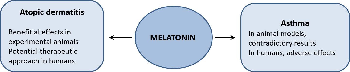
Marseglia L, D’Angelo G, Manti S, et al. (2014) Melatonin and atopy: role in atopic dermatitis and asthma. Int J Mol Sci 15: 13482–13493. doi: 10.3390/ijms150813482
![]() |
[97] |
Arnetz BB, Berg M (1996) Melatonin and adrenocorticotropic hormone levels in video display unit workers during work and leisure. J Occup Env Med 38: 1108–1110. doi: 10.1097/00043764-199611000-00010
![]() |
[98] |
Kimata H (2004) Reduction of allergen-specific IgE production by laughter. Eur J Clin Invest 34: 76–77. doi: 10.1111/j.1365-2362.2004.01294.x
![]() |
[99] | Chang Y-S, Lin M-H, Lee J-H, et al. (2015) Melatonin supplementation for children with atopic dermatitis and sleep disturbance: a randomized clinical trial. JAMA Pediatr 170: 1–8. |
[100] |
Maddox L, Schwartz DA (2002) The pathophysiology of asthma. Annu Rev Med 53: 477–498. doi: 10.1146/annurev.med.53.082901.103921
![]() |
[101] |
Sutherland ER (2005) Nocturnal asthma. J Allergy Clin Immunol 116: 1179–1186. doi: 10.1016/j.jaci.2005.09.028
![]() |
[102] | Karasu-Minareci E, Kaya Y, Belgin YF (2012) The achilles heel in melatonin: asthma. Iran J Allergy Asthma Immunol 11: 246–252. |
[103] |
Bowler RP, Crapo JD (2002) Oxidative stress in allergic respiratory diseases. J Allergy Clin Immunol 110: 349–356. doi: 10.1067/mai.2002.126780
![]() |
[104] |
Sackesen C, Ercan H, Dizdar E, et al. (2008) A comprehensive evaluation of the enzymatic and nonenzymatic antioxidant systems in childhood asthma. J Allergy Clin Immunol 122: 78–85. doi: 10.1016/j.jaci.2008.03.035
![]() |
[105] |
Chipps BE, Zeiger RS, Borish L, et al. (2012) Key findings and clinical implications from The Epidemiology and Natural History of Asthma: Outcomes and Treatment Regimens (TENOR) study. J Allergy Clin Immunol 130: 332–342. doi: 10.1016/j.jaci.2012.04.014
![]() |
[106] | Fei GH, Liu RY, Zhang ZH, et al. (2003) Relationships between melatonin and cortisol and the status of disease in patients with bronchial asthma. Zhonghua Jie He He Hu Xi Za Zhi 26: 679–682. |
[107] | Fei GH, Liu RY, Zhang ZH, et al. (2004) Alterations in circadian rhythms of melatonin and cortisol in patients with bronchial asthma. Acta Pharmacol Sin 25: 651–656. |
[108] | Evsiukova E V (1999) Melatonin effects on functional activity of platelets in patients with bronchial asthma. Ter Arkh 71: 35–37. |
[109] | Kos-Kudla B, Ostrowska Z, Marek B, et al. (2002) Circadian rhythm of melatonin in postmenopausal asthmatic women with hormone replacement therapy. Neuro Endocrinol Lett 23: 243–248. |
[110] |
Evsyukova H V (1999) The role of melatonin in pathogenesis of aspirin-sensitive asthma. Eur J Clin Invest 29: 563–567. doi: 10.1046/j.1365-2362.1999.00479.x
![]() |
[111] |
Evsyukova H V (2011) Expression of melatonin in platelets of patients with aspirin-induced asthma. Eur J Clin Invest 41: 781–784. doi: 10.1111/j.1365-2362.2011.02484.x
![]() |
[112] | Evsiukova E V, Okuneva EI, Zubzhitskaia LB, et al. (2008) Melatonin expression in nasal polyps in patients with asthmatic triad. Arkh Patol 70: 33–35. |
[113] | Evsyukova H V (2002) Aspirin-sensitive asthma due to diffuse neuroendocrine system pathology. Neuro Endocrinol Lett 23: 281–285. |
[114] |
Sutherland ER, Martin RJ, Ellison MC, et al. (2002) Immunomodulatory effects of melatonin in asthma. Am J Respir Crit Care Med 166: 1055–1061. doi: 10.1164/rccm.200204-356OC
![]() |
[115] |
Sutherland ER, Ellison MC, Kraft M, et al. (2003) Elevated serum melatonin is associated with the nocturnal worsening of asthma. J Allergy Clin Immunol 112: 513–517. doi: 10.1016/S0091-6749(03)01717-2
![]() |
[116] |
Gumral N, Caliskan S, Ozguner F, et al. (2009) Melatonin levels and enzymatic antioxidant defense system decrease in blood of patients with bronchial asthma. Toxicol Ind Heal 25: 411–416. doi: 10.1177/0748233709106625
![]() |
[117] |
Gumral N, Naziroglu M, Ongel K, et al. (2009) Antioxidant enzymes and melatonin levels in patients with bronchial asthma and chronic obstructive pulmonary disease during stable and exacerbation periods. Cell Biochem Funct 27: 276–283. doi: 10.1002/cbf.1569
![]() |
[118] |
Campos FL, da Silva-Junior FP, de V B, et al. (2004) Melatonin improves sleep in asthma: a randomized, double-blind, placebo-controlled study. Am J Respir Crit Care Med 170: 947–951. doi: 10.1164/rccm.200404-488OC
![]() |
[119] |
Martins E, Ligeiro de Oliveira AP, Fialho de Araujo AM, et al. (2001) Melatonin modulates allergic lung inflammation. J Pineal Res 31: 363–369. doi: 10.1034/j.1600-079X.2001.310412.x
![]() |
[120] |
Luo F, Liu X, Li S, et al. (2004) Melatonin promoted chemotaxins expression in lung epithelial cell stimulated with TNF-alpha. Respir Res 5: 20–28. doi: 10.1186/1465-9921-5-20
![]() |
[121] | Wang YT, Chen SL, Xu SY (2004) Effect of melatonin on the expression of nuclear factor-kappa B and airway inflammation in asthmatic rats. Zhonghua Er Ke Za Zhi 42: 94–97. |
[122] | Zhou L, Qian ZX, Li F, et al. (2007) The effect of melatonin on the regulation of collagen accumulation and matrix metalloproteinase-9 and tissue inhibitor of matrix metalloproteinase-1 mRNA and protein in a murine model of chronic asthma. Zhonghua Jie He He Hu Xi Za Zhi 30: 527–532. |
[123] |
Shin IS, Park JW, Shin NR, et al. (2014) Melatonin inhibits MUC5AC production via suppression of MAPK signaling in human airway epithelial cells. J Pineal Res 56: 398–407. doi: 10.1111/jpi.12127
![]() |
[124] |
Rioux JD, Abbas AK (2005) Paths to understanding the genetic basis of autoimmune disease. Nature 435: 584–589. doi: 10.1038/nature03723
![]() |
[125] |
Choy EH, Panayi GS (2001) Cytokine pathways and joint inflammation in rheumatoid arthritis. N Engl J Med 344: 907–916. doi: 10.1056/NEJM200103223441207
![]() |
[126] |
Imboden JB (2009) The immunopathogenesis of rheumatoid arthritis. Annu Rev Pathol 4: 417–434. doi: 10.1146/annurev.pathol.4.110807.092254
![]() |
[127] |
McInnes IB, Schett G (2011) The pathogenesis of rheumatoid arthritis. N Engl J Med 365: 2205–2219. doi: 10.1056/NEJMra1004965
![]() |
[128] |
Kalpakcioglu B, Senel K (2009) The role of melatonin in rheumatic diseases. Infect Disord Drug Targets 9: 453–456. doi: 10.2174/187152609788922546
![]() |
[129] |
Sanchez-Barcelo EJ, Mediavilla MD, Tan DX, et al. (2010) Clinical uses of melatonin: evaluation of human trials. Curr Med Chem 17: 2070–2095. doi: 10.2174/092986710791233689
![]() |
[130] |
Hansson I, Holmdahl R, Mattsson R (1990) Constant darkness enhances autoimmunity to type II collagen and exaggerates development of collagen-induced arthritis in DBA/1 mice. J Neuroimmunol 27: 79–84. doi: 10.1016/0165-5728(90)90139-E
![]() |
[131] | Hansson I, Holmdahl R, Mattsson R (1993) Pinealectomy ameliorates collagen II-induced arthritis in mice. Clin Exp Immunol 92: 432–436. |
[132] |
Hansson I, Holmdahl R, Mattsson R (1992) The pineal hormone melatonin exaggerates development of collagen-induced arthritis in mice. J Neuroimmunol 39: 23–30. doi: 10.1016/0165-5728(92)90171-G
![]() |
[133] |
Mattsson R, Hannsson I, Holmdahl R (1994) Pineal gland in autoimmunity: melatonin-dependent exaggeration of collagen-induced arthritis in mice. Autoimmunity 17: 83–86. doi: 10.3109/08916939409014661
![]() |
[134] |
Jimenez-Caliani AJ, Jimenez-Jorge S, Molinero P, et al. (2005) Dual effect of melatonin as proinflammatory and antioxidant in collagen-induced arthritis in rats. J Pineal Res 38: 93–99. doi: 10.1111/j.1600-079X.2004.00175.x
![]() |
[135] | Cano P, Cardinali DP, Chacon F, et al. (2002) Nighttime changes in norepinephrine and melatonin content and serotonin turnover in pineal glands of young and old rats injected with Freund’s adjuvant. Neuro Endocrinol Lett 23: 49–53. |
[136] |
Bang J, Chang HW, Jung HR, et al. (2012) Melatonin attenuates clock gene cryptochrome1, which may aggravate mouse anti-type II collagen antibody-induced arthritis. Rheumatol Int 32: 379–385. doi: 10.1007/s00296-010-1641-9
![]() |
[137] |
Chen Q, Wei W (2002) Effects and mechanisms of melatonin on inflammatory and immune responses of adjuvant arthritis rat. Int Immunopharmacol 2: 1443–1449. doi: 10.1016/S1567-5769(02)00088-7
![]() |
[138] |
Cutolo M, Balleari E, Giusti M, et al. (1988) Sex hormone status of male patients with rheumatoid arthritis: evidence of low serum concentrations of testosterone at baseline and after human chorionic gonadotropin stimulation. Arthritis Rheum 31: 1314–1317. doi: 10.1002/art.1780311015
![]() |
[139] |
Valenti S, Giusti M (2002) Melatonin participates in the control of testosterone secretion from rat testis: an overview of our experience. Ann N Y Acad Sci 966: 284–289. doi: 10.1111/j.1749-6632.2002.tb04228.x
![]() |
[140] |
Cutolo M, Maestroni GJ, Otsa K, et al. (2005) Circadian melatonin and cortisol levels in rheumatoid arthritis patients in winter time: a north and south Europe comparison. Ann Rheum Dis 64: 212–216. doi: 10.1136/ard.2004.023416
![]() |
[141] |
Arkema E V, Hart JE, Bertrand KA, et al. (2013) Exposure to ultraviolet-B and risk of developing rheumatoid arthritis among women in the Nurses’ Health Study. Ann Rheum Dis 72: 506–511. doi: 10.1136/annrheumdis-2012-202302
![]() |
[142] |
Brainard GC, Podolin PL, Leivy SW, et al. (1986) Near-ultraviolet radiation suppresses pineal melatonin content. Endocrinology 119: 2201–2205. doi: 10.1210/endo-119-5-2201
![]() |
[143] |
Holick MF (2007) Vitamin D deficiency. N Engl J Med 357: 266–281. doi: 10.1056/NEJMra070553
![]() |
[144] |
Becklund BR, Severson KS, Vang S V, et al. (2010) UV radiation suppresses experimental autoimmune encephalomyelitis independent of vitamin D production. Proc Natl Acad Sci U S A 107: 6418–6423. doi: 10.1073/pnas.1001119107
![]() |
[145] |
Skobowiat C, Slominski AT (2015) UVB Activates Hypothalamic-Pituitary-Adrenal Axis in C57BL/6 Mice. J Invest Dermatol 135: 1638–1648. doi: 10.1038/jid.2014.450
![]() |
[146] |
Cutolo M, Masi AT (2005) Circadian rhythms and arthritis. Rheum Dis Clin North Am 31: 115–129. doi: 10.1016/j.rdc.2004.09.005
![]() |
[147] |
Cutolo M (2012) Chronobiology and the treatment of rheumatoid arthritis. Curr Opin Rheumatol 24: 312–318. doi: 10.1097/BOR.0b013e3283521c78
![]() |
[148] |
Petrovsky N, Harrison LC (1998) The chronobiology of human cytokine production. Int Rev Immunol 16: 635–649. doi: 10.3109/08830189809043012
![]() |
[149] |
Cutolo M, Straub RH (2008) Circadian rhythms in arthritis: hormonal effects on the immune/inflammatory reaction. Autoimmun Rev 7: 223–228. doi: 10.1016/j.autrev.2007.11.019
![]() |
[150] |
El-Awady HM, El-Wakkad AS, Saleh MT, et al. (2007) Serum melatonin in juvenile rheumatoid arthritis: correlation with disease activity. Pak J Biol Sci 10: 1471–1476. doi: 10.3923/pjbs.2007.1471.1476
![]() |
[151] |
Afkhamizadeh M, Sahebari M, Seyyed-Hoseini SR (2014) Morning melatonin serum values do not correlate with disease activity in rheumatoid arthritis: a cross-sectional study. Rheumatol Int 34: 1145–1151. doi: 10.1007/s00296-013-2930-x
![]() |
[152] | West SK, Oosthuizen JM (1992) Melatonin levels are decreased in rheumatoid arthritis. J Basic Clin Physiol Pharmacol 3: 33–40. |
[153] |
Sulli A, Maestroni GJ, Villaggio B, et al. (2002) Melatonin serum levels in rheumatoid arthritis. Ann N Y Acad Sci 966: 276–283. doi: 10.1111/j.1749-6632.2002.tb04227.x
![]() |
[154] |
Forrest CM, Mackay GM, Stoy N, et al. (2007) Inflammatory status and kynurenine metabolism in rheumatoid arthritis treated with melatonin. Br J Clin Pharmacol 64: 517–526. doi: 10.1111/j.1365-2125.2007.02911.x
![]() |
[155] |
Maestroni GJ, Sulli A, Pizzorni C, et al. (2002) Melatonin in rheumatoid arthritis: synovial macrophages show melatonin receptors. Ann N Y Acad Sci 966: 271–275. doi: 10.1111/j.1749-6632.2002.tb04226.x
![]() |
[156] |
Cutolo M, Villaggio B, Candido F, et al. (1999) Melatonin influences interleukin-12 and nitric oxide production by primary cultures of rheumatoid synovial macrophages and THP-1 cells. Ann N Y Acad Sci 876: 246–254. doi: 10.1111/j.1749-6632.1999.tb07645.x
![]() |
[157] |
Kouri VP, Olkkonen J, Kaivosoja E, et al. (2013) Circadian timekeeping is disturbed in rheumatoid arthritis at molecular level. PLoS One 8: 54049–54059. doi: 10.1371/journal.pone.0054049
![]() |
[158] |
Nah SS, Won HJ, Park HJ, et al. (2009) Melatonin inhibits human fibroblast-like synoviocyte proliferation via extracellular signal-regulated protein kinase/P21(CIP1)/P27(KIP1) pathways. J Pineal Res 47: 70–74. doi: 10.1111/j.1600-079X.2009.00689.x
![]() |
[159] |
Liu X, Xu Y, Chen S, et al. (2014) Rescue of proinflammatory cytokine-inhibited chondrogenesis by the antiarthritic effect of melatonin in synovium mesenchymal stem cells via suppression of reactive oxygen species and matrix metalloproteinases. Free Radic Biol Med 68: 234–246. doi: 10.1016/j.freeradbiomed.2013.12.012
![]() |
[160] |
Lin GJ, Huang SH, Chen SJ, et al. (2013) Modulation by melatonin of the pathogenesis of inflammatory autoimmune diseases. Int J Mol Sci 14: 11742–11766. doi: 10.3390/ijms140611742
![]() |
[161] | Arushanian EB, Naumov SS, Ivanova VN, et al. (2014) Comparative study of the influence of melatonin and diclofenac on some hematologic indices of experimental rheumatoid arthritis in rats. Eksp Klin Farmakol 77: 13–15. |
[162] |
Steinman L (1996) Multiple sclerosis: a coordinated immunological attack against myelin in the central nervous system. Cell 85: 299–302. doi: 10.1016/S0092-8674(00)81107-1
![]() |
[163] |
Pugliatti M, Sotgiu S, Rosati G (2002) The worldwide prevalence of multiple sclerosis. Clin Neurol Neurosurg 104: 182–191. doi: 10.1016/S0303-8467(02)00036-7
![]() |
[164] |
Frohman EM, Racke MK, Raine CS (2006) Multiple sclerosis-the plaque and its pathogenesis. N Engl J Med 354: 942–955. doi: 10.1056/NEJMra052130
![]() |
[165] |
Lassmann H, van HJ (2011) The molecular basis of neurodegeneration in multiple sclerosis. FEBS Lett 585: 3715–3723. doi: 10.1016/j.febslet.2011.08.004
![]() |
[166] |
Glabinski A, Tawsek NS, Bartosz G (1993) Increased generation of superoxide radicals in the blood of MS patients. Acta Neurol Scand 88: 174–177. doi: 10.1111/j.1600-0447.1993.tb03434.x
![]() |
[167] |
Besler HT, Comoglu S (2003) Lipoprotein oxidation, plasma total antioxidant capacity and homocysteine level in patients with multiple sclerosis. Nutr Neurosci 6: 189–196. doi: 10.1080/1028415031000115945
![]() |
[168] |
MacMicking JD, Willenborg DO, Weidemann MJ, et al. (1992) Elevated secretion of reactive nitrogen and oxygen intermediates by inflammatory leukocytes in hyperacute experimental autoimmune encephalomyelitis: enhancement by the soluble products of encephalitogenic T cells. J Exp Med 176: 303–307. doi: 10.1084/jem.176.1.303
![]() |
[169] |
Hutter CD, Laing P (1996) Multiple sclerosis: sunlight, diet, immunology and aetiology. Med Hypotheses 46: 67–74. doi: 10.1016/S0306-9877(96)90002-X
![]() |
[170] |
Van der Mei IA, Ponsonby AL, Dwyer T, et al. (2003) Past exposure to sun, skin phenotype, and risk of multiple sclerosis: case-control study. BMJ 327: 316–322. doi: 10.1136/bmj.327.7410.316
![]() |
[171] |
Islam T, Gauderman WJ, Cozen W, et al. (2007) Childhood sun exposure influences risk of multiple sclerosis in monozygotic twins. Neurology 69: 381–388. doi: 10.1212/01.wnl.0000268266.50850.48
![]() |
[172] |
Mehta BK (2010) New hypotheses on sunlight and the geographic variability of multiple sclerosis prevalence. J Neurol Sci 292: 5–10. doi: 10.1016/j.jns.2010.02.004
![]() |
[173] | Ghorbani A, Salari M, Shaygannejad V, et al. (2013) The role of melatonin in the pathogenesis of multiple sclerosis: a case-control study. Int J Prev Med 4: S180–S184. |
[174] |
Kurtzke JF (1977) Geography in multiple sclerosis. J Neurol 215: 1–26. doi: 10.1007/BF00312546
![]() |
[175] |
Kurtzke JF (1967) On the fine structure of the distribution of multiple sclerosis. Acta Neurol Scand 43: 257–282. doi: 10.1111/j.1600-0404.1967.tb05733.x
![]() |
[176] |
Natarajan R, Einarsdottir E, Riutta A, et al. (2012) Melatonin pathway genes are associated with progressive subtypes and disability status in multiple sclerosis among Finnish patients. J Neuroimmunol 250: 106–110. doi: 10.1016/j.jneuroim.2012.05.014
![]() |
[177] |
Sandyk R (1993) Multiple sclerosis: the role of puberty and the pineal gland in its pathogenesis. Int J Neurosci 68: 209–225. doi: 10.3109/00207459308994277
![]() |
[178] |
Hedstrom AK, Akerstedt T, Hillert J, et al. (2011) Shift work at young age is associated with increased risk for multiple sclerosis. Ann Neurol 70: 733–741. doi: 10.1002/ana.22597
![]() |
[179] |
Brass SD, Duquette P, Proulx-Therrien J, et al. (2010) Sleep disorders in patients with multiple sclerosis. Sleep Med Rev 14: 121–129. doi: 10.1016/j.smrv.2009.07.005
![]() |
[180] | Barun B (2013) Pathophysiological background and clinical characteristics of sleep disorders in multiple sclerosis. Clin Neurol Neurosurg 115 Suppl1: S82–S85. |
[181] |
Sandyk R, Awerbuch GI (1992) Nocturnal plasma melatonin and alpha-melanocyte stimulating hormone levels during exacerbation of multiple sclerosis. Int J Neurosci 67: 173–186. doi: 10.3109/00207459208994783
![]() |
[182] | Gholipour T, Ghazizadeh T, Babapour S, et al. (2015) Decreased urinary level of melatonin as a marker of disease severity in patients with multiple sclerosis. Iran J Allergy, Asthma Immunol 14: 91–97. |
[183] |
Melamud L, Golan D, Luboshitzky R, et al. (2012) Melatonin dysregulation, sleep disturbances and fatigue in multiple sclerosis. J Neurol Sci 314: 37–40. doi: 10.1016/j.jns.2011.11.003
![]() |
[184] |
Escribano BM, Colin-Gonzalez AL, Santamaria A, et al. (2014) The role of melatonin in multiple sclerosis, Huntington’s disease and cerebral ischemia. CNS Neurol Disord Drug Targets 13: 1096–1119. doi: 10.2174/1871527313666140806160400
![]() |
[185] |
Damasceno A, Moraes AS, Farias A, et al. (2015) Disruption of melatonin circadian rhythm production is related to multiple sclerosis severity: A preliminary study. J Neurol Sci 353: 166–168. doi: 10.1016/j.jns.2015.03.040
![]() |
[186] |
Akpinar Z, Tokgoz S, Gokbel H, et al. (2008) The association of nocturnal serum melatonin levels with major depression in patients with acute multiple sclerosis. Psychiatry Res 161: 253–257. doi: 10.1016/j.psychres.2007.11.022
![]() |
[187] |
Sandyk R (1995) Diurnal variations in vision and relations to circadian melatonin secretion in multiple sclerosis. Int J Neurosci 83: 1–6. doi: 10.3109/00207459508986320
![]() |
[188] |
Anderson G, Rodriguez M (2011) Multiple sclerosis, seizures, and antiepileptics: role of IL-18, IDO, and melatonin. Eur J Neurol 18: 680–685. doi: 10.1111/j.1468-1331.2010.03257.x
![]() |
[189] |
Maestroni GJ (2001) The immunotherapeutic potential of melatonin. Expert Opin Investig Drugs 10: 467–476. doi: 10.1517/13543784.10.3.467
![]() |
[190] | Adamczyk-Sowa M, Pierzchala K, Sowa P, et al. (2014) Influence of melatonin supplementation on serum antioxidative properties and impact of the quality of life in multiple sclerosis patients. J Physiol Pharmacol 65: 543–550. |
[191] |
Emamgholipour S, Hossein-nezhad A, Sahraian MA, et al. (2016) Evidence for possible role of melatonin in reducing oxidative stress in multiple sclerosis through its effect on SIRT1 and antioxidant enzymes. Life Sci 145: 34–41. doi: 10.1016/j.lfs.2015.12.014
![]() |
[192] |
López-González A, álvarez-Sánchez N, Lardone PJ, et al. (2015) Melatonin treatment improves primary progressive multiple sclerosis: a case report. J Pineal Res 58: 173–177. doi: 10.1111/jpi.12203
![]() |
[193] |
Bahamonde C, Conde C, Aguera E, et al. (2014) Elevated melatonin levels in natalizumab-treated female patients with relapsing-remitting multiple sclerosis: relationship to oxidative stress. Eur J Pharmacol 730: 26–30. doi: 10.1016/j.ejphar.2014.02.020
![]() |
[194] | Kang JC, Ahn M, Kim YS, et al. (2001) Melatonin ameliorates autoimmune encephalomyelitis through suppression of intercellular adhesion molecule-1. J Vet Sci 2: 85–89. |
[195] |
Kashani IR, Rajabi Z, Akbari M, et al. (2014) Protective effects of melatonin against mitochondrial injury in a mouse model of multiple sclerosis. Exp Brain Res 232: 2835–2846. doi: 10.1007/s00221-014-3946-5
![]() |
[196] |
álvarez-Sánchez N, Cruz-Chamorro I, López-González A, et al. (2015) Melatonin controls experimental autoimmune encephalomyelitis by altering the T effector/regulatory balance. Brain Behav Immun 50: 101–114. doi: 10.1016/j.bbi.2015.06.021
![]() |
[197] | Chen S-J, Huang S-H, Chen J-W, et al. (2015) Melatonin enhances interleukin-10 expression and suppresses chemotaxis to inhibit inflammation in situ and reduce the severity of experimental autoimmune encephalomyelitis. Int Imunopharmacology 31: 169–177. |
[198] |
Farez MF, Mascanfroni ID, Méndez-Huergo SP, et al. (2015) Melatonin Contributes to the Seasonality of Multiple Sclerosis Relapses. Cell 162: 1338–1352. doi: 10.1016/j.cell.2015.08.025
![]() |
[199] |
Lee JS, Cua DJ (2015) Melatonin lulling Th17 cells to sleep. Cell 162: 1212–1214. doi: 10.1016/j.cell.2015.08.054
![]() |
[200] |
Constantinescu CS, Hilliard B, Ventura E, et al. (1997) Luzindole, a melatonin receptor antagonist, suppresses experimental autoimmune encephalomyelitis. Pathobiology 65: 190–194. doi: 10.1159/000164122
![]() |
[201] | Dubocovich ML, Yun K, Al-Ghoul WM, et al. (1998) Selective MT2 melatonin receptor antagonists block melatonin-mediated phase advances of circadian rhythms. FASEB J 12: 1211–1220. |
[202] |
Sandyk R (1997) Influence of the pineal gland on the expression of experimental allergic encephalomyelitis: possible relationship to the aquisition of multiple sclerosis. Int J Neurosci 90: 129–133. doi: 10.3109/00207459709000632
![]() |
[203] |
D’Cruz DP, Khamashta MA, Hughes GR (2007) Systemic lupus erythematosus. Lancet 369: 587–596. doi: 10.1016/S0140-6736(07)60279-7
![]() |
[204] |
Jancar S, Sanchez CM (2005) Immune complex-mediated tissue injury: a multistep paradigm. Trends Immunol 26: 48–55. doi: 10.1016/j.it.2004.11.007
![]() |
[205] | Kidd P (2003) Th1/Th2 balance: the hypothesis, its limitations, and implications for health and disease. Altern Med Rev 8: 223–246. |
[206] | Hayashi T (2010) Therapeutic strategies for SLE involving cytokines: mechanism-oriented therapies especially IFN-gamma targeting gene therapy. J Biomed Biotechnol 2010: 461641–461660. |
[207] |
Wong CK, Lit LC, Tam LS, et al. (2008) Hyperproduction of IL-23 and IL-17 in patients with systemic lupus erythematosus: implications for Th17-mediated inflammation in auto-immunity. Clin Immunol 127: 385–393. doi: 10.1016/j.clim.2008.01.019
![]() |
[208] |
Fairhurst AM, Wandstrat AE, Wakeland EK (2006) Systemic lupus erythematosus: multiple immunological phenotypes in a complex genetic disease. Adv Immunol 92: 1–69. doi: 10.1016/S0065-2776(06)92001-X
![]() |
[209] |
Tsokos GC (2011) Systemic lupus erythematosus. N Engl J Med 365: 2110–2121. doi: 10.1056/NEJMra1100359
![]() |
[210] |
Lenz SP, Izui S, Benediktsson H, et al. (1995) Lithium chloride enhances survival of NZB/W lupus mice: influence of melatonin and timing of treatment. Int J Immunopharmacol 17: 581–592. doi: 10.1016/0192-0561(95)00032-W
![]() |
[211] |
Lechner O, Dietrich H, Oliveira dos SA, et al. (2000) Altered circadian rhythms of the stress hormone and melatonin response in lupus-prone MRL/MP-fas(Ipr) mice. J Autoimmun 14: 325–333. doi: 10.1006/jaut.2000.0375
![]() |
[212] |
Yin Z, Bahtiyar G, Zhang N, et al. (2002) IL-10 regulates murine lupus. J Immunol 169: 2148–2155. doi: 10.4049/jimmunol.169.4.2148
![]() |
[213] |
Jimenez-Caliani AJ, Jimenez-Jorge S, Molinero P, et al. (2006) Sex-dependent effect of melatonin on systemic erythematosus lupus developed in Mrl/Mpj-Faslpr mice: it ameliorates the disease course in females, whereas it exacerbates it in males. Endocrinology 147: 1717–1724. doi: 10.1210/en.2005-0648
![]() |
[214] |
Jimenez-Caliani AJ, Jimenez-Jorge S, Molinero P, et al. (2008) Treatment with testosterone or estradiol in melatonin treated females and males MRL/MpJ-Faslpr mice induces negative effects in developing systemic lupus erythematosus. J Pineal Res 45: 204–211. doi: 10.1111/j.1600-079X.2008.00578.x
![]() |
[215] | Zhou LL, Wei W, Si JF, et al. (2010) Regulatory effect of melatonin on cytokine disturbances in the pristane-induced lupus mice. Mediat Inflamm 2010: 951210–951217. |
[216] |
Wu CC, Lu KC, Lin GJ, et al. (2012) Melatonin enhances endogenous heme oxygenase-1 and represses immune responses to ameliorate experimental murine membranous nephropathy. J Pineal Res 52: 460–469. doi: 10.1111/j.1600-079X.2011.00960.x
![]() |
[217] |
Haga HJ, Brun JG, Rekvig OP, et al. (1999) Seasonal variations in activity of systemic lupus erythematosus in a subarctic region. Lupus 8: 269–273. doi: 10.1191/096120399678847858
![]() |
[218] | Robeva R, Tanev D, Kirilov G, et al. (2013) Decreased daily melatonin levels in women with systemic lupus erythematosus - a short report. Balk Med J 30: 273–276. |
[219] | Kawasaki E, Abiru N, Eguchi K (2004) Prevention of type 1 diabetes: from the view point of beta cell damage. Diabetes Res Clin Pr 66 Suppl 1: S27–S32. |
[220] |
Daneman D (2006) Type 1 diabetes. Lancet 367: 847–858. doi: 10.1016/S0140-6736(06)68341-4
![]() |
[221] |
Ponsonby AL, Lucas RM, van Ider M (2005) UVR, vitamin D and three autoimmune diseases--multiple sclerosis, type 1 diabetes, rheumatoid arthritis. Photochem Photobiol 81: 1267–1275. doi: 10.1562/2005-02-15-IR-441
![]() |
[222] |
Aoki CA, Borchers AT, Ridgway WM, et al. (2005) NOD mice and autoimmunity. Autoimmun Rev 4: 373–379. doi: 10.1016/j.autrev.2005.02.002
![]() |
[223] |
Wicker LS, Todd JA, Peterson LB (1995) Genetic control of autoimmune diabetes in the NOD mouse. Annu Rev Immunol 13: 179–200. doi: 10.1146/annurev.iy.13.040195.001143
![]() |
[224] |
Liblau RS, Singer SM, McDevitt HO (1995) Th1 and Th2 CD4+ T cells in the pathogenesis of organ-specific autoimmune diseases. Immunol Today 16: 34–38. doi: 10.1016/0167-5699(95)80068-9
![]() |
[225] |
Hung JT, Liao JH, Lin YC, et al. (2005) Immunopathogenic role of TH1 cells in autoimmune diabetes: evidence from a T1 and T2 doubly transgenic non-obese diabetic mouse model. J Autoimmun 25: 181–192. doi: 10.1016/j.jaut.2005.08.010
![]() |
[226] |
Koarada S, Wu Y, Olshansky G, et al. (2002) Increased nonobese diabetic Th1: Th2 (IFN-gamma: IL-4) ratio is CD4+ T cell intrinsic and independent of APC genetic background. J Immunol 169: 6580–6587. doi: 10.4049/jimmunol.169.11.6580
![]() |
[227] |
Conti A, Maestroni GJ (1996) Role of the pineal gland and melatonin in the development of autoimmune diabetes in non-obese diabetic mice. J Pineal Res 20: 164–172. doi: 10.1111/j.1600-079X.1996.tb00253.x
![]() |
[228] |
Lin GJ, Huang SH, Chen YW, et al. (2009) Melatonin prolongs islet graft survival in diabetic NOD mice. J Pineal Res 47: 284–292. doi: 10.1111/j.1600-079X.2009.00712.x
![]() |
[229] |
Peschke E, Wolgast S, Bazwinsky I, et al. (2008) Increased melatonin synthesis in pineal glands of rats in streptozotocin induced type 1 diabetes. J Pineal Res 45: 439–448. doi: 10.1111/j.1600-079X.2008.00612.x
![]() |
[230] | Peschke E, Hofmann K, Bahr I, et al. (2011) The insulin-melatonin antagonism: studies in the LEW.1AR1-iddm rat (an animal model of human type 1 diabetes mellitus). Diabetologia 54: 1831–1840. |
[231] |
Peschke E, Hofmann K, Ponicke K, et al. (2012) Catecholamines are the key for explaining the biological relevance of insulin-melatonin antagonisms in type 1 and type 2 diabetes. J Pineal Res 52: 389–396. doi: 10.1111/j.1600-079X.2011.00951.x
![]() |
[232] |
Kor Y, Geyikli I, Keskin M, et al. (2014) Preliminary study: Evaluation of melatonin secretion in children and adolescents with type 1 diabetes mellitus. Indian J Endocrinol Metab 18: 565–568. doi: 10.4103/2230-8210.137521
![]() |
[233] |
Reyes-Toso CF, Roson MI, Albornoz LE, et al. (2002) Vascular reactivity in diabetic rats: effect of melatonin. J Pineal Res 33: 81–86. doi: 10.1034/j.1600-079X.2002.01886.x
![]() |
[234] |
Cavallo A, Daniels SR, Dolan LM, et al. (2004) Blood pressure-lowering effect of melatonin in type 1 diabetes. J Pineal Res 36: 262–266. doi: 10.1111/j.1600-079X.2004.00126.x
![]() |
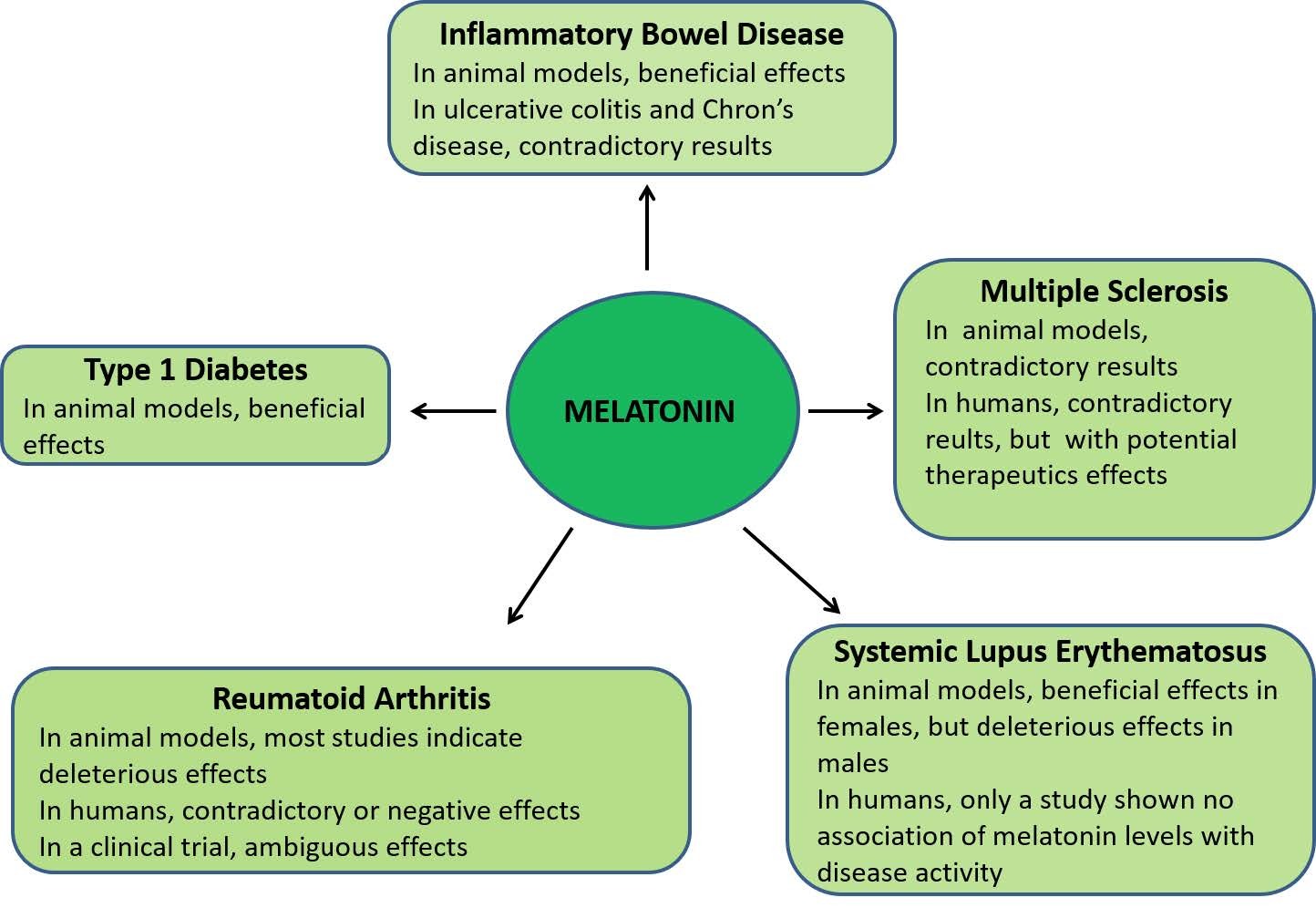
[235] | Abraham C, Cho JH (2009) Inflammatory bowel disease. N Engl J Med 19: 2066–2078. |
[236] |
Motilva V, Garcia-Maurino S, Talero E, et al. (2011) New paradigms in chronic intestinal inflammation and colon cancer: role of melatonin. J Pineal Res 51: 44–60. doi: 10.1111/j.1600-079X.2011.00915.x
![]() |
[237] |
Sartor RB (2006) Mechanisms of disease: pathogenesis of Crohn’s disease and ulcerative colitis. Nat Clin Pr Gastroenterol Hepatol 3: 390–407. doi: 10.1038/ncpgasthep0528
![]() |
[238] |
Neurath MF, Finotto S (2006) The many roads to inflammatory bowel diseases. Immunity 25: 189–191. doi: 10.1016/j.immuni.2006.08.005
![]() |
[239] | Fuss IJ, Neurath M, Boirivant M, et al. (1996) Disparate CD4+ lamina propria (LP) lymphokine secretion profiles in inflammatory bowel disease. Crohn’s disease LP cells manifest increased secretion of IFN-gamma, whereas ulcerative colitis LP cells manifest increased secretion of IL-5. J Immunol 157: 1261–1270. |
[240] |
Fujino S, Andoh A, Bamba S, et al. (2003) Increased expression of interleukin 17 in inflammatory bowel disease. Gut 52: 65–70. doi: 10.1136/gut.52.1.65
![]() |
[241] |
Hue S, Ahern P, Buonocore S, et al. (2006) Interleukin-23 drives innate and T cell-mediated intestinal inflammation. J Exp Med 203: 2473–2483. doi: 10.1084/jem.20061099
![]() |
[242] |
Elson CO, Cong Y, Weaver CT, et al. (2007) Monoclonal anti-interleukin 23 reverses active colitis in a T cell-mediated model in mice. Gastroenterology 132: 2359–2370. doi: 10.1053/j.gastro.2007.03.104
![]() |
[243] |
Barrett JC, Hansoul S, Nicolae DL, et al. (2008) Genome-wide association defines more than 30 distinct susceptibility loci for Crohn’s disease. Nat Genet 40: 955–962. doi: 10.1038/ng.175
![]() |
[244] |
Preuss F, Tang Y, Laposky AD, et al. (2008) Adverse effects of chronic circadian desynchronization in animals in a “challenging” environment. Am J Physiol Regul Integr Comp Physiol 295: R2034–R2040. doi: 10.1152/ajpregu.00118.2008
![]() |
[245] |
Tang Y, Preuss F, Turek FW, et al. (2009) Sleep deprivation worsens inflammation and delays recovery in a mouse model of colitis. Sleep Med 10: 597–603. doi: 10.1016/j.sleep.2008.12.009
![]() |
[246] |
Mickle A, Sood M, Zhang Z, et al. (2010) Antinociceptive effects of melatonin in a rat model of post-inflammatory visceral hyperalgesia: a centrally mediated process. Pain 149: 555–564. doi: 10.1016/j.pain.2010.03.030
![]() |
[247] |
Pentney PT, Bubenik GA (1995) Melatonin reduces the severity of dextran-induced colitis in mice. J Pineal Res 19: 31–39. doi: 10.1111/j.1600-079X.1995.tb00168.x
![]() |
[248] |
Cuzzocrea S, Mazzon E, Serraino I, et al. (2001) Melatonin reduces dinitrobenzene sulfonic acid-induced colitis. J Pineal Res 30: 1–12. doi: 10.1034/j.1600-079X.2001.300101.x
![]() |
[249] |
Trivedi PP, Jena GB (2013) Melatonin reduces ulcerative colitis-associated local and systemic damage in mice: investigation on possible mechanisms. Dig Dis Sci 58: 3460–3474. doi: 10.1007/s10620-013-2831-6
![]() |
[250] | Park Y-S, Chung S-H, Lee S-K, et al. (2015) Melatonin improves experimental colitis with sleep deprivation. Int J Mol Med 35: 979–986. |
[251] |
Dong WG, Mei Q, Yu JP, et al. (2003) Effects of melatonin on the expression of iNOS and COX-2 in rat models of colitis. World J Gastroenterol 9: 1307–1311. doi: 10.3748/wjg.v9.i6.1307
![]() |
[252] |
Tahan G, Gramignoli R, Marongiu F, et al. (2011) Melatonin expresses powerful anti-inflammatory and antioxidant activities resulting in complete improvement of acetic-acid-induced colitis in rats. Dig Dis Sci 56: 715–720. doi: 10.1007/s10620-010-1364-5
![]() |
[253] |
Akcan A, Kucuk C, Sozuer E, et al. (2008) Melatonin reduces bacterial translocation and apoptosis in trinitrobenzene sulphonic acid-induced colitis of rats. World J Gastroenterol 14: 918–924. doi: 10.3748/wjg.14.918
![]() |
[254] |
Esposito E, Mazzon E, Riccardi L, et al. (2008) Matrix metalloproteinase-9 and metalloproteinase-2 activity and expression is reduced by melatonin during experimental colitis. J Pineal Res 45: 166–173. doi: 10.1111/j.1600-079X.2008.00572.x
![]() |
[255] |
Mazzon E, Esposito E, Crisafulli C, et al. (2006) Melatonin modulates signal transduction pathways and apoptosis in experimental colitis. J Pineal Res 41: 363–373. doi: 10.1111/j.1600-079X.2006.00378.x
![]() |
[256] |
Li JH, Yu JP, Yu HG, et al. (2005) Melatonin reduces inflammatory injury through inhibiting NF-kappaB activation in rats with colitis. Mediat Inflamm 2005: 185–193. doi: 10.1155/MI.2005.185
![]() |
[257] |
Tasdemir S, Parlakpinar H, Vardi N, et al. (2013) Effect of endogen-exogenous melatonin and erythropoietin on dinitrobenzene sulfonic acid-induced colitis. Fundam Clin Pharmacol 27: 299–307. doi: 10.1111/j.1472-8206.2011.01016.x
![]() |
[258] |
Mann S (2003) Melatonin for ulcerative colitis? Am J Gastroenterol 98: 232–233. doi: 10.1111/j.1572-0241.2003.07190.x
![]() |
[259] |
Calvo JR, Guerrero JM, Osuna C, et al. (2002) Melatonin triggers Crohn’s disease symptoms. J Pineal Res 32: 277–278. doi: 10.1034/k.1600-079X.2002.01881.x
![]() |
[260] |
Maldonado MD, Calvo JR (2008) Melatonin usage in ulcerative colitis: a case report. J Pineal Res 45: 339–340. doi: 10.1111/j.1600-079X.2008.00584.x
![]() |
[261] | Rakhimova OI (2010) Use of melatonin in combined treatment for inflammatory bowel diseases. Ter Arkh 82: 64–68. |
[262] |
Mozaffari S, Abdollahi M (2011) Melatonin, a promising supplement in inflammatory bowel disease: a comprehensive review of evidences. Curr Pharm Des 17: 4372–4378. doi: 10.2174/138161211798999357
![]() |
[263] |
Jena G, Trivedi PP (2014) A review of the use of melatonin in ulcerative colitis: experimental evidence and new approaches. Inflamm Bowel Dis 20: 553–563. doi: 10.1097/01.MIB.0000436962.32164.6e
![]() |
[264] | Chojnacki C, Wisniewska-Jarosinska M, Walecka-Kapica E, et al. (2011) Evaluation of melatonin effectiveness in the adjuvant treatment of ulcerative colitis. J Physiol Pharmacol 62: 327–334. |
[265] |
Yang J, Carra S, Zhu WG, et al. (2013) The regulation of the autophagic network and its implications for human disease. Int J Biol Sci 9: 1121–1133. doi: 10.7150/ijbs.6666
![]() |
[266] |
Cheng Y, Ren X, Hait WN, et al. (2013) Therapeutic targeting of autophagy in disease: biology and pharmacology. Pharmacol Rev 65: 1162–1197. doi: 10.1124/pr.112.007120
![]() |
[267] | Ryter SW, Mizumura K, Choi AM (2014) The impact of autophagy on cell death modalities. Int J Cell Biol 2014: 502676–502688. |
[268] |
Talero E, Garcia-Maurino S, Motilva V (2014) Melatonin, autophagy and intestinal bowel disease. Curr Pharm Des 20: 4816–4827. doi: 10.2174/1381612819666131119110835
![]() |
[269] |
Swanson GR, Burgess HJ, Keshavarzian A (2011) Sleep disturbances and inflammatory bowel disease: a potential trigger for disease flare? Expert Rev Clin Immunol 7: 29–36. doi: 10.1586/eci.10.83
![]() |
[270] | Takagi T, Inada Y, Naito Y (2013) Circadian rhythm and inflammatory bowel disease. Nihon Rinsho 71: 2165–2170. |
[271] |
Chung SH, Park YS, Kim OS, et al. (2014) Melatonin attenuates dextran sodium sulfate induced colitis with sleep deprivation: possible mechanism by microarray analysis. Dig Dis Sci 59: 1134–1141. doi: 10.1007/s10620-013-3013-2
![]() |
[272] |
Anderson G, Rodriguez M (2015) Multiple sclerosis: the role of melatonin and N-acetylserotonin. Mult Scler Relat Disord 4: 112–123. doi: 10.1016/j.msard.2014.12.001
![]() |
[273] | Miller E, Morel A, Saso L, et al. (2015) Melatonin redox activity. Its potential clinical applications in neurodegenerative disorders. Curr Top Med Chem 15: 163–169. |
[274] |
Carrillo-Vico A, Guerrero JM, Lardone PJ, et al. (2005) A review of the multiple actions of melatonin on the immune system. Endocrine 27: 189–200. doi: 10.1385/ENDO:27:2:189
![]() |
[275] |
Pandi-Perumal SR, Srinivasan V, Maestroni GJM, et al. (2006) Melatonin: Nature’s most versatile biological signal? FEBS J 273: 2813–2838. doi: 10.1111/j.1742-4658.2006.05322.x
![]() |
1. | Andrzej T. Slominski, Ruediger Hardeland, Michal A. Zmijewski, Radomir M. Slominski, Russel J. Reiter, Ralf Paus, Melatonin: A Cutaneous Perspective on its Production, Metabolism, and Functions, 2018, 138, 0022202X, 490, 10.1016/j.jid.2017.10.025 | |
2. | Yunho Jin, Yoo Jin Choi, Kyu Heo, Seong Joon Park, Melatonin as an Oncostatic Molecule Based on Its Anti-Aromatase Role in Breast Cancer, 2021, 22, 1422-0067, 438, 10.3390/ijms22010438 | |
3. | Gunhyuk Park, Seung Hoon Lee, Dal-Seok Oh, Yong-ung Kim, Melatonin inhibits neuronal dysfunction-associated with neuroinflammation by atopic psychological stress in NC/Nga atopic-like mouse models, 2017, 63, 07423098, e12420, 10.1111/jpi.12420 | |
4. | Lucreția Anghel, Liliana Baroiu, Corina Popazu, Diana Pătraș, Silvia Fotea, Alexandru Nechifor, Anamaria Ciubara, Luiza Nechita, Carmina Mușat, Ioana Stefanopol, Alin Tatu, Alexandru Ciubara, Benefits and adverse events of melatonin use in the elderly (Review), 2022, 23, 1792-0981, 10.3892/etm.2022.11142 | |
5. | Ahmed Wahhab Mohammed Khafaji, Adeeb Ahmed Kadhim Al-Zubaidy, Iqbal Ghalib Farhood, Hayder Ridha Salman, Ameliorative effects of topical ramelteon on imiquimod-induced psoriasiform inflammation in mice, 2024, 397, 0028-1298, 6231, 10.1007/s00210-024-03017-7 | |
6. | Juan R. Calvo, María D. Maldonado, Immunoregulatory properties of melatonin in the humoral immune system: A narrative review, 2024, 269, 01652478, 106901, 10.1016/j.imlet.2024.106901 |