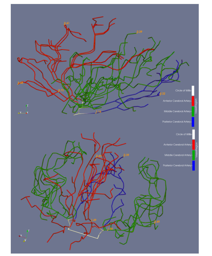
Vascular endothelial growth factor (VEGF) is a key protein involved in the process of angiogenesis. VEGF is of particular interest after a traumatic brain injury (TBI), as it re-establishes the cerebral vascular network in effort to allow for proper cerebral blood flow and thereby oxygenation of damaged brain tissue. For this reason, angiogenesis is critical in the progression and recovery of TBI patients in the days and weeks post injury. Although well established experimental work has led to advances in our understanding of TBI and the progression of angiogenisis, many constraints still exist with existing methods, especially when considering patient progression in the days following injury. To better understand the healing process on the proposed time scales, we develop a computational model that quickly simulates vessel growth and recovery by coupling VEGF and its interactions with its associated receptors to a physiologically inspired fractal model of the microvascular re-growth. We use this model to clarify the role that diffusivity, receptor kinetics and location of the TBI play in overall blood volume restoration in the weeks post injury and show that proper therapeutic angiogenesis, or vasculogenic therapies, could speed recovery of the patient as a function of the location of injury.
Citation: Austin Baird, Laura Oelsner, Charles Fisher, Matt Witte, My Huynh. A multiscale computational model of angiogenesis after traumatic brain injury, investigating the role location plays in volumetric recovery[J]. Mathematical Biosciences and Engineering, 2021, 18(4): 3227-3257. doi: 10.3934/mbe.2021161
Vascular endothelial growth factor (VEGF) is a key protein involved in the process of angiogenesis. VEGF is of particular interest after a traumatic brain injury (TBI), as it re-establishes the cerebral vascular network in effort to allow for proper cerebral blood flow and thereby oxygenation of damaged brain tissue. For this reason, angiogenesis is critical in the progression and recovery of TBI patients in the days and weeks post injury. Although well established experimental work has led to advances in our understanding of TBI and the progression of angiogenisis, many constraints still exist with existing methods, especially when considering patient progression in the days following injury. To better understand the healing process on the proposed time scales, we develop a computational model that quickly simulates vessel growth and recovery by coupling VEGF and its interactions with its associated receptors to a physiologically inspired fractal model of the microvascular re-growth. We use this model to clarify the role that diffusivity, receptor kinetics and location of the TBI play in overall blood volume restoration in the weeks post injury and show that proper therapeutic angiogenesis, or vasculogenic therapies, could speed recovery of the patient as a function of the location of injury.

| [1] | M. Faul, M. M. Wald, L. Xu, V. G. Coronado, Traumatic brain injury in the United States; emergency department visits, hospitalizations, and deaths, 2002-2006, 2010. |
| [2] |
D. W. Marion, J. Darby, H. Yonas, Acute regional cerebral blood flow changes caused by severe head injuries, J. Neurosurg., 74 (1991), 407-414. doi: 10.3171/jns.1991.74.3.0407

|
| [3] |
B. K. Giri, I. K. Krishnappa, R. M. Bryan, C. Robertson, Regional cerebral blood flow after cortical impact injury complicated by a secondary insult in rats, Stroke, 31 (2000), 961-967. doi: 10.1161/01.STR.31.4.961
|
| [4] |
E. E. Abrahamson, L. M. Foley, S. T. DeKosky, T. K. Hitchens, C. Ho, P. M. Kochanek, et al., Cerebral blood flow changes after brain injury in human amyloid-beta knock-in mice, J. Cerebr. Blood F. Met., 33 (2013), 826-833. doi: 10.1038/jcbfm.2013.24
|
| [5] |
D. F. Kelly, N. A. Martin, R. Kordestani, G. Counelis, D. A. Hovda, M. Bergsneider, et al., Cerebral blood flow as a predictor of outcome following traumatic brain injury, J. Neurosurg., 86 (1997), 633-641. doi: 10.3171/jns.1997.86.4.0633
|
| [6] | G. Shiina, T. Onuma, M. Kameyama, Y. Shimosegawa, K. Ishii, R. Shirane, et al., Sequential assessment of cerebral blood flow in diffuse brain injury by 123I-iodoamphetamine single-photon emission CT, Am. J. Neuroradiol., 19 (1998), 297-302. |
| [7] |
L. CHERIAN, C. S. Robertson, J. C. GOODMAN, Secondary insults increase injury after controlled cortical impact in rats, J. Neurotraum., 13 (1996), 371-383. doi: 10.1089/neu.1996.13.371
|
| [8] | A. Trofimov, G. Kalent'ev, D. Agarkova, Cerebrovascular resistance in patients with severe combined traumatic brain injury, Zhurnal voprosy neirokhirurgii imeni NN Burdenko, 79 (2015), 28-33. |
| [9] |
K. N. Corps, T. L. Roth, D. B. McGavern, Inflammation and neuroprotection in traumatic brain injury, JAMA Neurol., 72 (2015), 355-362. doi: 10.1001/jamaneurol.2014.3558
|
| [10] |
S. L. Aungst, S. V. Kabadi, S. M. Thompson, B. A. Stoica, A. I. Faden, Repeated mild traumatic brain injury causes chronic neuroinflammation, changes in hippocampal synaptic plasticity, and associated cognitive deficits, J. Cerebr. Blood F. Met., 34 (2014), 1223-1232. doi: 10.1038/jcbfm.2014.75
|
| [11] |
P. Dore-Duffy, X. Wang, A. Mehedi, C. W. Kreipke, J. A. Rafols, Differential expression of capillary VEGF isoforms following traumatic brain injury, Neurolog. Res., 29 (2007), 395-403. doi: 10.1179/016164107X204729
|
| [12] |
L. S. Angelo, R. Kurzrock, Vascular endothelial growth factor and its relationship to inflammatory mediators, Clin. Cancer Res., 13 (2007), 2825-2830. doi: 10.1158/1078-0432.CCR-06-2416
|
| [13] |
N. Papadopoulos, J. Martin, Q. Ruan, A. Rafique, M. P. Rosconi, E. Shi, et al., Binding and neutralization of vascular endothelial growth factor (VEGF) and related ligands by VEGF Trap, ranibizumab and bevacizumab, Angiogenesis, 15 (2012), 171-185. doi: 10.1007/s10456-011-9249-6
|
| [14] |
N. Draoui, P. de Zeeuw, P. Carmeliet, Angiogenesis revisited from a metabolic perspective: role and therapeutic implications of endothelial cell metabolism, Open Biol., 7 (2017), 170219. doi: 10.1098/rsob.170219
|
| [15] |
M. Unbekandt, P. M. del Moral, F. G. Sala, S. Bellusci, D. Warburton, V. Fleury, Tracheal occlusion increases the rate of epithelial branching of embryonic mouse lung via the FGF10-FGFR2b-Sprouty2 pathway, Mech. Dev., 125 (2008), 314-324. doi: 10.1016/j.mod.2007.10.013
|
| [16] |
R. D. Travasso, E. C. Poiré, M. Castro, J. C. Rodrguez-Manzaneque, A. Hernández-Machado, Tumor angiogenesis and vascular patterning: a mathematical model, PloS one, 6 (2011), e19989. doi: 10.1371/journal.pone.0019989
|
| [17] |
M. O. Kim, A. Adji, M. F. O'Rourke, A. P. Avolio, P. Smielewski, J. D. Pickard, et al., Principles of cerebral hemodynamics when intracranial pressure is raised: lessons from the peripheral circulation, J. Hypertens., 33 (2015), 1233. doi: 10.1097/HJH.0000000000000539
|
| [18] |
A. Pries, B. Reglin, T. Secomb, Structural adaptation of microvascular networks: functional roles of adaptive responses, Am. J. Physiol. Heart Circ. Physiol., 281 (2001), H1015-H1025. doi: 10.1152/ajpheart.2001.281.3.H1015
|
| [19] |
S. R. McDougall, A. R. Anderson, M. A. Chaplain, Mathematical modelling of dynamic adaptive tumour-induced angiogenesis: clinical implications and therapeutic targeting strategies, J. Theor. Biol., 241 (2006), 564-589. doi: 10.1016/j.jtbi.2005.12.022
|
| [20] |
M. R. Owen, T. Alarcón, P. K. Maini, H. M. Byrne, Angiogenesis and vascular remodelling in normal and cancerous tissues, J. Math. Biol., 58 (2009), 689. doi: 10.1007/s00285-008-0213-z
|
| [21] |
A. R. Anderson, M. Chaplain, Continuous and discrete mathematical models of tumor-induced angiogenesis, Bull. Math. Biol., 60 (1998), 857-899. doi: 10.1006/bulm.1998.0042
|
| [22] | A. Anderson, M. A. Chaplain, A mathematical model for capillary network formation in the absence of endothelial cell proliferation, Appl. Math. Lett., 11 (1998), 109-114. |
| [23] |
F. Mac Gabhann, A. S. Popel, Differential binding of VEGF isoforms to VEGF receptor 2 in the presence of neuropilin-1: a computational model, Am. J. Physiol. Heart Circ. Physiol., 288 (2005), H2851-H2860. doi: 10.1152/ajpheart.01218.2004
|
| [24] |
W. W. Yuen, N. R. Du, C. H. Chan, E. A. Silva, D. J. Mooney, Mimicking nature by codelivery of stimulant and inhibitor to create temporally stable and spatially restricted angiogenic zones, Proc. Natl. Acad. Sci. U.S.A., 107 (2010), 17933-17938. doi: 10.1073/pnas.1001192107
|
| [25] |
H. A. Levine, B. D. Sleeman, M. Nilsen-Hamilton, Mathematical modeling of the onset of capillary formation initiating angiogenesis, J. Math. Biol., 42 (2001), 195-238. doi: 10.1007/s002850000037
|
| [26] |
S. Maggelakis, A. Savakis, A mathematical model of growth factor induced capillary growth in the retina, Math. Computer Model., 24 (1996), 33-41. doi: 10.1016/0895-7177(96)00124-0
|
| [27] |
F. Mac Gabhann, M. T. Yang, A. S. Popel, Monte Carlo simulations of VEGF binding to cell surface receptors in vitro, Biochim. Biophys. Acta Mol. Cell Res., 1746 (2005), 95-107. doi: 10.1016/j.bbamcr.2005.09.004
|
| [28] |
F. Mac Gabhann, J. W. Ji, A. S. Popel, Computational model of vascular endothelial growth factor spatial distribution in muscle and pro-angiogenic cell therapy, PLoS Comput. Biol., 2 (2006), e127. doi: 10.1371/journal.pcbi.0020127
|
| [29] |
A. Myers, J. Kovach, S. Vuk-Pavlović, Binding, internalization, and intracellular processing of protein ligands. Derivation of rate constants by computer modeling, J. Biol. Chem., 262 (1987), 6494-6499. doi: 10.1016/S0021-9258(18)48269-5
|
| [30] |
J. W. Baish, Y. Gazit, D. A. Berk, M. Nozue, L. T. Baxter, R. K. Jain, Role of tumor vascular architecture in nutrient and drug delivery: an invasion percolation-based network model, Microvasc. Res., 51 (1996), 327-346. doi: 10.1006/mvre.1996.0031
|
| [31] |
D. Warburton, Developmental biology: order in the lung, Nature, 453 (2008), 733. doi: 10.1038/453733a
|
| [32] | D. Hunt, V. M. Savage, Asymmetries arising from the space-filling nature of vascular networks, Phys. Rev. E, 93 (2016), 062305. |
| [33] |
J. Keelan, E. M. Chung, J. P. Hague, Simulated annealing approach to vascular structure with application to the coronary arteries, Royal Soc. Open Sci., 3 (2016), 150431. doi: 10.1098/rsos.150431
|
| [34] |
Y. Huo, G. S. Kassab, Intraspecific scaling laws of vascular trees, J. R. Soc. Interface, 9 (2012), 190-200. doi: 10.1098/rsif.2011.0270
|
| [35] |
T. Araújo, A. M. Mendonça, A. Campilho, Parametric model fitting-based approach for retinal blood vessel caliber estimation in eye fundus images, PloS one, 13 (2018), e0194702. doi: 10.1371/journal.pone.0194702
|
| [36] |
B. Xiong, A. Li, Y. Lou, S. Chen, B. Long, J. Peng, et al., Precise cerebral vascular atlas in stereotaxic coordinates of whole mouse brain, Front. Neuroanat., 11 (2017), 128. doi: 10.3389/fnana.2017.00128
|
| [37] |
G. Hartung, C. Vesel, R. Morley, A. Alaraj, J. Sled, D. Kleinfeld, et al., Simulations of blood as a suspension predicts a depth dependent hematocrit in the circulation throughout the cerebral cortex, PLoS Comput. Biol., 14 (2018), e1006549. doi: 10.1371/journal.pcbi.1006549
|
| [38] | A. Linninger, N. Vaicaitis, Computational Modeling of Cerebral Vasculature, Computational Modeling of Cerebral Vasculature, Laboratory for Product and Process Design, Department of Bioengineering, University of Illinois at Chicago, (2011). |
| [39] | L. Gagnon, A. F. Smith, D. A. Boas, A. Devor, T. W. Secomb, S. Sakadžić, Modeling of cerebral oxygen transport based on in vivo microscopic imaging of microvascular network structure, blood flow, and oxygenation, Front. Comput. Neurosci., 10 (2016), 82. |
| [40] |
W. L. Nowinski, I. Volkau, Y. Marchenko, A. Thirunavuukarasuu, T. T. Ng, V. M. Runge, A 3D model of human cerebrovasculature derived from 3T magnetic resonance angiography, Neuroinformatics, 7 (2009), 23-36. doi: 10.1007/s12021-008-9028-8
|
| [41] |
E. Bullitt, D. Zeng, G. Gerig, S. Aylward, S. Joshi, J. K. Smith, et al., Vessel tortuosity and brain tumor malignancy: a blinded study1, Acad. Radiol., 12 (2005), 1232-1240. doi: 10.1016/j.acra.2005.05.027
|
| [42] | R. Kikinis, S. D. Pieper, K. G. Vosburgh, 3D Slicer: a platform for subject-specific image analysis, visualization, and clinical support, In: Intraoperative imaging and image-guided therapy, Springer (2014). 277-289. |
| [43] |
A. Fedorov, R. Beichel, J. Kalpathy-Cramer, J. Finet, J. C. Fillion-Robin, S. Pujol, et al., 3D Slicer as an image computing platform for the Quantitative Imaging Network, Magn. Reson. Imaging, 30 (2012), 1323-1341. doi: 10.1016/j.mri.2012.05.001
|
| [44] | D. Purves, G. Augustine, D. Fitzpatrick, L. Katz, A. LaMantia, J. McNamara, et al., The blood supply of the brain and spinal cord, Neuroscience, 2 (2001). |
| [45] | A. Tameem, H. Krovvidi, Cerebral physiology, Continuing Education in Anaesthesia Critical Care & Pain, 13 (2013), 113-118. |
| [46] | K. L. Monson, M. I. Converse, G. T. Manley, Cerebral blood vessel damage in traumatic brain injury, Clin. Biomech., 64 (2019) 98-113. |
| [47] | K. L. Monson, M. Converse, G. T. Manley, Cerebral blood vessel damage in traumatic brain injury, Clin. Biomech., (2018). |
| [48] |
M. O. Stefanini, F. T. Wu, F. Mac Gabhann, A. S. Popel, A compartment model of VEGF distribution in blood, healthy and diseased tissues, BMC Syst. Biol., 2 (2008), 77. doi: 10.1186/1752-0509-2-77
|
| [49] |
P. Perdikaris, L. Grinberg, G. E. Karniadakis, An effective fractal-tree closure model for simulating blood flow in large arterial networks, Ann. Biomed. Eng., 43 (2015), 1432-1442. doi: 10.1007/s10439-014-1221-3
|
| [50] |
M. S. Olufsen, Structured tree outflow condition for blood flow in larger systemic arteries, Am. J. Physiol. Heart Circ. Physiol., 276 (1999), H257-H268. doi: 10.1152/ajpheart.1999.276.1.H257
|
| [51] |
M. Zamir, On fractal properties of arterial trees, J. Theor. Biol., 197 (1999), 517-526. doi: 10.1006/jtbi.1998.0892
|
| [52] |
R. Prakash, S. T. Carmichael, Blood-brain barrier breakdown and neovascularization processes after stroke and traumatic brain injury, Curr. Opin. Neurol., 28 (2015), 556. doi: 10.1097/WCO.0000000000000248
|
| [53] |
C. D. Murray, The physiological principle of minimum work: I. The vascular system and the cost of blood volume, Proc. Natl. Acad. Sci. U.S.A., 12 (1926), 207-214. doi: 10.1073/pnas.12.3.207
|
| [54] | J. A. Adam, Blood vessel branching: beyond the standard calculus problem. Math. Mag., 84 (2011), 196-207. |
| [55] |
M. Zamir, Arterial branching within the confines of fractal L-system formalism, J. Gen. Physiol., 118 (2001), 267-276. doi: 10.1085/jgp.118.3.267
|
| [56] |
Y. Huo, G. Finet, T. Lefevre, Y. Louvard, I. Moussa, G. S. Kassab, Which diameter and angle rule provides optimal flow patterns in a coronary bifurcation?, J. Biomech., 45 (2012), 1273-1279. doi: 10.1016/j.jbiomech.2012.01.033
|
| [57] |
B. L. Krock, N. Skuli, M. C. Simon, Hypoxia-induced angiogenesis: good and evil, Genes cancer, 2 (2011), 1117-1133. doi: 10.1177/1947601911423654
|
| [58] |
A. Salehi, J. H. Zhang, A. Obenaus, Response of the cerebral vasculature following traumatic brain injury, J. Cerebr. Blood F. Met., 37 (2017), 2320-2339. doi: 10.1177/0271678X17701460
|
| [59] |
J. M. Isner, T. Asahara, Angiogenesis and vasculogenesis as therapeutic strategies for postnatal neovascularization, J. Clin. Investig., 103 (1999), 1231-1236. doi: 10.1172/JCI6889
|
| [60] |
F. M. Gabhann, A. S. Popel, Interactions of VEGF isoforms with VEGFR-1, VEGFR-2, and neuropilin in vivo: a computational model of human skeletal muscle, Am. J. Physiol. Heart Circ. Physiol., 292 (2007), H459-H474. doi: 10.1152/ajpheart.00637.2006
|
| [61] |
C. J. Peach, L. E. Kilpatrick, R. Friedman-Ohana, K. Zimmerman, M. B. Robers, K. V. Wood, et al., Real-time ligand binding of fluorescent VEGF-A isoforms that discriminate between VEGFR2 and NRP1 in living cells, Cell Chem. Biol., 25 (2018), 1208-1218. doi: 10.1016/j.chembiol.2018.06.012
|
| [62] | M. Teran, M. A. Nugent, Characterization of receptor binding kinetics for vascular endothelial growth factor-A using SPR, Anal. Biochem., 564 (2019), 21-31. |
| [63] |
P. M. Shore, E. K. Jackson, S. R. Wisniewski, R. S. B. Clark, P. D. Adelson, P. M. Kochanek, {Vascular Endothelial Growth Factor Is Increased in Cerebrospinal Fluid after Traumatic Brain Injury in Infants and Children}, Neurosurgery, 54 (2004), 605-612. doi: 10.1227/01.NEU.0000108642.88724.DB
|
| [64] |
P. Mellergård, F. Sjögren, J. Hillman, Release of VEGF and FGF in the extracellular space following severe subarachnoidal haemorrhage or traumatic head injury in humans, British J. Neurosurg., 24 (2010), 276-282. doi: 10.3109/02688690903521605
|
| [65] |
M. K. Sköld, C. V. Gertten, A. C. Sandbergnordqvist, T. Mathiesen, S. Holmin, VEGF and VEGF receptor expression after experimental brain contusion in rat, J. Neurotraum., 22 (2005), 353-367. doi: 10.1089/neu.2005.22.353
|
| [66] | Y. Liu, D. Wang, X. Chen, J. Yuan, H. Zhang, J. Fu, et al., Effect of hydrogen-rich water on the angiogenesis in lesion boundary brain tissue of traumatic brain injury-challenged rats, Int. J. Clin. Exp. Pathol., 10 (2017), 3807-3815. |
| [67] |
A. Chodobski, I. Chung, E. Koźniewska, T. Ivanenko, W. Chang, J. Harrington, et al., Early neutrophilic expression of vascular endothelial growth factor after traumatic brain injury, Neuroscience, 122 (2003), 853-867. doi: 10.1016/j.neuroscience.2003.08.055
|
| [68] | J. M. Taylor, Improving Traumatic Brain Injury Outcomes Through Gene Therapy and Exercise, University of Kansas, 2015. |
| [69] |
S. Guo, Y. Zhen, A. Wang, Transplantation of bone mesenchymal stem cells promotes angiogenesis and improves neurological function after traumatic brain injury in mouse, Neuropsychiatr. Dis. Treat., 13 (2017), 2757. doi: 10.2147/NDT.S141534
|
| [70] |
H. Wu, H. Jiang, D. Lu, C. Qu, Y. Xiong, D. Zhou, et al., Induction of angiogenesis and modulation of vascular endothelial growth factor receptor-2 by simvastatin after traumatic brain injury, Neurosurgery, 68 (2011), 1363-1371. doi: 10.1227/NEU.0b013e31820c06b9
|
| [71] |
O. O. Adeoye, S. M. Butler, M. C. Hubbell, A. Semotiuk, J. M. Williams, W. J. Pearce, Contribution of increased VEGF receptors to hypoxic changes in fetal ovine carotid artery contractile proteins, Am. J. Physiol., Cell Physiol., 304 (2013), C656-C665. doi: 10.1152/ajpcell.00110.2012
|
| [72] |
P. Reeson, K. A. Tennant, K. Gerrow, J. Wang, S. W. Novak, K. Thompson, et al., Delayed inhibition of VEGF signaling after stroke attenuates blood-brain barrier breakdown and improves functional recovery in a comorbidity-dependent manner, J. Neurosci., 35 (2015), 5128-5143. doi: 10.1523/JNEUROSCI.2810-14.2015
|
| [73] |
S. Nag, J. Manias, J. H. Eubanks, D. J. Stewart, Increased Expression of Vascular Endothelial Growth Factor-D Following Brain Injury, Int. J. Mol. Sci., 20 (2019), 1594. doi: 10.3390/ijms20071594
|
| [74] | Y. Liu, Q. Lan, D. Wang, X. Chen, J. Yuan, H. Zhang, Effect of hydrogen-rich water on the angiogenesis in lesion boundary brain tissue of traumatic brain injury-challenged rats, Int. J. Clin. Exp. Pathol., 10 (2017), 3807-3815. |
| [75] |
E. J. Neuberger, B. Swietek, L. Corrubia, A. Prasanna, V. Santhakumar, Enhanced dentate neurogenesis after brain injury undermines long-term neurogenic potential and promotes seizure susceptibility, Stem Cell Rep., 9 (2017), 972-984. doi: 10.1016/j.stemcr.2017.07.015
|
| [76] |
D. Giulian, J. Chen, J. Chen, J. George, M. Noponen, The role of mononuclear phagocytes in wound healing after traumatic injury to adult mammalian brain, J. Neurosci., 9 (1989), 4416-4429. doi: 10.1523/JNEUROSCI.09-12-04416.1989
|
| [77] |
P. Assis-Nascimento, Y. Tsenkina, D. J. Liebl, EphB3 signaling induces cortical endothelial cell death and disrupts the blood-brain barrier after traumatic brain injury, Cell Death Dis., 9 (2018), 1-15. doi: 10.1038/s41419-017-0012-9
|
| [78] | M. Simons, E. Gordon, L. Claesson-Welsh, Mechanisms and regulation of endothelial VEGF receptor signalling, Nat. Rev. Mol. Cell Biol., 17 (2016), 611. |
| [79] |
M. M. van Eijck, G. G. Schoonman, J. van der Naalt, J. de Vries, G. Roks, Diffuse axonal injury after traumatic brain injury is a prognostic factor for functional outcome: a systematic review and meta-analysis, Brain Injury, 32 (2018), 395-402. doi: 10.1080/02699052.2018.1429018
|
| [80] |
A. Chodobski, B. J. Zink, J. Szmydynger-Chodobska, Blood-brain barrier pathophysiology in traumatic brain injury, Transl. Stroke. Res., 2 (2011), 492-516. doi: 10.1007/s12975-011-0125-x
|
| [81] |
J. Lafuente, E. Argandona, B. Mitre, VEGFR-2 expression in brain injury: its distribution related to brain-blood barrier markers, J. Neural Transm., 113 (2006), 487-496. doi: 10.1007/s00702-005-0407-0
|


Figures(17) / Tables(1)

Austin Baird, Laura Oelsner, Charles Fisher, Matt Witte, My Huynh. A multiscale computational model of angiogenesis after traumatic brain injury, investigating the role location plays in volumetric recovery[J]. Mathematical Biosciences and Engineering, 2021, 18(4): 3227-3257. doi: 10.3934/mbe.2021161


 DownLoad: