Citation: Chen Ma, Zhihao Yao, Qinran Zhang, Xiufen Zou. Quantitative integration of radiomic and genomic data improves survival prediction of low-grade glioma patients[J]. Mathematical Biosciences and Engineering, 2021, 18(1): 727-744. doi: 10.3934/mbe.2021039

| [1] | E. B. Claus, K. M. Walsh, J. K. Wiencke, A. M. Molinaro, J. L. Wiemels, J. M. Schildkraut, et al., Survival and low-grade glioma: the emergence of genetic information, Neurosurg. Focus, 38 (2015), E6. |
| [2] | K. Lote, T. Egeland, B. Hager, B. Stenwig, K. Skullerud, J. Berg-Johnsen, et al., Survival, prognostic factors, and therapeutic efficacy in low-grade glioma: a retrospective study in 379 patients, J. Clin. Oncol., 15 (1997), 3129–3140. |
| [3] |
D. Schiff, P. D. Brown, C. Giannini, Outcome in adult low-grade glioma: the impact of prognostic factors and treatment, Neurology, 69 (2007), 1366–1373. doi: 10.1212/01.wnl.0000277271.47601.a1

|
| [4] | Z. P. Liang, P. C. Lauterbur, Principles of Magnetic Resonance Imaging: A Signal Processing Perspective, SPIE Optical Engineering Press, 2000. |
| [5] | F. Pignatti, M. V. Den Bent, D. Curran, C. Debruyne, R. Sylvester, P. Therasse, et al., Prognostic factors for survival in adult patients with cerebral low-grade glioma, J. Clin. Oncol., 20 (2002), 2076–2084. |
| [6] | T. C. Wang, Y. H. Huang, C. S. Huang, J. H. Chen, G. Y. Huang, Y. C. Chang et al., Computeraided diagnosis of breast dce-mri using pharmacokinetic model and 3-d morphology analysis, Magn. Reson. Imaging, 32 (2014), 197–205. |
| [7] | R. R. Agravat, M. S. Raval, Prediction of overall survival of brain tumor patients, TENCON 2019-2019 IEEE Region 10 Conference (TENCON), 2019. |
| [8] | Z. A. Shboul, L. Vidyaratne, M. Alam, K. M. Iftekharuddin, Glioblastoma and survival prediction, International MICCAI Brainlesion Workshop, 2017. |
| [9] | A. Jungo, R. Mckinley, R. Meier, U. Knecht, L. Vera, J. Perez-Beteta, et al., Towards uncertaintyassisted brain tumor segmentation and survival prediction, International MICCAI Brainlesion Workshop, 2017. |
| [10] |
J. Sachdeva, V. Kumar, I. Gupta, N. Khandelwal, C. K. Ahuja, Segmentation, feature extraction, and multiclass brain tumor classification, J. Digital Imaging, 26 (2013), 1141–1150. doi: 10.1007/s10278-013-9600-0
|
| [11] | L. Chato, S. Latifi, Machine learning and deep learning techniques to predict overall survival of brain tumor patients using mri images, in 2017 IEEE 17th International Conference on Bioinformatics and Bioengineering (BIBE), 2017. |
| [12] |
S. D. Kahn, On the future of genomic data, Science, 331 (2011), 728–729. doi: 10.1126/science.1197891
|
| [13] | H. J. Aerts, E. R. Velazquez, R. T. Leijenaar, C. Parmar, P. Lambin, Decoding tumour phenotype by noninvasive imaging using a quantitative radiomics approach, Nat. Commun., 5 (2014), 1–9. |
| [14] |
P. Grossmann, O. Stringfield, N. El-Hachem, M. M. Bui, E. R. Velazquez, C. Parmar, et al., Defining the biological basis of radiomic phenotypes in lung cancer, Elife, 6 (2017), e23421. doi: 10.7554/eLife.23421
|
| [15] | W. Xia, Y. Chen, R. Zhang, Z. Yan, X. Zhou, B. Zhang, et al., Radiogenomics of hepatocellular carcinoma: multiregion analysis-based identification of prognostic imaging biomarkers by integrating gene data—a preliminary study, Phys. Med. Biol., 63 (2018), 035044. |
| [16] | S. Bakas, H. Akbari, A. Sotiras, M. Bilello, M. Rozycki, J. Kirby, et al., Segmentation labels and radiomic features for the pre-operative scans of the tcga-lgg collection, Cancer Imaging Arch., 286 (2017). |
| [17] | S. Bakas, H. Akbari, A. Sotiras, M. Bilello, M. Rozycki, J. Kirby, et al., Advancing the cancer genome atlas glioma mri collections with expert segmentation labels and radiomic features, Sci. Data, 4 (2017), 170117. |
| [18] | K. Clark, B. Vendt, K. Smith, J. Freymann, J. Kirby, P. Koppel, et al., The cancer imaging archive (tcia): Maintaining and operating a public information repository, J. Digital Imaging, 26 (2013), 1045–1057. |
| [19] | P. Langfelder, S. Horvath, Wgcna: an r package for weighted correlation network analysis, BMC Bioinf., 9 (2008), 559. |
| [20] | M. Fan, P. Xia, B. Liu, L. Zhang, Y. Wang, X. Gao, et al., Tumour heterogeneity revealed by unsupervised decomposition of dynamic contrast-enhanced magnetic resonance imaging is associated with underlying gene expression patterns and poor survival in breast cancer patients, Breast Cancer Res., 21 (2019), 112. |
| [21] | D. R. Cox, Regression models and life tables, J. R. Stat. Soc., 34 (1972), 187–202. |
| [22] | F. E. Harrell Jr, K. L. Lee, D. B. Mark, Multivariable prognostic models: issues in developing models, evaluating assumptions and adequacy, and measuring and reducing errors, Stat. Med., 15 (1996), 361–387. |
| [23] |
F. Santosa, W. W. Symes, Linear inversion of band-limited reflection seismograms, SIAM J. Sci. Stat. Comput., 7 (1986), 1307–1330. doi: 10.1137/0907087
|
| [24] | R. Tibshirani, Regression shrinkage and selection via the lasso, J. R. Stat. Soc. Ser. B Methodol., 58 (1996), 267–288. |
| [25] | C. Cortes, V. Vapnik, Support-vector networks, Mach. Learn., 20 (1995), 273–297. |
| [26] | N. Cristianini, J. Shawe-Taylor, An Introduction to Support Vector Machines and Other KernelBased Learning Methods, Cambridge university press, 2000. |
| [27] | R. E. Fan, P. H. Chen, C. J. Lin, Working set selection using second order information for training support vector machines, J. Mach. Learn. Res., 6 (2005), 1889–1918. |
| [28] |
I. Guyon, A. J. Weston, S. Barnhill, V. Vapnik, Gene selection for cancer classification using svm, Mach. Learn. J., 46 (2002), 389–422. doi: 10.1023/A:1012487302797
|
| [29] | J. Kennedy, R. Eberhart, Particle swarm optimization, Proceedings of ICNN'95-International Conference on Neural Networks, 1995. |
| [30] | Y. Zhou, B. Zhou, L. Pache, M. Chang, A. H. Khodabakhshi, O. Tanaseichuk, et al., Metascape provides a biologist-oriented resource for the analysis of systems-level datasets, Nat. Commun., 10 (2019), 1–10. |
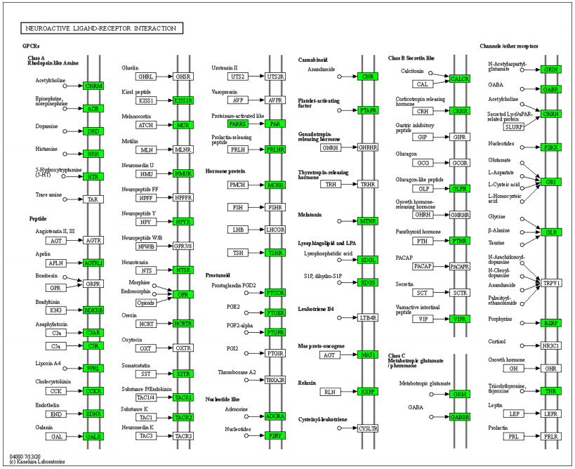
| [31] | J. Pal, V. Patil, A. Kumar, K. Kaur, C. Sarkar, K. Somasundaram, Genetic landscape of glioma reveals defective neuroactive ligand receptor interaction pathway as a poor prognosticator in glioblastoma patients, AACR, 77 (2017), 2454–2454. |
| [32] | R. Wang, J. Wei, Z. Li, Y. Tian, C. Du, Bioinformatical analysis of gene expression signatures of different glioma subtypes, Oncol. Lett., 15 (2018), 2807–2814. |
| [33] |
P. J. Heagerty, T. Lumley, M. S. Pepe, Time-dependent roc curves for censored survival data and a diagnostic marker, Biometrics, 56 (2000), 337–344. doi: 10.1111/j.0006-341X.2000.00337.x
|
 Supplementary Table S1.xlsx Supplementary Table S1.xlsx |
 |
| Supplementary Table S2.xlsx |
|
| Supplementary Table S3.xlsx |
|
| Supplementary Table S4.xlsx |
|
| Supplementary Table S5.xlsx |
|
| Supplementary Table S6.xlsx |
|
| Supplementary Table S7.xlsx |
|
Figures(11) / Tables(5)

Chen Ma, Zhihao Yao, Qinran Zhang, Xiufen Zou. Quantitative integration of radiomic and genomic data improves survival prediction of low-grade glioma patients[J]. Mathematical Biosciences and Engineering, 2021, 18(1): 727-744. doi: 10.3934/mbe.2021039


 DownLoad: