Citation: Zhen Liu, Pande Zhang, Ming Yan, Yimin Xie, Guangzhi Huang. Additive manufacturing of specific ankle-foot orthoses for persons after stroke: A preliminary study based on gait analysis data[J]. Mathematical Biosciences and Engineering, 2019, 16(6): 8134-8143. doi: 10.3934/mbe.2019410

| [1] | E. Cakar, O. Durmus, L. Tekin, et al., The ankle-foot orthosis improves balance and reduces fall risk of chronic spastic hemiparetic patients, Eur. J. Phy. Rehab. Med., 46 (2010), 363-368. |
| [2] | S. F. Tyson, E. Sadeghi-Demneh and C. J. Nester, A systematic review and meta-analysis of the effect of an ankle-foot orthosis on gait biomechanics after stroke, Clin. Rehabil., 27 (2013), 879-891. |
| [3] | V. Bouchalová, E. Houben, D. Tancsik, et al., The influence of an ankle-foot orthosis on the spatiotemporal gait parameters and functional balance in chronic stroke patients, J. Phys. Ther. Sci., 28 (2016), 1621-1628. |
| [4] | S. Telfer, J. Pallari, J. Munguia, et al., Embracing additive manufacture: Implications for foot and ankle orthosis design, BMC Musculoskelet Disord., 13 (2012), 84. |
| [5] | S. Milusheva, D. Tochev, L. Stefanova, et al., Virtual models and prototype of individual ankle foot orthosis. in ISB XXth Congress—ASB29th Annual Meeting, 2005, Cleveland, Ohio. |
| [6] | A. S. Salles and D. E. Gyi, An evaluation of personalised insoles developed using additive manufacturing, J. Sports Sci., 31 (2013), 442-450. |
| [7] | C. E. Dombroski, M. E. Balsdon and A. Froats, The use of a low cost 3D scanning and printing tool in the manufacture of custom-made foot orthoses: A preliminary study, BMC Res. Notes, 7 (2014), 443. |
| [8] | C. Lunsford, G. Grindle, B. Salatin, et al., Innovations with 3-Dimensional printing in physical medicine and rehabilitation: A review of the literature, PMR, 8 (2016), 1201-1212. |
| [9] | E. Wojciechowski, A.Y. Chang, D. Balassone, et al., Feasibility of designing, manufacturing and delivering 3D printed ankle-foot orthoses: A systematic review, J. Foot Ankle Res., 12 (2019), 11. |
| [10] | G. B. Kim, S. Lee, H. Kim, et al., Three-dimensional printing: Basic principles and applications in medicine and radiology, Korean J. Radiol, 17 (2016), 182-197. |
| [11] | M. C. Faustini, R. R. Neptune, R. H. Crawford et al., Manufacture of passive dynamic ankle-foot orthoses using selective laser sintering. IEEE Trans. Biomed. Eng., 55 (2008), 784-790. |
| [12] | E. S. Schrank and S. J. Stanhope, Dimensional accuracy of ankle-foot orthoses constructed by rapid customization and manufacturing framework, J. Rehabil. Res. Dev., 48 (2011), 31-42. |
| [13] | C. Mavroidis, R. G. Ranky, M.L. Sivak, et al., Patient specific ankle-foot orthoses using rapid prototyping, J. Neuroeng. Rehabil., 8 (2011),1. |
| [14] | E. S. Schrank, L. Hitch, K. Wallace, et al., Assessment of a virtual functional prototyping process for rapid manufacture of passive-dynamic ankle-foot orthoses, J. Biomech. Eng., 135 (2013), 101011-101017. |
| [15] | V. Creylman, L. Muraru, J. Pallari, et al., Gait assessment during the initial fitting of customized selective laser sintering ankle foot orthoses insubjects with drop foot, Prosthet. Orthot. Int., 37 (2013), 132-138. |
| [16] | R. K. Chen, Y. Jin, J. Wensman, et al., Additive manufacturing of custom orthoses and prostheses-A review, Addit. Manufact., 12 (2016), 77-89. |
| [17] | G. N. Levy, R. Schindel and J. P. Kruth, Rapid manufacturing and rapid tooling with layer manufacture (LM) technologies, state of the art and future perspectives, CIRP-Ann-Manuf Techn., 52 (2003), 589-609. |
| [18] | HP Inc.: Products-PA12 [www8.hp.com/us/en/printers/3d-printers/materials.html]. |
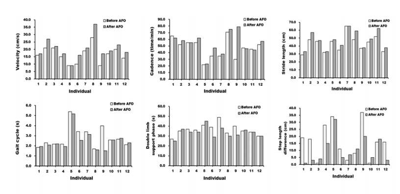
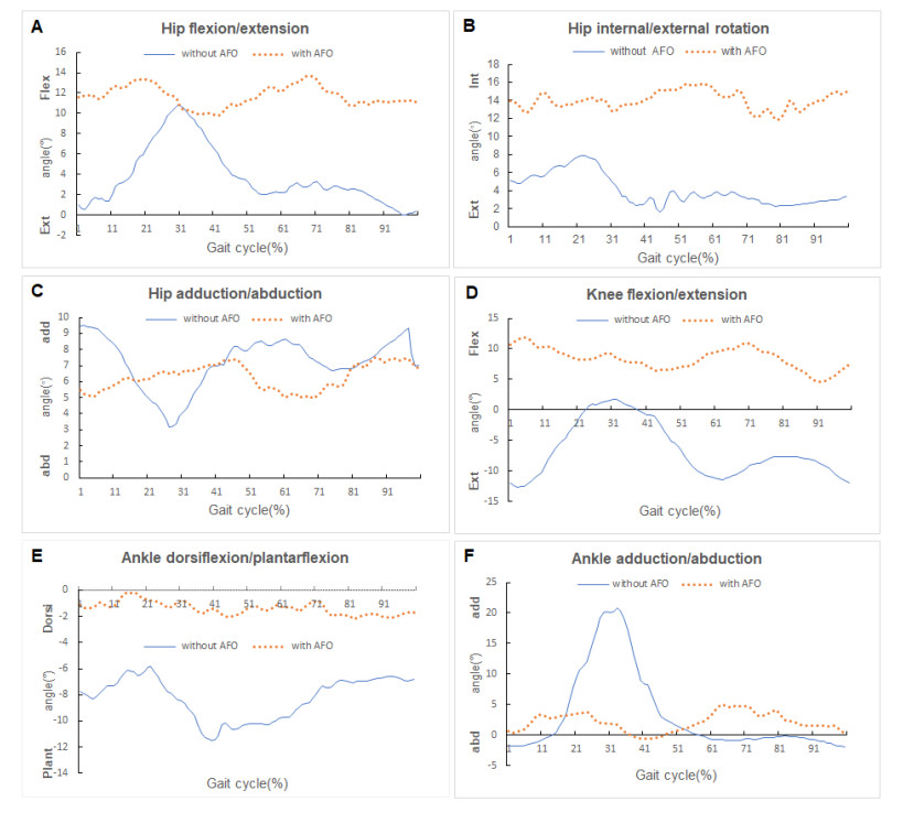
| [19] | K. K. Patterson, W. H. Gage, D. Brooks, et al., Evaluation of gait symmetry after stroke: A comparison of current methods and recommendations for standardization, Gait Posture., 31 (2010), 241-246. |
| [20] | M. Drużbicki, A. Guzik, G. Przysada, et al., Changes in gait symmetry after training on a treadmill with biofeedback in chronic stroke patients: A 6-month follow-up from a randomized controlled trial, Med. Sci. Monit., 22 (2016), 4859-4868. |
| [21] | P. Tack, J. Victor, P. Gemmel, et al., 3D-printing techniques in a medical setting: A systematic literature review, Biomed. Eng. Online, 15 (2016), 115. |
| [22] | C. L. Ventola, Medical applications for 3D printing: Current and projected uses, 39 (2014), 704-711. |
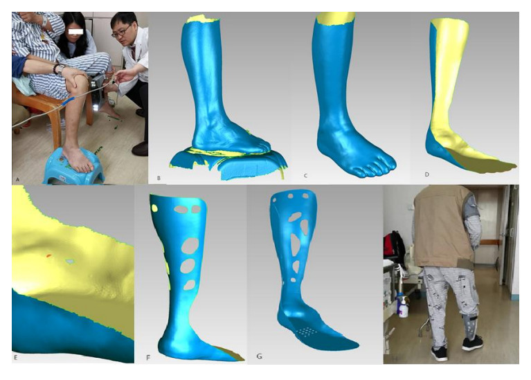
Figures(3) / Tables(2)

Zhen Liu, Pande Zhang, Ming Yan, Yimin Xie, Guangzhi Huang. Additive manufacturing of specific ankle-foot orthoses for persons after stroke: A preliminary study based on gait analysis data[J]. Mathematical Biosciences and Engineering, 2019, 16(6): 8134-8143. doi: 10.3934/mbe.2019410


 DownLoad: