Spatangoid heart urchins are dominant bioturbators in marine soft-sediment ecosystems worldwide. Their repeated sediment reworking prevents biogeochemical sediment stratification and colonization by other species, with implications for sedimentary reaction processes that affect the local sediment–seawater solute exchange. Here, we used a simple exclusion experiment to investigate how a subtidal Echinocardium cordatum population (18.2 ± 6.7 individuals m−2), foraging at an individual speed of ~45 cm per day affects the sediment–seawater solute exchange. To do so, we removed all heart urchins from eight one-meter-diameter areas of the 10-m deep seafloor of Man O'War Bay, Hauraki Gulf, New Zealand, and prevented recolonization and thus sediment reworking for 56 days. Subsequently, we measured the sediment–seawater exchange of O2, NO3–, NO2–, NH4+, and N2 both within and outside the exclusion areas, under light or dark conditions, and found no difference. The absence of a legacy effect of foraging E. cordatum after their removal suggests that, at least in this habitat, their influence on the sediment–seawater solute exchange may be limited to sediment being displaced in the immediate surrounding of the urchin. This unexpected result underlines the importance of evaluating the influence of bioturbators on the sediment–seawater solute exchange in the context of local environmental conditions, animal behavior, and population characteristics.
Citation: Roen McLeod, Michelle N. Simone, Kay Vopel. Effects of sediment disturbance by the heart urchin Echinocardium cordatum on the sediment–seawater solute exchange: An exclusion experiment[J]. AIMS Geosciences, 2024, 10(3): 484-497. doi: 10.3934/geosci.2024025
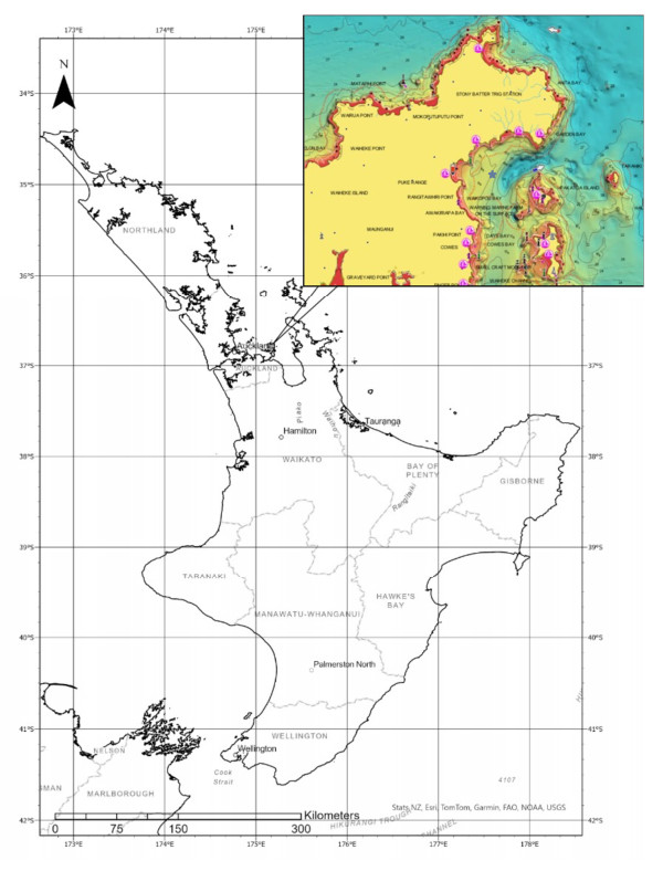
Spatangoid heart urchins are dominant bioturbators in marine soft-sediment ecosystems worldwide. Their repeated sediment reworking prevents biogeochemical sediment stratification and colonization by other species, with implications for sedimentary reaction processes that affect the local sediment–seawater solute exchange. Here, we used a simple exclusion experiment to investigate how a subtidal Echinocardium cordatum population (18.2 ± 6.7 individuals m−2), foraging at an individual speed of ~45 cm per day affects the sediment–seawater solute exchange. To do so, we removed all heart urchins from eight one-meter-diameter areas of the 10-m deep seafloor of Man O'War Bay, Hauraki Gulf, New Zealand, and prevented recolonization and thus sediment reworking for 56 days. Subsequently, we measured the sediment–seawater exchange of O2, NO3–, NO2–, NH4+, and N2 both within and outside the exclusion areas, under light or dark conditions, and found no difference. The absence of a legacy effect of foraging E. cordatum after their removal suggests that, at least in this habitat, their influence on the sediment–seawater solute exchange may be limited to sediment being displaced in the immediate surrounding of the urchin. This unexpected result underlines the importance of evaluating the influence of bioturbators on the sediment–seawater solute exchange in the context of local environmental conditions, animal behavior, and population characteristics.

| [1] |
Aller RC, Aller JY (1998) The effect of biogenic irrigation intensity and solute exchange on diagenetic reaction rates in marine sediments. J Mar Res 56: 905–936. https://doi.org/10.1357/002224098321667413 doi: 10.1357/002224098321667413

|
| [2] |
Banta G, Holmer M, Jensen M, et al. (1999) Effects of two polychaete worms, Nereis diversicolor and Arenicola marina, on aerobic and anaerobic decomposition in a sandy marine sediment. Aquat Microb Ecol 19: 189–204. https://doi.org/10.3354/ame019189 doi: 10.3354/ame019189
|
| [3] | Solan M, Wigham BD (2005) Biogenic particle reworking and bacterial–invertebrate interactions in marine sediments. In: Kristensen E, Haese RR, Kostka JE Eds., Coastal and Estuarine Studies, Vol. 60,105–124. American Geophysical Union. https://doi.org/10.1029/CE060p0105 |
| [4] |
Kristensen E, Penha-Lopes G, Delefosse M, et al. (2012) What is bioturbation? The need for a precise definition for fauna in aquatic sciences. Mar Ecol Prog Ser 446: 285–302. https://doi.org/10.3354/meps09506 doi: 10.3354/meps09506
|
| [5] |
Forster S, Graf G, Kitlar J, et al. (1995) Effects of bioturbation in oxic and hypoxic conditions: A microcosm experiment with a North Sea sediment community. Mar Ecol Prog Ser 116: 153–161. https://doi.org/10.3354/meps116153 doi: 10.3354/meps116153
|
| [6] |
Lohrer AM, Thrush SF, Gibbs MM (2004) Bioturbators enhance ecosystem function through complex biogeochemical interactions. Nature 431: 1092–1095. https://doi.org/10.1038/nature03042 doi: 10.1038/nature03042
|
| [7] |
Volkenborn N, Hedtkamp SIC, van Beusekom JEE, et al. (2007) Effects of bioturbation and bioirrigation by lugworms (Arenicola marina) on physical and chemical sediment properties and implications for intertidal habitat succession. Estuarine Coastal Shelf Sci 74: 331–343. https://doi.org/10.1016/j.ecss.2007.05.001 doi: 10.1016/j.ecss.2007.05.001
|
| [8] |
Osinga R, Kop AJ, Malschaert JFP, et al. (1997) Effects of the sea urchin Echinocardium cordatum on bacterial production and carbon flow in experimental benthic systems under increasing organic loading. J Sea Res 37: 109–121. https://doi.org/10.1016/S1385-1101(97)00003-8 doi: 10.1016/S1385-1101(97)00003-8
|
| [9] |
Lohrer AM, Thrush SF, Hunt L, et al. (2005) Rapid reworking of subtidal sediments by burrowing spatangoid urchins. J Exp Mar Biol Ecol 321: 155–169. https://doi.org/10.1016/j.jembe.2005.02.002 doi: 10.1016/j.jembe.2005.02.002
|
| [10] |
Lohrer AM, Thrush SF, Hewitt JE, et al. (2015) The up-scaling of ecosystem functions in a heterogeneous world. Sci Rep 5: 10349. https://doi.org/10.1038/srep10349 doi: 10.1038/srep10349
|
| [11] |
Nakamura Y (2001) Autoecology of the heart urchin, Echinocardium cordatum, in the muddy sediment of the Seto Inland Sea, Japan. J Mar Biol Assoc U K 81: 289–297. https://doi.org/10.1017/S0025315401003769 doi: 10.1017/S0025315401003769
|
| [12] | De Ridder C, Saucède T (2020) Echinocardium cordatum. In: Developments in Aquaculture and Fisheries Science, Vol. 43,337–357. Elsevier. https://doi.org/10.1016/B978-0-12-819570-3.00020-2 |
| [13] |
Buchanan JB (1966) The biology of Echinocardium cordatum[Echinodermata: Spatangoidea] from different habitats. J Mar Biol Assoc U K 46: 97–114. https://doi.org/10.1017/S0025315400017574 doi: 10.1017/S0025315400017574
|
| [14] |
Foster-Smith RL (1978) An analysis of water flow in tube-living animals. J Exp Mar Biol Ecol 34: 73–95. https://doi.org/10.1016/0022-0981(78)90058-8 doi: 10.1016/0022-0981(78)90058-8
|
| [15] |
Vopel K, Vopel A, Thistle D, Hancock N (2007) Effects of spatangoid heart urchins on O2 supply into coastal sediment. Mar Ecol Prog Ser 333: 161–171. https://doi.org/10.3354/meps333161 doi: 10.3354/meps333161
|
| [16] |
Smith AB (1980) The structure and arrangement of echinoid tubercles. Philos Trans R Soc B 289: 1–54. https://doi.org/10.1098/rstb.1980.0026 doi: 10.1098/rstb.1980.0026
|
| [17] |
Boon AR, Duineveld GCA (2012) Phytopigments and fatty acids in the gut of the deposit-feeding heart urchin Echinocardium cordatum in the southern North Sea: Selective feeding and its contribution to the benthic carbon budget. J Sea Res 67: 77–84. https://doi.org/10.1016/j.seares.2011.10.004 doi: 10.1016/j.seares.2011.10.004
|
| [18] | De Ridder C, Jangoux M, Impe E (2020) Food selection and absorption efficiency in the spatangoid echinoid, Echinocardium cordatum (Echinodermata). In: Keegan BF, O'Connor BDS, Echinodermata 1st ed., 245–251. CRC Press. https://doi.org/10.1201/9781003079224-48 |
| [19] |
Hollertz K, Duchêne JC (2001) Burrowing behaviour and sediment reworking in the heart urchin Brissopsis lyrifera Forbes (Spatangoida). Mar Biol 139: 951–957. https://doi.org/10.1007/s002270100629 doi: 10.1007/s002270100629
|
| [20] |
Kanazawa K (1995) How spatangoids produce their traces: Relationship between burrowing mechanism and trace structure. Lethaia 28: 211–219. https://doi.org/10.1111/j.1502-3931.1995.tb01424.x doi: 10.1111/j.1502-3931.1995.tb01424.x
|
| [21] | Aller RC (1982) The Effects of macrobenthos on chemical properties of marine sediment and overlying water. In: McCall PL, Tevesz MJS, Animal-Sediment Relations Vol. 100, 53–102. Springer US. https://doi.org/10.1007/978-1-4757-1317-6_2 |
| [22] |
Glud RN (2008) Oxygen dynamics of marine sediments. Mar Biol Res 4:243–289. https://doi.org/10.1080/17451000801888726 doi: 10.1080/17451000801888726
|
| [23] |
Braeckman U, Provoost P, Gribsholt B, et al. (2010) Role of macrofauna functional traits and density in biogeochemical fluxes and bioturbation. Mar Ecol Prog Ser 399: 173–186. https://doi.org/10.3354/meps08336 doi: 10.3354/meps08336
|
| [24] |
Wilson PS, Vopel K (2012) Estimating the in situ distribution of acid volatile sulfides from sediment profile images. Limnol Oceanogr Methods 10: 1070–1077. https://doi.org/10.4319/lom.2012.10.1070 doi: 10.4319/lom.2012.10.1070
|
| [25] |
Wong KC, O'Shea S (2010) Sediment macrobenthos off eastern Waiheke Island, Hauraki Gulf, New Zealand. N Z J Mar Freshwater Res 44: 149–165. https://doi.org/10.1080/00288330.2010.498088 doi: 10.1080/00288330.2010.498088
|
| [26] |
Rho T, Coverly S, Kim ES, et al. (2016) Practical considerations for the segmented-flow analysis of nitrate and ammonium in seawater and the avoidance of matrix effects. Ocean Sci J 50: 709–720. https://doi.org/10.1007/s12601-015-0064-7 doi: 10.1007/s12601-015-0064-7
|
| [27] | R Core Team (2022) R: A language and environment for statistical computing. [Computer software]. R Foundation for Statistical Computing. Available from: https://www.R-project.org/. |
| [28] |
Needham HR, Pilditch CA, Lohrer AM, Thrush SF (2011) Context-specific bioturbation mediates changes to ecosystem functioning. Ecosystems 14: 1096–1109. https://doi.org/10.1007/s10021-011-9468-0 doi: 10.1007/s10021-011-9468-0
|
| [29] |
Seike K, Sassa S, Shirai K, et al. (2022) Sediment hardness and water temperature affect the burrowing of Echinocardium cordatum: Implications for mass mortality during the 2011 earthquake–liquefaction–tsunami disaster. Estuarine Coastal Shelf Sci 267: 107763. https://doi.org/10.1016/j.ecss.2022.107763 doi: 10.1016/j.ecss.2022.107763
|
| [30] |
Aoki L, McGlathery K (2019) High rates of N fixation in seagrass sediments measured via a direct 30N2 push-pull method. Mar Ecol Prog Ser 616: 1–11. https://doi.org/10.3354/meps12961 doi: 10.3354/meps12961
|
| [31] |
Fulweiler RW, Nixon SW, Buckley BA, et al. (2007) Reversal of the net dinitrogen gas flux in coastal marine sediments. Nature 448: 180–182. https://doi.org/10.1038/nature05963 doi: 10.1038/nature05963
|
| [32] |
Newell SE, McCarthy MJ, Gardner WS, et al. (2016) Sediment nitrogen fixation: A call for re-evaluating coastal N budgets. Estuaries Coasts 39: 1626–1638. https://doi.org/10.1007/s12237-016-0116-y doi: 10.1007/s12237-016-0116-y
|
| [33] |
Whiting GJ, Gandy EL, Yoch DC (1986) Tight coupling of root-associated nitrogen fixation and plant photosynthesis in the salt marsh grass Spartina alterniflora and carbon dioxide enhancement of nitrogenase activity. Appl Environ Microbiol 52: 108–113. https://doi.org/10.1128/aem.52.1.108-113.1986 doi: 10.1128/aem.52.1.108-113.1986
|
| [34] |
Bertics VJ, Löscher CR, Salonen I, et al. (2013) Occurrence of benthic microbial nitrogen fixation coupled to sulfate reduction in the seasonally hypoxic Eckernförde Bay, Baltic Sea. Biogeosciences 10: 1243–1258. https://doi.org/10.5194/bg-10-1243-2013 doi: 10.5194/bg-10-1243-2013
|
| [35] |
Joshi HM, Tabita FR (1996) A global two component signal transduction system that integrates the control of photosynthesis, carbon dioxide assimilation, and nitrogen fixation. Proc Natl Acad Sci 93: 14515–14520. https://doi.org/10.1073/pnas.93.25.14515 doi: 10.1073/pnas.93.25.14515
|
| [36] |
Bombar D, Paerl RW, Riemann L (2016) Marine non-cyanobacterial diazotrophs: Moving beyond molecular detection. Trends Microbiol 24: 916–927. https://doi.org/10.1016/j.tim.2016.07.002 doi: 10.1016/j.tim.2016.07.002
|
| [37] | Stal LJ (2015) Nitrogen Fixation in Cyanobacteria. In: Encyclopedia of Life Sciences, 1st ed., 1–9. Wiley. https://doi.org/10.1002/9780470015902.a0021159.pub2 |


Figures(3) / Tables(2)

Roen McLeod, Michelle N. Simone, Kay Vopel. Effects of sediment disturbance by the heart urchin Echinocardium cordatum on the sediment–seawater solute exchange: An exclusion experiment[J]. AIMS Geosciences, 2024, 10(3): 484-497. doi: 10.3934/geosci.2024025


 DownLoad: