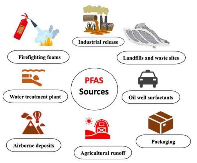
Emerging per and poly-fluoroalkyl substances (PFASs) are very resistant to degradation and have negative impacts on human and environmental health at very low concentrations. The initial stage in removing PFASs from contaminated locations is their detection and quantification. Particularly utilized in this context are Gas Chromatography-Mass Spectrometry (GC-MS), Liquid Chromatography-Tandem Mass Spectrometry (LC-MS/MS), and High-Performance Liquid Chromatography (HPLC). In this review, we seek to contribute to our understanding of the state-of-the-art in emerging PFAS by offering a complete analysis of PFAS in environments, taking into consideration their sources, classes, and properties. Polyfluoroalkyl ether substances (PFAES), short-chain PFA, replacement PFA, and fluorotelomer-based substances can bioaccumulate in living species, making their detection even more necessary. We intend to provide researchers with an overview of the current state of research on PFASs in environments, encompassing the toxicological effects and their detection and quantification methods, serving as a guide for current and future studies.
Citation: Eliasu Issaka, Mabruk Adams, Enock Adjei Agyekum, Josephine Baffoe, Blessing Tornyeava. Per- and poly-fluoroalkyl substances: A review of sources, properties, chromatographic detection, and toxicological implications[J]. AIMS Environmental Science, 2025, 12(2): 321-351. doi: 10.3934/environsci.2025015
Emerging per and poly-fluoroalkyl substances (PFASs) are very resistant to degradation and have negative impacts on human and environmental health at very low concentrations. The initial stage in removing PFASs from contaminated locations is their detection and quantification. Particularly utilized in this context are Gas Chromatography-Mass Spectrometry (GC-MS), Liquid Chromatography-Tandem Mass Spectrometry (LC-MS/MS), and High-Performance Liquid Chromatography (HPLC). In this review, we seek to contribute to our understanding of the state-of-the-art in emerging PFAS by offering a complete analysis of PFAS in environments, taking into consideration their sources, classes, and properties. Polyfluoroalkyl ether substances (PFAES), short-chain PFA, replacement PFA, and fluorotelomer-based substances can bioaccumulate in living species, making their detection even more necessary. We intend to provide researchers with an overview of the current state of research on PFASs in environments, encompassing the toxicological effects and their detection and quantification methods, serving as a guide for current and future studies.

| [1] | Elmore SA, Boorman GA (2023) Environmental Toxicologic Pathology and Human Health. Haschek and Rousseaux's Handbook of Toxicologic Pathology, Volume 3: Environmental Toxicologic Pathology and Major Toxicant Classes 3–32. https://doi.org/10.1016/B978-0-443-16153-7.00001-0 |
| [2] |
Sadia M, Nollen I, Helmus R, et al. (2023) Occurrence, Fate, and Related Health Risks of PFAS in Raw and Produced Drinking Water. Environ Sci Technol 57: 3062–3074. https://doi.org/10.1021/acs.est.2c06015 doi: 10.1021/acs.est.2c06015

|
| [3] |
Lohmann R, Cousins IT, Dewitt JC, et al. (2020) Are Fluoropolymers Really of Low Concern for Human and Environmental Health and Separate from Other PFAS? Environ Sci Technol 54: 12820–12828. https://doi.org/10.1021/acs.est.0c03244 doi: 10.1021/acs.est.0c03244
|
| [4] |
Yan S, Murtadha B, Foster G, et al. (2023) Development and testing of novel functionalized polymeric thin-films for equilibrium passive sampling of PFAS compounds in water. Chemical Engineering Journal 477. https://doi.org/10.1016/j.cej.2023.146734 doi: 10.1016/j.cej.2023.146734
|
| [5] |
Bayode AA, Emmanuel SS, Akinyemi AO, et al. (2024) Innovative techniques for combating a common enemy forever chemicals: A comprehensive approach to mitigating per- and polyfluoroalkyl substances (PFAS) contamination. Environ Res 261. https://doi.org/10.1016/j.envres.2024.119719 doi: 10.1016/j.envres.2024.119719
|
| [6] |
Bayode AA, Emmanuel SS, Akinyemi AO, et al. (2024) Innovative techniques for combating a common enemy forever chemicals: A comprehensive approach to mitigating per- and polyfluoroalkyl substances (PFAS) contamination. Environ Res 261. https://doi.org/10.1016/j.envres.2024.119719 doi: 10.1016/j.envres.2024.119719
|
| [7] |
Park M, Wu S, Lopez IJ, et al. (2020) Adsorption of perfluoroalkyl substances (PFAS) in groundwater by granular activated carbons: Roles of hydrophobicity of PFAS and carbon characteristics. Water Res 170: 115364. https://doi.org/10.1016/j.watres.2019.115364 doi: 10.1016/j.watres.2019.115364
|
| [8] |
Tian Y, Zhou Q, Zhang L, et al. (2023) In utero exposure to per-/polyfluoroalkyl substances (PFASs): Preeclampsia in pregnancy and low birth weight for neonates. Chemosphere 313: 137490. https://doi.org/10.1016/j.chemosphere.2022.137490 doi: 10.1016/j.chemosphere.2022.137490
|
| [9] |
Ashley-Martin J, Hammond J, Velez MP (2024) Assessing preconception exposure to environmental chemicals and fecundity: Strategies, challenges, and research priorities. Reproductive Toxicology 125. https://doi.org/10.1016/j.reprotox.2024.108578 doi: 10.1016/j.reprotox.2024.108578
|
| [10] |
Teymoorian T, Munoz G, Sauvé S (2025) PFAS contamination in tap water: Target and suspect screening of zwitterionic, cationic, and anionic species across Canada and beyond. Environ Int 195. https://doi.org/10.1016/j.envint.2025.109250 doi: 10.1016/j.envint.2025.109250
|
| [11] |
Merino N, Qu Y, Deeb RA, et al. (2016) Degradation and Removal Methods for Perfluoroalkyl and Polyfluoroalkyl Substances in Water. Environ Eng Sci 33: 615–649. https://doi.org/10.1016/j.envint.2025.109250 doi: 10.1016/j.envint.2025.109250
|
| [12] |
Berhanu A, Mutanda I, Taolin J, et al. (2023) A review of microbial degradation of per- and polyfluoroalkyl substances (PFAS): Biotransformation routes and enzymes. Science of the Total Environment 859. https://doi.org/10.1016/j.scitotenv.2022.160010 doi: 10.1016/j.scitotenv.2022.160010
|
| [13] |
Kuppan N, Padman M, Mahadeva M, et al. (2024) A comprehensive review of sustainable bioremediation techniques: Eco friendly solutions for waste and pollution management. Waste Management Bulletin 2: 154–171. https://doi.org/10.1016/j.wmb.2024.07.005 doi: 10.1016/j.wmb.2024.07.005
|
| [14] |
Chukwuonye GN, Alqattan ZA, Jones M, et al. (2024) Toxic layering and compound extremes: Per- and polyfluoroalkyl substances (PFAS) exposure in rural, environmental justice copper mining communities. Science of the Total Environment 957. https://doi.org/10.1016/j.scitotenv.2024.177767 doi: 10.1016/j.scitotenv.2024.177767
|
| [15] | Wang F, Xiang L, Sze-Yin Leung K, et al. (2024) Emerging contaminants: A One Health perspective. Innovation 5. |
| [16] |
Groffen T, Bervoets L, Jeong Y, et al. (2021) A rapid method for the detection and quantification of legacy and emerging per- and polyfluoroalkyl substances (PFAS) in bird feathers using UPLC-MS/MS. Journal of Chromatography B 1172: 122653. https://doi.org/10.1016/j.jchromb.2021.122653 doi: 10.1016/j.jchromb.2021.122653
|
| [17] |
Dodds JN, Kirkwood-Donelson KI, Boatman AK, et al. (2024) Evaluating Solid Phase Adsorption Toxin Tracking (SPATT) for passive monitoring of per- and polyfluoroalkyl substances (PFAS) with Ion Mobility Spectrometry-Mass Spectrometry (IMS-MS). Science of The Total Environment 947: 174574. https://doi.org/10.1016/j.scitotenv.2024.174574 doi: 10.1016/j.scitotenv.2024.174574
|
| [18] |
Manojkumar Y, Pilli S, Rao PV, et al. (2023) Sources, occurrence and toxic effects of emerging per- and polyfluoroalkyl substances (PFAS). Neurotoxicol Teratol 97: 107174. https://doi.org/10.1016/j.ntt.2023.107174 doi: 10.1016/j.ntt.2023.107174
|
| [19] |
De Silva AO, Armitage JM, Bruton TA, et al. (2021) PFAS Exposure Pathways for Humans and Wildlife: A Synthesis of Current Knowledge and Key Gaps in Understanding. Environ Toxicol Chem 40: 631–657. https://doi.org/10.1002/etc.4935 doi: 10.1002/etc.4935
|
| [20] |
Gaines LGT (2023) Historical and current usage of per- and polyfluoroalkyl substances (PFAS): A literature review. Am J Ind Med 66: 353–378. https://doi.org/10.1002/ajim.23362 doi: 10.1002/ajim.23362
|
| [21] |
Brunn H, Arnold G, Kö rner W, et al. (2023) PFAS: forever chemicals—persistent, bioaccumulative and mobile. Reviewing the status and the need for their phase out and remediation of contaminated sites. Environ Sci Eur 35. https://doi.org/10.1186/s12302-023-00721-8 doi: 10.1186/s12302-023-00721-8
|
| [22] |
Cookson ES, Detwiler RL (2022) Global patterns and temporal trends of perfluoroalkyl substances in municipal wastewater: A meta-analysis. Water Res 221. https://doi.org/10.1016/j.watres.2022.118784 doi: 10.1016/j.watres.2022.118784
|
| [23] |
Jha G, Kankarla V, McLennon E, et al. (2021) Per- and Polyfluoroalkyl Substances (PFAS) in Integrated Crop–Livestock Systems: Environmental Exposure and Human Health Risks. Int J Environ Res Public Health 18: 12550. https://doi.org/10.3390/ijerph182312550 doi: 10.3390/ijerph182312550
|
| [24] |
Lin H, Lao JY, Wang Q, et al. (2022) Per- and polyfluoroalkyl substances in the atmosphere of waste management infrastructures: Uncovering secondary fluorotelomer alcohols, particle size distribution, and human inhalation exposure. Environ Int 167. https://doi.org/10.1016/j.envint.2022.107434 doi: 10.1016/j.envint.2022.107434
|
| [25] |
Wang P, Zhang M, Li Q, et al. (2021) Atmospheric diffusion of perfluoroalkyl acids emitted from fluorochemical industry and its associated health risks. Environ Int 146. https://doi.org/10.1016/j.envint.2020.106247 doi: 10.1016/j.envint.2020.106247
|
| [26] |
Buck RC, Korzeniowski SH, Laganis E, et al. (2021) Identification and classification of commercially relevant per- and poly-fluoroalkyl substances (PFAS). Integr Environ Assess Manag 17: 1045–1055. https://doi.org/10.1002/ieam.4450 doi: 10.1002/ieam.4450
|
| [27] |
Zhang X, Liu J, Zhang H, et al. (2025) Highly selective guanidine-linked covalent organic framework for efficient removal of perfluoroalkyl carboxylic acids from water samples. Sep Purif Technol 357: 130039. https://doi.org/10.1016/j.seppur.2024.130039 doi: 10.1016/j.seppur.2024.130039
|
| [28] |
Xu C, Yin S, Liu Y, et al. (2019) Prenatal exposure to chlorinated polyfluoroalkyl ether sulfonic acids and perfluoroalkyl acids: Potential role of maternal determinants and associations with birth outcomes. J Hazard Mater 380. https://doi.org/10.1016/j.jhazmat.2019.120867 doi: 10.1016/j.jhazmat.2019.120867
|
| [29] |
Brendel S, Fetter É, Staude C, et al. (2018) Short-chain perfluoroalkyl acids: environmental concerns and a regulatory strategy under REACH. Environ Sci Eur 30: 1–11. https://doi.org/10.1186/s12302-018-0134-4 doi: 10.1186/s12302-018-0134-4
|
| [30] |
Marques E, Pfohl M, Wei W, et al. (2022) Replacement per- and polyfluoroalkyl substances (PFAS) are potent modulators of lipogenic and drug metabolizing gene expression signatures in primary human hepatocytes. Toxicol Appl Pharmacol 442. https://doi.org/10.1016/j.taap.2022.115991 doi: 10.1016/j.taap.2022.115991
|
| [31] |
Roth K, Yang Z, Agarwal M, et al. (2021) Exposure to a mixture of legacy, alternative, and replacement per- and polyfluoroalkyl substances (PFAS) results in sex-dependent modulation of cholesterol metabolism and liver injury. Environ Int 157. https://doi.org/10.1016/j.envint.2021.106843 doi: 10.1016/j.envint.2021.106843
|
| [32] |
Pan Z, Miao W, Wang C, et al. (2021) 6:2 Cl-PFESA has the potential to cause liver damage and induce lipid metabolism disorders in female mice through the action of PPAR-γ. Environmental Pollution 287: 117329. https://doi.org/10.1016/j.envpol.2021.117329 doi: 10.1016/j.envpol.2021.117329
|
| [33] |
Pervez MN, Jiang T, Mahato JK, et al. (2024) Surface Modification of Graphene Oxide for Fast Removal of Per- and Polyfluoroalkyl Substances (PFAS) Mixtures from River Water. ACS ES and T Water 4: 2968–2980. https://doi.org/10.1021/acsestwater.4c00187 doi: 10.1021/acsestwater.4c00187
|
| [34] |
Xu C, Yin S, Liu Y, et al. (2019) Prenatal exposure to chlorinated polyfluoroalkyl ether sulfonic acids and perfluoroalkyl acids: Potential role of maternal determinants and associations with birth outcomes. J Hazard Mater 380. https://doi.org/10.1016/j.jhazmat.2019.120867 doi: 10.1016/j.jhazmat.2019.120867
|
| [35] |
Gomez-Ruiz B, Gómez-Lavín S, Diban N, et al. (2017) Boron doped diamond electrooxidation of 6:2 fluorotelomers and perfluorocarboxylic acids. Application to industrial wastewaters treatment. Journal of Electroanalytical Chemistry 798: 51–57. https://doi.org/10.1016/j.jelechem.2017.05.033 doi: 10.1016/j.jelechem.2017.05.033
|
| [36] | Daramola O (2021) Metabolism of 6: 2 Fluorotelomer Alcohol by CYP 2A6. |
| [37] |
Riedel TP, Wallace MAG, Shields EP, et al. (2021) Low temperature thermal treatment of gas-phase fluorotelomer alcohols by calcium oxide. Chemosphere 272. https://doi.org/10.1016/j.chemosphere.2021.129859 doi: 10.1016/j.chemosphere.2021.129859
|
| [38] |
Kassotis CD, Vandenberg LN, Demeneix BA, et al. (2020) Endocrine-disrupting chemicals: economic, regulatory, and policy implications. Lancet Diabetes Endocrinol 8: 719–730. https://doi.org/10.1016/S2213-8587(20)30128-5 doi: 10.1016/S2213-8587(20)30128-5
|
| [39] |
Manojkumar Y, Pilli S, Rao PV, et al. (2023) Sources, occurrence and toxic effects of emerging per- and polyfluoroalkyl substances (PFAS). Neurotoxicol Teratol 97. https://doi.org/10.1016/j.ntt.2023.107174 doi: 10.1016/j.ntt.2023.107174
|
| [40] |
Thompson D, Zolfigol N, Xia Z, et al. (2024) Recent progress in per- and polyfluoroalkyl substances (PFAS) sensing: A critical mini-review. Sensors and Actuators Reports 100189. https://doi.org/10.1016/j.snr.2024.100189 doi: 10.1016/j.snr.2024.100189
|
| [41] |
Sharma N, Kumar V, Sugumar V, et al. (2024) A comprehensive review on the need for integrated strategies and process modifications for per- and polyfluoroalkyl substances (PFAS) removal: Current insights and future prospects. Case Studies in Chemical and Environmental Engineering 9. https://doi.org/10.1016/j.cscee.2024.100623 doi: 10.1016/j.cscee.2024.100623
|
| [42] |
Lu Y, Gao J, Nguyen HT, et al. (2021) Occurrence of per- and polyfluoroalkyl substances (PFASs) in wastewater of major cities across China in 2014 and 2016. Chemosphere 279. https://doi.org/10.1016/j.chemosphere.2021.130590 doi: 10.1016/j.chemosphere.2021.130590
|
| [43] |
Valencia A, Ordonez D, Sadmani AHMA, et al. (2023) Comparing the removal and fate of long and short chain per- and polyfluoroalkyl substances (PFAS) during surface water treatment via specialty adsorbents. Journal of Water Process Engineering 56. https://doi.org/10.1016/j.jwpe.2023.104345 doi: 10.1016/j.jwpe.2023.104345
|
| [44] |
Giannelli Moneta B, Feo ML, Torre M, et al. (2023) Occurrence of per- and polyfluorinated alkyl substances in wastewater treatment plants in Northern Italy. Science of the Total Environment 894. https://doi.org/10.1016/j.scitotenv.2023.165089 doi: 10.1016/j.scitotenv.2023.165089
|
| [45] |
Zhang YH, Ding TT, Huang ZY, et al. (2023) Environmental exposure and ecological risk of perfluorinated substances (PFASs) in the Shaying River Basin, China. Chemosphere 339. https://doi.org/10.1016/j.chemosphere.2023.139537 doi: 10.1016/j.chemosphere.2023.139537
|
| [46] |
Jeong Y, Da Silva KM, Iturrospe E, et al. (2022) Occurrence and contamination profile of legacy and emerging per- and polyfluoroalkyl substances (PFAS) in Belgian wastewater using target, suspect and non-target screening approaches. J Hazard Mater 437. https://doi.org/10.1016/j.jhazmat.2022.129378 doi: 10.1016/j.jhazmat.2022.129378
|
| [47] |
Zarębska M, Bajkacz S, Hordyjewicz-Baran Z (2024) Assessment of legacy and emerging PFAS in the Oder River: Occurrence, distribution, and sources. Environ Res 118608. https://doi.org/10.1016/j.envres.2024.118608 doi: 10.1016/j.envres.2024.118608
|
| [48] |
Labine LM, Oliveira Pereira EA, Kleywegt S, et al. (2022) Comparison of sub-lethal metabolic perturbations of select legacy and novel perfluorinated alkyl substances (PFAS) in Daphnia magna. Environ Res 212. https://doi.org/10.1016/j.envres.2022.113582 doi: 10.1016/j.envres.2022.113582
|
| [49] | EPA- USA (2024) United States Environmental Protection Agency, Per- and Polyfluoroalkyl Substances (PFAS) | US EPA, 2024. Available from: https://www.epa.gov/pfas. |
| [50] |
Brunn H, Arnold G, Kö rner W, et al. (2023) PFAS: forever chemicals—persistent, bioaccumulative and mobile. Reviewing the status and the need for their phase out and remediation of contaminated sites. Environ Sci Eur 35. https://doi.org/10.1186/s12302-023-00721-8 doi: 10.1186/s12302-023-00721-8
|
| [51] |
Moretti S, Brambilla G, Maffucci F, et al. (2024) Occurrence and pattern of legacy and emerging per- and Poly-FluoroAlkyl substances (PFAS) in eggs of loggerhead turtle Caretta caretta from western Mediterranean. Environmental Pollution 343. https://doi.org/10.1016/j.envpol.2023.123257 doi: 10.1016/j.envpol.2023.123257
|
| [52] |
Ankley GT, Cureton P, Hoke RA, et al. (2021) Assessing the Ecological Risks of Per- and Polyfluoroalkyl Substances: Current State-of-the Science and a Proposed Path Forward. Environ Toxicol Chem 40: 564–605. https://doi.org/10.1002/etc.4869 doi: 10.1002/etc.4869
|
| [53] | Cousins I, DeWitt J, Glüge J, et al. Strategies for grouping per-and polyfluoroalkyl substances (PFAS) to protect human and environmental health. pubs.rsc.org. |
| [54] |
Guelfo JL, Korzeniowski S, Mills MA, et al. (2021) Environmental Sources, Chemistry, Fate, and Transport of Per- and Polyfluoroalkyl Substances: State of the Science, Key Knowledge Gaps, and Recommendations Presented at the August 2019 SETAC Focus Topic Meeting. Environ Toxicol Chem 40: 3234–3260. https://doi.org/10.1002/etc.5182 doi: 10.1002/etc.5182
|
| [55] |
McCord J, Strynar M (2019) Identification of Per- and Polyfluoroalkyl Substances in the Cape Fear River by High Resolution Mass Spectrometry and Nontargeted Screening. Environ Sci Technol 53: 4717–4727. https://doi.org/10.1021/acs.est.8b06017 doi: 10.1021/acs.est.8b06017
|
| [56] |
Strynar M, Dagnino S, McMahen R, et al. (2015) Identification of Novel Perfluoroalkyl Ether Carboxylic Acids (PFECAs) and Sulfonic Acids (PFESAs) in Natural Waters Using Accurate Mass Time-of-Flight Mass Spectrometry (TOFMS). Environ Sci Technol 49: 11622–11630. https://doi.org/10.1021/acs.est.5b01215 doi: 10.1021/acs.est.5b01215
|
| [57] |
Tsianou M, Bedrov D, Alexandridis P (2023) Surfactants in the Environment: Self-Assembly of PFAS Pollutants in Solution and at Interfaces. ACS Symposium Series 1457: 443–462. https://doi.org/10.1021/bk-2023-1457.ch016 doi: 10.1021/bk-2023-1457.ch016
|
| [58] |
Yao J, Sheng N, Guo Y, et al. (2022) Nontargeted Identification and Temporal Trends of Per- and Polyfluoroalkyl Substances in a Fluorochemical Industrial Zone and Adjacent Taihu Lake. Environ Sci Technol 56: 7986–7996. https://doi.org/10.1021/acs.est.2c00891 doi: 10.1021/acs.est.2c00891
|
| [59] |
Jin B, Zhu Y, Zhao W, et al. (2023) Aerobic Biotransformation and Defluorination of Fluoroalkylether Substances (ether PFAS): Substrate Specificity, Pathways, and Applications. Environ Sci Technol Lett 10: 755–761. https://doi.org/10.1021/acs.estlett.3c00411 doi: 10.1021/acs.estlett.3c00411
|
| [60] |
Frigerio G, Cafagna S, Polledri E, et al. (2022) Development and validation of an LC–MS/MS method for the quantitation of 30 legacy and emerging per- and polyfluoroalkyl substances (PFASs) in human plasma, including HFPO-DA, DONA, and cC6O4. Anal Bioanal Chem 414: 1259–1278. https://doi.org/10.1007/s00216-021-03762-1 doi: 10.1007/s00216-021-03762-1
|
| [61] | Liu J, international SA-E, 2013 undefined (2013) Microbial degradation of polyfluoroalkyl chemicals in the environment: a review. Elsevier. https://doi.org/10.1016/j.envint.2013.08.022 |
| [62] | Cousins I, DeWitt J, Glüge J, et al. The high persistence of PFAS is sufficient for their management as a chemical class. pubs.rsc.org. |
| [63] | Meegoda J, Kewalramani J, … BL-… journal of environmental, et al. A review of the applications, environmental release, and remediation technologies of per-and polyfluoroalkyl substances. mdpi.com. |
| [64] | Walkowiak-Kulikowska J (2022) Poly/Perfluorinated Alkyl Substances (PFASs)–Synthetic Methods, Properties and Applications. https://doi.org/10.1039/9781839167591-00022 |
| [65] |
Olsen GW (2015) PFAS Biomonitoring in Higher Exposed Populations. Molecular and Integrative Toxicology 77–125. https://doi.org/10.1007/978-3-319-15518-0_4 doi: 10.1007/978-3-319-15518-0_4
|
| [66] |
Guelfo JL, Korzeniowski S, Mills MA, et al. (2021) Environmental sources, chemistry, Fate, and transport of per‐and Polyfluoroalkyl Substances: State of the science, key knowledge gaps, and recommendations. Wiley Online Library 40: 3234–3260. https://doi.org/10.1002/etc.5182 doi: 10.1002/etc.5182
|
| [67] |
Yu X, Nishimura F, Hidaka T (2018) Effects of microbial activity on perfluorinated carboxylic acids (PFCAs) generation during aerobic biotransformation of fluorotelomer alcohols in activated sludge. Science of the Total Environment 610–611: 776–785. https://doi.org/10.1016/j.scitotenv.2017.08.075 doi: 10.1016/j.scitotenv.2017.08.075
|
| [68] |
Manojkumar Y, Pilli S, Rao PV, et al. (2023) Sources, occurrence and toxic effects of emerging per- and polyfluoroalkyl substances (PFAS). Neurotoxicol Teratol 97. https://doi.org/10.1016/j.ntt.2023.107174 doi: 10.1016/j.ntt.2023.107174
|
| [69] |
Choi YJ, Helbling DE, Liu J, et al. (2022) Microbial biotransformation of aqueous film-forming foam derived polyfluoroalkyl substances. Science of the Total Environment 824. https://doi.org/10.1016/j.scitotenv.2022.153711 doi: 10.1016/j.scitotenv.2022.153711
|
| [70] |
Sharma N, Kumar V, Sugumar V, et al. (2024) A comprehensive review on the need for integrated strategies and process modifications for per- and polyfluoroalkyl substances (PFAS) removal: Current insights and future prospects. Case Studies in Chemical and Environmental Engineering 9. https://doi.org/10.1016/j.cscee.2024.100623 doi: 10.1016/j.cscee.2024.100623
|
| [71] |
Dong S, Yan PF, Liu C, et al. (2023) Assessing aerobic biotransformation of 8:2 fluorotelomer alcohol in aqueous film-forming foam (AFFF)-impacted soils: Pathways and microbial community dynamics. J Hazard Mater 446. https://doi.org/10.1016/j.jhazmat.2022.130629 doi: 10.1016/j.jhazmat.2022.130629
|
| [72] |
Fiedler H, Kennedy T, Henry BJ (2021) A Critical Review of a Recommended Analytical and Classification Approach for Organic Fluorinated Compounds with an Emphasis on Per- and Polyfluoroalkyl Substances. Integr Environ Assess Manag 17: 331–351. https://doi.org/10.1002/ieam.4352 doi: 10.1002/ieam.4352
|
| [73] |
Guelfo JL, Korzeniowski S, Mills MA, et al. (2021) Environmental Sources, Chemistry, Fate, and Transport of Per- and Polyfluoroalkyl Substances: State of the Science, Key Knowledge Gaps, and Recommendations Presented at the August 2019 SETAC Focus Topic Meeting. Environ Toxicol Chem 40: 3234–3260. https://doi.org/10.1002/etc.5182 doi: 10.1002/etc.5182
|
| [74] |
Dasu K, Xia X, Siriwardena D, et al. (2022) Concentration profiles of per- and polyfluoroalkyl substances in major sources to the environment. J Environ Manage 301. https://doi.org/10.1016/j.jenvman.2021.113879 doi: 10.1016/j.jenvman.2021.113879
|
| [75] |
Ritscher A, Wang Z, Scheringer M, et al. (2018) Zürich Statement on Future Actions on Per- and Polyfluoroalkyl Substances (PFASs). Environ Health Perspect 126. https://doi.org/10.1289/EHP4158 doi: 10.1289/EHP4158
|
| [76] | Issaka E, Adams M, Baffoe J, et al. (2024) Covalent organic frameworks: a review of synthesis methods, properties and applications for per- and poly-fluoroalkyl substances removal. Clean Technologies and Environmental Policy 2024 1–28. |
| [77] |
Holmquist H, Schellenberger S, van der Veen I, et al. (2016) Properties, performance and associated hazards of state-of-the-art durable water repellent (DWR) chemistry for textile finishing. Environ Int 91: 251–264. https://doi.org/10.1016/j.envint.2016.02.035 doi: 10.1016/j.envint.2016.02.035
|
| [78] |
Li YF, Ho CY, Liu YJ, et al. (2024) Enhance electrocoagulation-flotation (ECF) removal efficiency perfluorohexanoic acid (PFHxA) by adding surfactants. J Environ Chem Eng 12. https://doi.org/10.1016/j.jece.2023.111773 doi: 10.1016/j.jece.2023.111773
|
| [79] |
Manouras E, Ioannou D, Zeniou A, et al. (2024) Superhydrophobic and oleophobic Nylon, PES and PVDF membranes using plasma nanotexturing: Empowering membrane distillation and contributing to PFAS free hydrophobic membranes. Micro and Nano Engineering 24. https://doi.org/10.1016/j.mne.2024.100269 doi: 10.1016/j.mne.2024.100269
|
| [80] |
Kourtchev I, Sebben BG, Bogush A, et al. (2023) Per- and polyfluoroalkyl substances (PFASs) in urban PM2.5 samples from Curitiba, Brazil. Atmos Environ 309. https://doi.org/10.1016/j.atmosenv.2023.119911 doi: 10.1016/j.atmosenv.2023.119911
|
| [81] |
Ji B, Zhao Y, Yang Y, et al. (2023) Curbing per- and polyfluoroalkyl substances (PFASs): First investigation in a constructed wetland-microbial fuel cell system. Water Res 230. https://doi.org/10.1016/j.watres.2022.119530 doi: 10.1016/j.watres.2022.119530
|
| [82] |
Yamijala SSRKC, Shinde R, Hanasaki K, et al. (2022) Photo-induced degradation of PFASs: Excited-state mechanisms from real-time time-dependent density functional theory. J Hazard Mater 423. https://doi.org/10.1016/j.jhazmat.2021.127026 doi: 10.1016/j.jhazmat.2021.127026
|
| [83] |
Yadav S, Ibrar I, Al-Juboori RA, et al. (2022) Updated review on emerging technologies for PFAS contaminated water treatment. Chemical Engineering Research and Design 182: 667–700. https://doi.org/10.1016/j.cherd.2022.04.009 doi: 10.1016/j.cherd.2022.04.009
|
| [84] |
Ducrotoy J-P (2024) Chemical Introductions to the Systems: Point Source Pollution (Persistent Chemicals). Reference Module in Earth Systems and Environmental Sciences. https://doi.org/10.1016/B978-0-323-90798-9.00083-4 doi: 10.1016/B978-0-323-90798-9.00083-4
|
| [85] |
Popli S, Badgujar PC, Agarwal T, et al. (2022) Persistent organic pollutants in foods, their interplay with gut microbiota and resultant toxicity. Science of the Total Environment 832. https://doi.org/10.1016/j.scitotenv.2022.155084 doi: 10.1016/j.scitotenv.2022.155084
|
| [86] |
Krafft MP, Riess JG (2015) Per- and polyfluorinated substances (PFASs): Environmental challenges. Curr Opin Colloid Interface Sci 20: 192–212. https://doi.org/10.1016/j.cocis.2015.07.004 doi: 10.1016/j.cocis.2015.07.004
|
| [87] |
Koskue V, Monetti J, Rossi N, et al. (2022) Fate of pharmaceuticals and PFASs during the electrochemical generation of a nitrogen-rich nutrient product from real reject water. J Environ Chem Eng 10. https://doi.org/10.1016/j.jece.2022.107284 doi: 10.1016/j.jece.2022.107284
|
| [88] |
Ducrotoy J-P (2024) Chemical Introductions to the Systems: Point Source Pollution (Persistent Chemicals). Reference Module in Earth Systems and Environmental Sciences. https://doi.org/10.1016/B978-0-323-90798-9.00083-4 doi: 10.1016/B978-0-323-90798-9.00083-4
|
| [89] |
Liu F, Pignatello JJ, Sun R, et al. (2024) A Comprehensive Review of Novel Adsorbents for Per- and Polyfluoroalkyl Substances in Water. ACS ES & T Water. https://doi.org/10.1021/acsestwater.3c00569 doi: 10.1021/acsestwater.3c00569
|
| [90] |
Oliver DP, Navarro DA, Baldock J, et al. (2020) Sorption behaviour of per- and polyfluoroalkyl substances (PFASs) as affected by the properties of coastal estuarine sediments. Science of The Total Environment 720: 137263. https://doi.org/10.1016/j.scitotenv.2020.137263 doi: 10.1016/j.scitotenv.2020.137263
|
| [91] |
Mikhael E, Bouazza A, Gates WP, et al. (2024) Unlocking the sorption mechanism of perfluoroalkyl acids (PFAAs) on geosynthetics: Case of the geotextile components of geosynthetic clay liners. Geotextiles and Geomembranes 52: 59–71. https://doi.org/10.1016/j.geotexmem.2023.09.002 doi: 10.1016/j.geotexmem.2023.09.002
|
| [92] |
Umeh AC, Hassan M, Egbuatu M, et al. (2023) Multicomponent PFAS sorption and desorption in common commercial adsorbents: Kinetics, isotherm, adsorbent dose, pH, and index ion and ionic strength effects. Science of the Total Environment 904. https://doi.org/10.1016/j.scitotenv.2023.166568 doi: 10.1016/j.scitotenv.2023.166568
|
| [93] |
Qi Y, Cao H, Pan W, et al. (2022) The role of dissolved organic matter during Per- and Polyfluorinated Substance (PFAS) adsorption, degradation, and plant uptake: A review. J Hazard Mater 436. https://doi.org/10.1016/j.jhazmat.2022.129139 doi: 10.1016/j.jhazmat.2022.129139
|
| [94] |
Issaka E, Wariboko MA, Mohammed A, et al. (2023) Trends in enzyme mimics for enhanced catalytic cascade systems for bio-sensing of environmental pollutants -A review. Chemical Engineering Journal Advances 15. https://doi.org/10.1016/j.ceja.2023.100510 doi: 10.1016/j.ceja.2023.100510
|
| [95] |
M.B. B, Rhakho N, Jena SR, et al. (2023) Detection of PFAS via surface-enhanced Raman scattering: Challenges and future perspectives. Sustainable Chemistry for the Environment 3: 100031. https://doi.org/10.1016/j.scenv.2023.100031 doi: 10.1016/j.scenv.2023.100031
|
| [96] |
Deng Z-H, Cheng C-G, Wang X-L, et al. (2018) Preconcentration and Determination of Perfluoroalkyl Substances (PFASs) in Water Samples by Bamboo Charcoal-Based Solid-Phase Extraction Prior to Liquid Chromatography–Tandem Mass Spectrometry. Molecules 23: 902. https://doi.org/10.3390/molecules23040902 doi: 10.3390/molecules23040902
|
| [97] |
Miserli K, Athanasiou V, Boti V, et al. (2023) Determination of PFAS in wastewaters and natural waters by solid phase extraction and UHPLC LTQ/Orbitrap MS for assessing occurrence and removals. Case Studies in Chemical and Environmental Engineering 8: 100505. https://doi.org/10.1016/j.cscee.2023.100505 doi: 10.1016/j.cscee.2023.100505
|
| [98] |
Tang S, Qin X, Lv Y, et al. (2022) Adsorption of three perfluoroalkyl sulfonate compounds from environmental water and human serum samples using cationic porous covalent organic framework as adsorbents and detection combination with MALDI-TOF MS. Appl Surf Sci 601: 154224. https://doi.org/10.1016/j.apsusc.2022.154224 doi: 10.1016/j.apsusc.2022.154224
|
| [99] |
Nassazzi W, Lai FY, Ahrens L (2022) A novel method for extraction, clean-up and analysis of per- and polyfluoroalkyl substances (PFAS) in different plant matrices using LC-MS/MS. Journal of Chromatography B 1212: 123514. https://doi.org/10.1016/j.jchromb.2022.123514 doi: 10.1016/j.jchromb.2022.123514
|
| [100] |
Piva E, Fais P, Cecchetto G, et al. (2021) Determination of perfluoroalkyl substances (PFAS) in human hair by liquid chromatography-high accurate mass spectrometry (LC-QTOF). Journal of Chromatography B 1172: 122651. https://doi.org/10.1016/j.jchromb.2021.122651 doi: 10.1016/j.jchromb.2021.122651
|
| [101] |
Dvorakova D, Jurikova M, Svobodova V, et al. (2023) Complex monitoring of perfluoroalkyl substances (PFAS) from tap drinking water in the Czech Republic. Water Res 247: 120764. https://doi.org/10.1016/j.watres.2023.120764 doi: 10.1016/j.watres.2023.120764
|
| [102] |
Forster ALB, Zhang Y, Westerman DC, et al. (2023) Improved total organic fluorine methods for more comprehensive measurement of PFAS in industrial wastewater, river water, and air. Water Res 235: 119859. https://doi.org/10.1016/j.watres.2023.119859 doi: 10.1016/j.watres.2023.119859
|
| [103] |
Simon F, Gehrenkemper L, von der Au M, et al. (2022) A fast and simple PFAS extraction method utilizing HR–CS–GFMAS for soil samples. Chemosphere 295: 133922. https://doi.org/10.1016/j.chemosphere.2022.133922 doi: 10.1016/j.chemosphere.2022.133922
|
| [104] |
Wang Y, Munir U, Huang Q (2023) Occurrence of per- and polyfluoroalkyl substances (PFAS) in soil: Sources, fate, and remediation. Soil and Environmental Health 1. https://doi.org/10.1016/j.seh.2023.100004 doi: 10.1016/j.seh.2023.100004
|
| [105] |
Lukić Bilela L, Matijošytė I, Krutkevičius J, et al. (2023) Impact of per- and polyfluorinated alkyl substances (PFAS) on the marine environment: Raising awareness, challenges, legislation, and mitigation approaches under the One Health concept. Mar Pollut Bull 194: 115309. https://doi.org/10.1016/j.marpolbul.2023.115309 doi: 10.1016/j.marpolbul.2023.115309
|
| [106] |
Joerss H, Menger F (2023) The complex 'PFAS world' - How recent discoveries and novel screening tools reinforce existing concerns. Curr Opin Green Sustain Chem 40. https://doi.org/10.1016/j.cogsc.2023.100775 doi: 10.1016/j.cogsc.2023.100775
|
| [107] |
Ganesan S, Chawengkijwanich C, Gopalakrishnan M, et al. (2022) Detection methods for sub-nanogram level of emerging pollutants – Per and polyfluoroalkyl substances. Food and Chemical Toxicology 168: 113377. https://doi.org/10.1016/j.fct.2022.113377 doi: 10.1016/j.fct.2022.113377
|
| [108] |
Orazbayeva D, Muratuly A, Bektassov M, et al. (2022) Chromatographic determination of pesticides in soil: Current trends in analysis and sample preparation. Trends in Environmental Analytical Chemistry 35: e00174. https://doi.org/10.1016/j.teac.2022.e00174 doi: 10.1016/j.teac.2022.e00174
|
| [109] |
Fontanals N, Pocurull E, Borrull F, et al. (2021) Clean-up techniques in the pressurized liquid extraction of abiotic environmental solid samples. Trends in Environmental Analytical Chemistry 29. https://doi.org/10.1016/j.teac.2020.e00111 doi: 10.1016/j.teac.2020.e00111
|
| [110] |
Oflu S, Erarpat S, Zaman BT, et al. (2023) Quantification of trace fenuron in waste water samples by matrix matching calibration strategy and gas chromatography–mass spectrometry after simultaneous derivatization and preconcentration. Environ Monit Assess 195. https://doi.org/10.1007/s10661-023-11575-1 doi: 10.1007/s10661-023-11575-1
|
| [111] |
Hassan MdT-A, Chen X, Fnu PIJ, et al. (2024) Rapid detection of per- and polyfluoroalkyl substances (PFAS) using paper spray-based mass spectrometry. J Hazard Mater 465: 133366. https://doi.org/10.1016/j.jhazmat.2023.133366 doi: 10.1016/j.jhazmat.2023.133366
|
| [112] |
Kourtchev I, Hellebust S, Heffernan E, et al. (2022) A new on-line SPE LC-HRMS method for the analysis of Perfluoroalkyl and Polyfluoroalkyl Substances (PFAS) in PM2.5 and its application for screening atmospheric particulates from Dublin and Enniscorthy, Ireland. Science of The Total Environment 835: 155496. https://doi.org/10.1016/j.scitotenv.2022.155496 doi: 10.1016/j.scitotenv.2022.155496
|
| [113] |
Maciel-Silva FW, Viganó J, Castro LEN, et al. (2022) Pressurized liquid extraction coupled in-line with SPE and on-line with HPLC (PLE-SPExHPLC) for the recovery and purification of anthocyanins from SC-CO2 semi-defatted Aç aí (Euterpe oleracea). Food Research International 160. https://doi.org/10.1016/j.foodres.2022.111711 doi: 10.1016/j.foodres.2022.111711
|
| [114] |
Gurrani S, Prakasham K, Zii Ying JL, et al. (2023) A low-cost eco-friendly fast drug extraction (FaDEx) technique for environmental and bio-monitoring of psychoactive drug in urban water and sports-persons' urine samples. Environ Res 217. https://doi.org/10.1016/j.envres.2022.114787 doi: 10.1016/j.envres.2022.114787
|
| [115] |
van Leeuwen SPJ, de Boer J (2007) Extraction and clean-up strategies for the analysis of poly- and perfluoroalkyl substances in environmental and human matrices. J Chromatogr A 1153: 172–185. https://doi.org/10.1016/j.chroma.2007.02.069 doi: 10.1016/j.chroma.2007.02.069
|
| [116] |
Omidoyin KC, Jho EH (2024) Environmental occurrence and ecotoxicological risks of plastic leachates in aquatic and terrestrial environments. Science of the Total Environment 954. https://doi.org/10.1016/j.scitotenv.2024.176728 doi: 10.1016/j.scitotenv.2024.176728
|
| [117] |
Xie Z, Hu Y, Lin J, et al. (2023) Calix[4]arene-based covalent organic frameworks with host-guest recognition for selective adsorption of six per- and polyfluoroalkyl substances in food followed by UHPLC-MS/MS detection. J Hazard Mater 459: 132198. https://doi.org/10.1016/j.jhazmat.2023.132198 doi: 10.1016/j.jhazmat.2023.132198
|
| [118] |
Chow YN, Foo KY (2023) Insights into the per- and substances-contaminated paper mill processing discharge: Detection, phytotoxicity, bioaccumulative profiling, and health risk verification. J Clean Prod 384: 135478. https://doi.org/10.1016/j.jclepro.2022.135478 doi: 10.1016/j.jclepro.2022.135478
|
| [119] |
Al Amin Md, Sobhani Z, Chadalavada S, et al. (2020) Smartphone-based / Fluoro-SPE for selective detection of PFAS at ppb level. Environ Technol Innov 18: 100778. https://doi.org/10.1016/j.eti.2020.100778 doi: 10.1016/j.eti.2020.100778
|
| [120] |
G H, G S, R.S. R, et al. (2023) Early detection of emerging persistent perfluorinated alkyl substances (PFAS) along the east coast of India. Science of The Total Environment 902: 166155. https://doi.org/10.1016/j.scitotenv.2023.166155 doi: 10.1016/j.scitotenv.2023.166155
|
| [121] |
Choi H, Bae I-A, Choi JC, et al. (2018) Perfluorinated compounds in food simulants after migration from fluorocarbon resin-coated frying pans, baking utensils, and non-stick baking papers on the Korean market. Food Additives & Contaminants: Part B 11: 264–272. https://doi.org/10.1080/19393210.2018.1499677 doi: 10.1080/19393210.2018.1499677
|
| [122] |
Park N, Kho Y, Kim J (2021) Levels of Perfluorinated Compounds in Liquid Milk Products in Korea. Journal of Food Hygiene and Safety 36: 310–315. https://doi.org/10.13103/JFHS.2021.36.4.310 doi: 10.13103/JFHS.2021.36.4.310
|
| [123] |
Wang J, Wang H (2018) One-step fabrication of coating-free mesh with underwater superoleophobicity for highly efficient oil/water separation. Surf Coat Technol 340: 1–7. https://doi.org/10.1016/j.surfcoat.2018.02.036 doi: 10.1016/j.surfcoat.2018.02.036
|
| [124] |
Souza MCO, Saraiva MCP, Honda M, et al. (2020) Exposure to per- and polyfluorinated alkyl substances in pregnant Brazilian women and its association with fetal growth. Environ Res 187. https://doi.org/10.1016/j.envres.2020.109585 doi: 10.1016/j.envres.2020.109585
|
| [125] |
Chen MH, Ng S, Hsieh CJ, et al. (2017) The impact of prenatal perfluoroalkyl substances exposure on neonatal and child growth. Science of the Total Environment 607–608: 669–675. https://doi.org/10.1016/j.scitotenv.2017.06.273 doi: 10.1016/j.scitotenv.2017.06.273
|
| [126] |
Cao W, Liu X, Liu X, et al. (2018) Perfluoroalkyl substances in umbilical cord serum and gestational and postnatal growth in a Chinese birth cohort. Environ Int 116: 197–205. https://doi.org/10.1016/j.envint.2018.04.015 doi: 10.1016/j.envint.2018.04.015
|
| [127] |
Liu B, Lu X, Jiang A, et al. (2024) Influence of maternal endocrine disrupting chemicals exposure on adverse pregnancy outcomes: A systematic review and meta-analysis. Ecotoxicol Environ Saf 270. https://doi.org/10.1016/j.ecoenv.2023.115851 doi: 10.1016/j.ecoenv.2023.115851
|
| [128] |
Souza MCO, Saraiva MCP, Honda M, et al. (2020) Exposure to per- and polyfluorinated alkyl substances in pregnant Brazilian women and its association with fetal growth. Environ Res 187. https://doi.org/10.1016/j.envres.2020.109585 doi: 10.1016/j.envres.2020.109585
|
| [129] | Kumara MK, Bhattacharyya D (2022) Per- and poly-fluoroalkyl substances (PFASs) in drinking water and related health effects. Current Developments in Biotechnology and Bioengineering: Sustainable Treatment Technologies for Per- and Poly-fluoroalkyl Substances 71–103. https://doi.org/10.1016/B978-0-323-99906-9.00016-4 |
| [130] | Temkin AM, Hocevar BA, Andrews DQ, et al. (2020) Application of the Key Characteristics of Carcinogens to Per and Polyfluoroalkyl Substances. International Journal of Environmental Research and Public Health 2020, Vol 17, Page 1668 17: 1668. https://doi.org/10.3390/ijerph17051668 |
| [131] |
Girardi P, Merler E (2019) A mortality study on male subjects exposed to polyfluoroalkyl acids with high internal dose of perfluorooctanoic acid. Environ Res 179. https://doi.org/10.1016/j.envres.2019.108743 doi: 10.1016/j.envres.2019.108743
|
| [132] |
Yang Z, Liu H yu, Yang Q yun, et al. (2022) Associations between exposure to perfluoroalkyl substances and birth outcomes: A meta-analysis. Chemosphere 291. https://doi.org/10.1016/j.chemosphere.2021.132909 doi: 10.1016/j.chemosphere.2021.132909
|
| [133] | UNEP Stockholm Convention (2019) UNEP, The New POPs under the Stockholm Convention, 2019. Available from: http://www.pops.int/SearchResults/tabid/37/Default.aspx?Search = PFOA http://www.pops.int/TheConvention/ThePOPs/TheNewPOPs/tabid/2511/Default.asp. |
| [134] |
Sinclair GM, Long SM, Jones OAH (2020) What are the effects of PFAS exposure at environmentally relevant concentrations? Chemosphere 258: 127340. https://doi.org/10.1016/j.chemosphere.2020.127340 doi: 10.1016/j.chemosphere.2020.127340
|
| [135] |
Wanninayake DM (2021) Comparison of currently available PFAS remediation technologies in water: A review. J Environ Manage 283: 111977. https://doi.org/10.1016/j.jenvman.2021.111977 doi: 10.1016/j.jenvman.2021.111977
|
| [136] |
Brendel S, Fetter É, Staude C, et al. (2018) Short-chain perfluoroalkyl acids: environmental concerns and a regulatory strategy under REACH. Environ Sci Eur 30: 9. https://doi.org/10.1186/s12302-018-0134-4 doi: 10.1186/s12302-018-0134-4
|
| [137] |
Brusseau ML (2018) Assessing the potential contributions of additional retention processes to PFAS retardation in the subsurface. Science of The Total Environment 613–614: 176–185. https://doi.org/10.1016/j.scitotenv.2017.09.065 doi: 10.1016/j.scitotenv.2017.09.065
|


Figures(6) / Tables(1)

Eliasu Issaka, Mabruk Adams, Enock Adjei Agyekum, Josephine Baffoe, Blessing Tornyeava. Per- and poly-fluoroalkyl substances: A review of sources, properties, chromatographic detection, and toxicological implications[J]. AIMS Environmental Science, 2025, 12(2): 321-351. doi: 10.3934/environsci.2025015


 DownLoad: