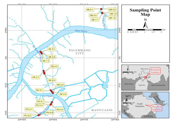
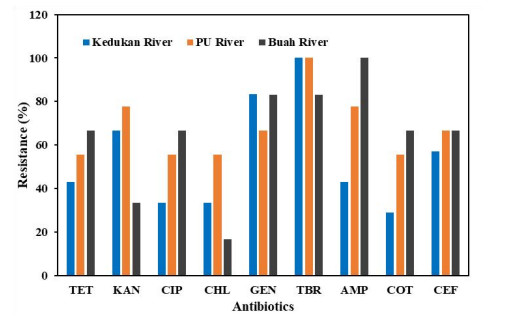
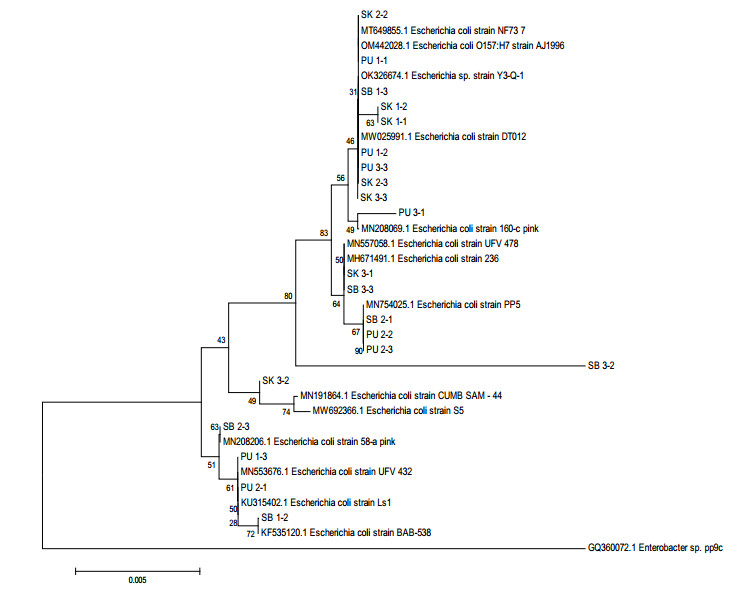
One of the indicators of water pollution is the presence of coliform bacteria, such as Escherichia coli (E. coli). The development of resistance properties to several antibiotics by this pathogen is a serious health problem. Therefore, this study aims to isolate and identify antibiotic-resistant E. coli using the 16S rRNA gene. Samples were taken along the Kedukan, PU, and Buah rivers in Palembang City, South Sumatra, Indonesia, using the purposive sampling method. Each river consists of 9 sampling points, namely, the lower, middle and the upstream regions. The water quality result for the three rivers show that several water quality parameters do not meet the water quality standards: namely, BOD levels in the Kedukan and Buah rivers, Fe levels in the Kedukan and PU rivers, and chlorine in the three rivers. The results showed that E. coli isolates from the Kedukan, PU and Buah rivers were sensitive to 9 antibiotics. The highest resistance (100%) of E. coli isolates to tobramycin was in the Kedukan and PU rivers, while those from the Buah river were resistant to ampicillin. The phylogenetic tree analysis showed genetic diversity. Two main groups were formed from E. coli, namely, A and B, which consist of 17 and 4 isolates, respectively. Furthermore, the water quality analysis results of the Kedukan, PU, and Buah rivers in Palembang City, South Sumatra, revealed that they are polluted.
Citation: Riri Novita Sunarti, Sri Budiarti, Marieska Verawaty, Bayo Alhusaeri Siregar, Poedji Loekitowati Hariani. Diversity of Antibiotic-Resistant Escherichia coli from Rivers in Palembang City, South Sumatra, Indonesia[J]. AIMS Environmental Science, 2022, 9(5): 721-734. doi: 10.3934/environsci.2022041
One of the indicators of water pollution is the presence of coliform bacteria, such as Escherichia coli (E. coli). The development of resistance properties to several antibiotics by this pathogen is a serious health problem. Therefore, this study aims to isolate and identify antibiotic-resistant E. coli using the 16S rRNA gene. Samples were taken along the Kedukan, PU, and Buah rivers in Palembang City, South Sumatra, Indonesia, using the purposive sampling method. Each river consists of 9 sampling points, namely, the lower, middle and the upstream regions. The water quality result for the three rivers show that several water quality parameters do not meet the water quality standards: namely, BOD levels in the Kedukan and Buah rivers, Fe levels in the Kedukan and PU rivers, and chlorine in the three rivers. The results showed that E. coli isolates from the Kedukan, PU and Buah rivers were sensitive to 9 antibiotics. The highest resistance (100%) of E. coli isolates to tobramycin was in the Kedukan and PU rivers, while those from the Buah river were resistant to ampicillin. The phylogenetic tree analysis showed genetic diversity. Two main groups were formed from E. coli, namely, A and B, which consist of 17 and 4 isolates, respectively. Furthermore, the water quality analysis results of the Kedukan, PU, and Buah rivers in Palembang City, South Sumatra, revealed that they are polluted.

| [1] | Ministry of Health of the Republic of Indonesia. Indonesia health profile 2018[Indonesia Health Statistics 2018]. Available from https://pusdatin.kemkes.go.id/resources/download/pusdatin/profil-kesehatan-indonesia/PROFIL_KESEHATAN_2018_1.pdf |
| [2] |
Rosyada, Amrina, Putri, et al. (2018). Investigation of diarrhea cases in toddlers in Palembang city in 2015-2016 with a geographic information system approach. Andalas Journal of Public Health 12: 90–96. https://doi.org/10.24893/jkma.v12i2.359 doi: 10.24893/jkma.v12i2.359

|
| [3] |
Farthing M, Salam MA, Lindberg G, et al. (2013) Acute diarrhea in adults and children a global perspective. J Clin Gastroenterol 47: 12–20. http://10.1097/MCG.0b013e31826df662 doi: 10.1097/MCG.0b013e31826df662
|
| [4] | Mellmann A, Harmsen D, Cummings CA, et al. (2011) Prospective genomic characterization of the german enterohemorrhagic Escherichia coli O104: H4 outbreak by rapid next-generation sequencing technology. Plos One 6: 1–9. https://doi.org/10.1371/journal.pone.002275 |
| [5] |
Frank C, Weber D, Cramer JP, et al. (2011) Epidemic profile of Shiga-toxin–producing Escherichia coli O104:H4 outbreak in Germany. N Engl J Med 365: 1771–780. https://doi.org/10.1056/NEJMoa1106483 doi: 10.1056/NEJMoa1106483
|
| [6] |
Poolman JT, Wacker M (2016) Extraintestinal pathogenic Escherichia coli, a common human pathogen: challenges for vaccine development and progress in the field. J Infect Dis 213: 1–8. https:/doi.org/10.1093/infdis/jiv429 doi: 10.1093/infdis/jiv429
|
| [7] |
Vejborg RM, Hancock V, Schembri MA, et al. (2011) Comparative Genomics of Escherichia coli Strains Causing Urinary Tract Infections. Appl Environ Microbiol 77: 3268–3278. https:/doi.org/10.1128/AEM.02970-10 doi: 10.1128/AEM.02970-10
|
| [8] |
Christine G, Budiarti S, Astuti RI (2018) Diversity of urinary tract infection bacteria in children in Indonesia based on metagenomics. Biodiversity 19: 1375–1381. https://doi.org/10.13057/biodiv/d190425 doi: 10.13057/biodiv/d190425
|
| [9] |
Lee DS, Lee SJ, Choe HS (2018) Community-acquired urinary tract infection by Escherichia coli in the era of antibiotic resistance. Biomed Res Int 2018: 1–4. https://doi.org/10.1155/2018/7656752 doi: 10.1155/2018/7656752
|
| [10] |
Moissenet D, Salauze B, Clermont O, et al. (2010) Meningitis caused by Escherichia coli producing TEM-52 extended-spectrum -lactamase within an extensive outbreak in a neonatal ward: epidemiological investigation and characterization of the strain. J Clin Microbiol 48: 2459–2463. https://doi.org/10.1128/JCM.00529-10 doi: 10.1128/JCM.00529-10
|
| [11] |
Kim KS (2016) Human Meningitis-Associated Escherichia coli. EcoSal Plus 7: 1–15. http://10.1128/ecosalplus.ESP-0015-2015 doi: 10.1128/ecosalplus.ESP-0015-2015
|
| [12] |
Liu Y, Zhu M, Fu X, et al. (2021) Escherichia coli causing neonatal meningitis during 2001–2020: a study in eastern China. Int J Gen Med 14: 3007–3016. https://doi.org/10.2147/IJGM.S317299 doi: 10.2147/IJGM.S317299
|
| [13] |
Alvarez CAR, Bernal E, Tello JA, et al. 2019. Severe sepsis and septic shock by Escherichia coli, clinical and microbiological analysis in Medellin, Colombia. Rev Chilena Infectol 36: 447–454. http://10.4067/S0716-10182019000400447 doi: 10.4067/S0716-10182019000400447
|
| [14] |
Austin, CB, Wright MS, Stepanauskas R, et al. (2006) Co-selection of antibiotics and metal resistance. Trends Microbiol 14: 176–182. http://10.1016/j.tim.2006.02.006 doi: 10.1016/j.tim.2006.02.006
|
| [15] |
Payus C, Ying TS, Kui WN (2016) Effect of heavy metal contamination on the DNA mutation on Nepenthes plant from an abandoned mine. Res J Environ Toxicol 10: 193–204. https://doi.org/10.3923/rjet.2016.193.204 doi: 10.3923/rjet.2016.193.204
|
| [16] |
Li M, Yuning He, Jing S, et al. (2019) Chronic exposure to an environmentally relevant triclosan concentration induces persistent triclosan resistance but reversible antibiotic tolerance in Escherichia coli. Environ Sci Technol 53: 3277−3286. http://10.1021/acs.est.8b06763 doi: 10.1021/acs.est.8b06763
|
| [17] |
Jin M, Liu L, Wang D, et al. (2020) Chlorine disinfection promotes the exchange of antibiotic resistance genes across bacterial genera by natural transformation. ISME Journal 14: 1847–1856. https://doi.org/10.1038/s41396-020-0656-9 doi: 10.1038/s41396-020-0656-9
|
| [18] |
Tong C, Hu H, Chen G, et al. (2021) Chlorine disinfectants promote microbial resistance in Pseudomonas sp. Environ Res 199: 1–8. https://doi.org/10.1016/j.envres.2021.111296 doi: 10.1016/j.envres.2021.111296
|
| [19] |
Spring S, Scheuner C, Goker M, et al. (2015) A taxonomic framework for emerging groups of ecologically important marine gammaproteobacteria based on the reconstruction of evolutionary relationships using genome-scale data. Front Microbiol 6: 1–17. http://10.3389/fmicb.2015.00281 doi: 10.3389/fmicb.2015.00281
|
| [20] |
Tsukuda M, Kitahara K, Miyazaki K (2017) Comparative RNA function analysis reveals high functional similarity between distantly related bacterial 16 S rRNAs. Scientific Reports 7: 1–8. http://10.1038/s41598-017-10214-3 doi: 10.1038/s41598-017-10214-3
|
| [21] |
Idris AB, Hassan HG, Ali MAS, et al. (2020). Molecular phylogenetic analysis of 16S rRNA sequences identified two lineages of helicobacter pylori strains detected from different regions in Sudan suggestive of differential evolution. Int J Microbiol 2020: 1–12. https://doi.org/10.1155/2020/8825718 doi: 10.1155/2020/8825718
|
| [22] | SNI (2009) Indonesian National Standard (SNI 6989.72: 2009); Water and wastewater – Section 72: Biochemical Oxygen Demand (BOD) testing methods. National Standardization Agency, Jakarta. http://sispk.bsn.go.id/sni/DetailSNI/8217 |
| [23] | SNI (2019) Indonesian National Standard (SNI 6989.2: 2009); Water and wastewater- Part 2: Test method for chemical oxygen demand (COD) with closed reflux spectrophotometrically. National Standardization Agency, Jakarta. http://sispk.bsn.go.id/sni/DetailSNI/7861 |
| [24] | SNI (2004) Indonesian National Standard (SNI 06-6989.3: 2004); Water and wastewater- Part 3: Test method for total suspended solids (TSS) gravimetrically. National Standardization Agency, Jakarta. http://sispk.bsn.go.id/sni/DetailSNI/6725 |
| [25] | SNI (2009) Indonesian National Standard (SNI 6989.4: 2009); Water and wastewater- Part 4: Method of the assay for iron (Fe) by Atomic Absorption Spectrophotometry (AAS)-flame. National Standardization Agency, Jakarta. http://sispk.bsn.go.id/sni/DetailSNI/7864 |
| [26] | SNI (2005) Indonesian National Standard (SNI 06-6989.38: 2005); Water and wastewater- Part 38: Method of testing the levels of Cadmium (Cd) with an Atomic Absorption Spectrophotometer (AAS) in a carbon furnace. National Standardization Agency, Jakarta. http://sispk.bsn.go.id/sni/DetailSNI/7011 |
| [27] | SNI (2009) Indonesian National Standard (SNI 6989.8: 2009); Water and wastewater- Part 8: Method of the assay for Lead (Pb) by Atomic Absorption Spectrophotometry (AAS) – flame. National Standardization Agency, Jakarta. http://sispk.bsn.go.id/sni/DetailSNI/8199 |
| [28] | SNI (2009) Indonesian National Standard (SNI 6989.19: 2009); Water and wastewater – Part 19: Method of chloride (Cl-) test by the argentometric method. National Standardization Agency, Jakarta. http://sispk.bsn.go.id/sni/DetailSNI/7862 |
| [29] | SNI (2008) Indonesian National Standard (SNI 2897: 2008); Methods for testing microbial contamination in meat, eggs, and milk, as well as their processed products. National Standardization Agency, Jakarta. http://sispk.bsn.go.id/sni/DetailSNI/7504 |
| [30] | SNI (1992) Indonesian National Standard (SNI 01-2897: 1992); How to Test Microbial Contamination. National Standardization Agency, Jakarta. http://sispk.bsn.go.id/sni/DetailSNI/3267 |
| [31] | Performance Standards for Antimicrobial Susceptibility Testing. Twenty-Second Informational Supplement. (2012). Vol. 32, Issue 3. Wayne, USA. https://clsi.org/standards/products/microbiology/documents/m100/ |
| [32] | CLSI (Clinical and Laboratory Standards Institute). (2017). M100 Performance Standards for Antimicrobials. file:///C:/Users/USER/Downloads/CLSI2013.pdf |
| [33] |
Jiang H, Dong H, Zhang G, et al. (2006) Microbial diversity in water and sediment of Lake Chaka, an Athalassohaline lake in Northwestern China. Appl Environ Microbiol 72: 3832–2845. https://doi.org/10.1128/AEM.02869-05. doi: 10.1128/AEM.02869-05
|
| [34] | Chapman D, Kimstach V (1996) Selection of water quality variables. W of biota, sediments, and water quality assessments: a guide to the use of biota. In: sediments and water in environmental monitoring, 2nd Edition, Chapman Edition, E & FN Spon, London, 59–126. https://doi.org/10.4324/9780203476710 |
| [35] |
Clifford ND, Roxanna I, Candelaria-Ley, et al. (2015) Extreme water quality degradation following a catastrophic forest fire. Freshwater Biology 60: 2584–2599. https://doi.org/10.1111/fwb.12548 doi: 10.1111/fwb.12548
|
| [36] |
Jiang HL, Zhi HW, Xiao JZ, et al. (2015) Health effects from swimming training in chlorinated pools and the corresponding metabolic stress pathways. Plos One 10: 1–14. https://doi.org/10.1371/journal.pone.0119241 doi: 10.1371/journal.pone.0119241
|
| [37] |
Mohsen IH, Mohsen AH, Zaidan HK (2019) Health effects of chlorinated water: a review article. Pak J Biotechnol 16: 263–267. https://doi.org/10.34016/pjbt.2019.16.3.24 doi: 10.34016/pjbt.2019.16.3.24
|
| [38] |
Seo M, Lee H, Kim Y (2019) Relationship between Coliform bacteria and water quality factors at weir stations in the Nakdong River South Korea. Water 11: 1–16. https://doi.org/10.3390/w11061171 doi: 10.3390/w11061171
|
| [39] |
Carene N, I Wayan S, I Nyoman S. (2019) Isolated hemolysis profile of Streptococcus Sp. isolation results from swine's tonsil in a slaughterhouse at Punggul and Bongkasa village. Vet. Anim. Sci 2 (2): 46–51. https://doi.org/10.24843/JVAS.2019.v02.i02.p01 doi: 10.24843/JVAS.2019.v02.i02.p01
|
| [40] | Baker AC, Wright MS, Stepanauskas R, et al. (2006) Co-selection of antibiotics and metal resistance. Trends Microbiol 14: 176–182. |
| [41] |
Mignard S, Flandrois JP (2006) 16S rRNA sequencing in routine bacterial identification: A 30-month experiment. J Microbiol Methods 67: 574–581. http://doi.org/10.1016/j.mimet.2006.05.009 doi: 10.1016/j.mimet.2006.05.009
|
| [42] |
Vetrovsky T, Baldrian P (2013) The variability of the 16S rRNA gene in bacterial genomes and its consequences for bacterial community analyses. Plos One 8: 1–10. https://doi.org/10.1371/journal.pone.0057923 doi: 10.1371/journal.pone.0057923
|
| [43] |
Kuhnert P, Boena MK, Stephan R, et al. (2009) Phylogeny and prediction of genetic similarity of Cronobacter and related taxa by multilocus sequence analysis (MLSA). Int J Food Microbiol 136: 152–158. https://doi.org/10.1016/j.ijfoodmicro.2009.02.022 doi: 10.1016/j.ijfoodmicro.2009.02.022
|
Figures(4) / Tables(4)

Riri Novita Sunarti, Sri Budiarti, Marieska Verawaty, Bayo Alhusaeri Siregar, Poedji Loekitowati Hariani. Diversity of Antibiotic-Resistant Escherichia coli from Rivers in Palembang City, South Sumatra, Indonesia[J]. AIMS Environmental Science, 2022, 9(5): 721-734. doi: 10.3934/environsci.2022041


 DownLoad: