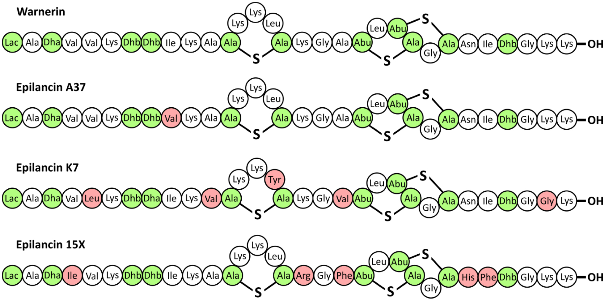
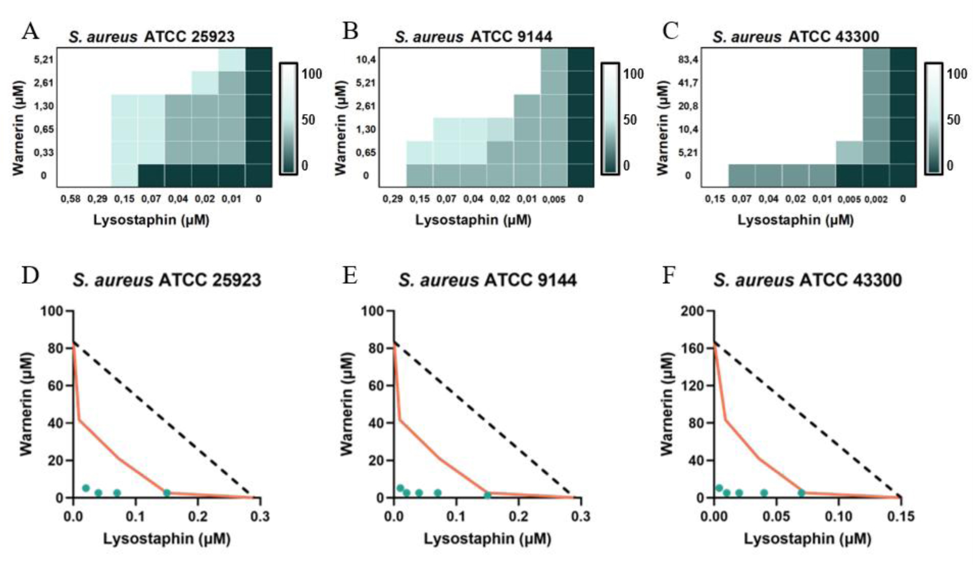
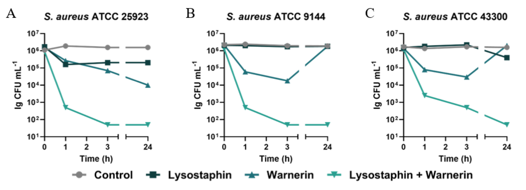
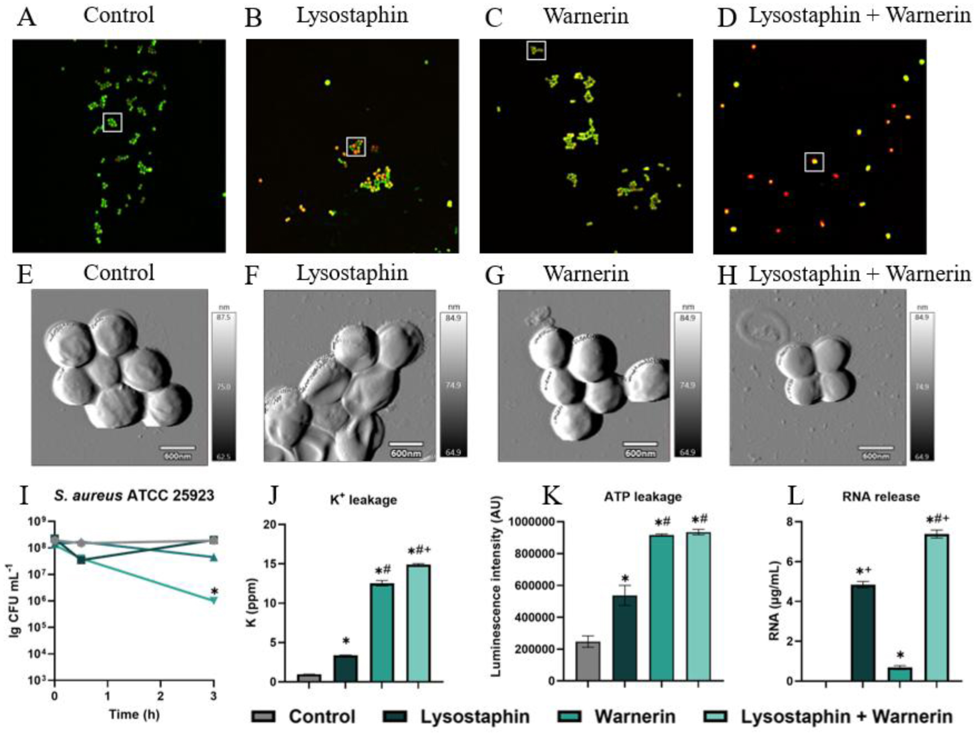
Whole-genome sequencing of Staphylococcus warneri KL-1 (GenBank GCA_056393345.1) confirmed that this strain harbors a biosynthetic gene cluster encoding epilancins, a class I antimicrobial peptide known as a lantibiotic. The peptide warnerin produced by this strain belongs to the epilancin group and has a high degree of homology with epilancin A37, with a single substitution at position 8 (Val→Ile). Three studied Staphylococcus aureus strains, including MRSA, exhibited low sensitivity to warnerin (MICs 83.38–166.75 µM) and moderate sensitivity to lysostaphin (LS) (MICs 0.15–0.29 µM). However, the combination of LS and warnerin reduced the MIC of warnerin by 16-fold and that of LS by 16–32-fold, depending on the strain. Compared with untreated cells, exposure to subMIC warnerin did not affect cell viability but increased the release of K+, ATP, and RNA, indicating disruption of membrane integrity. The synergistic interaction between warnerin and LS was confirmed by the fractional inhibitory concentration index and isobologram analyses and was associated with enhanced cell damage. The bactericidal effect of the combination was demonstrated using a time-kill assay. Morphological changes in S. aureus cells under the influence of warnerin, LS, and their combination were demonstrated via confocal microscopy and AFM.
Citation: Aleksandr Lobanov, Ekaterina Yaburova, Daria Eroshenko, Tatyana Polyudova. The synergism of the antibacterial action of lysostaphin and warnerin, an antibacterial peptide of the epilancin group, against Staphylococcus aureus[J]. AIMS Microbiology, 2026, 12(4): 774-788. doi: 10.3934/microbiol.2026033
Whole-genome sequencing of Staphylococcus warneri KL-1 (GenBank GCA_056393345.1) confirmed that this strain harbors a biosynthetic gene cluster encoding epilancins, a class I antimicrobial peptide known as a lantibiotic. The peptide warnerin produced by this strain belongs to the epilancin group and has a high degree of homology with epilancin A37, with a single substitution at position 8 (Val→Ile). Three studied Staphylococcus aureus strains, including MRSA, exhibited low sensitivity to warnerin (MICs 83.38–166.75 µM) and moderate sensitivity to lysostaphin (LS) (MICs 0.15–0.29 µM). However, the combination of LS and warnerin reduced the MIC of warnerin by 16-fold and that of LS by 16–32-fold, depending on the strain. Compared with untreated cells, exposure to subMIC warnerin did not affect cell viability but increased the release of K+, ATP, and RNA, indicating disruption of membrane integrity. The synergistic interaction between warnerin and LS was confirmed by the fractional inhibitory concentration index and isobologram analyses and was associated with enhanced cell damage. The bactericidal effect of the combination was demonstrated using a time-kill assay. Morphological changes in S. aureus cells under the influence of warnerin, LS, and their combination were demonstrated via confocal microscopy and AFM.

This research was carried out on the basis of the Center for Collective Use “Research of Materials and Substances” of the Perm Federal Research Center, Ural Branch of the Russian Academy of Sciences. The authors acknowledge the Interdisciplinary Center for Shared Facilities of Kazan Federal University for performing whole-genome sequencing.
This work was supported by the Ministry of Science and Higher Education of the Russian Federation (124020500028-4).
The authors declare no conflict of interest.
Aleksandr Lobanov: Writing—original draft, Writing—review & editing, Investigation, Formal analysis, Data curation, Visualization. Ekaterina Yaburova: Writing—review & editing, Formal analysis. Daria Eroshenko: Writing—review & editing, Conceptualization. Tatyana Polyudova: Writing—original draft, Writing—review & editing, Supervision, Resources, Methodology, Investigation.
| [1] |
Bier K, Schittek B (2021) Beneficial effects of coagulase-negative Staphylococci on Staphylococcus aureus skin colonization. Exp Dermatol 30: 1442-1452. https://doi.org/10.1111/exd.14381 
|
| [2] |
Fernández-Fernández R, Lozano C, Eguizábal P, et al. (2022) Bacteriocin-like inhibitory substances in staphylococci of different origins and species with activity against relevant pathogens. Front Microbiol 13. https://doi.org/10.3389/fmicb.2022.870510
|
| [3] |
Polyudova TV, Lemkina LM, Antip'eva MV, et al. (2024) Prevalence of production of antagonism peptide factors among coagulase-negative staphylococci. Microbiology 93: 847-854. https://doi.org/10.1134/S0026261724606626
|
| [4] |
Gründling A, Missiakas DM, Schneewind O (2006) Staphylococcus aureus mutants with increased lysostaphin resistance. J Bacteriol 188: 6286-6297. https://doi.org/10.1128/JB.00457-06
|
| [5] |
Yang XY, Li CR, Lou RH, et al. (2007) In vitro activity of recombinant lysostaphin against Staphylococcus aureus isolates from hospitals in Beijing, China. J Med Microbiol 56: 71-76. https://doi.org/10.1099/jmm.0.46788-0
|
| [6] |
Szweda P, Schielmann M, Frankowska A, et al. (2014) Antibiotic resistance in Staphylococcus aureus strains isolated from cows with mastitis in eastern Poland and analysis of susceptibility of resistant strains to alternative nonantibiotic agents: Lysostaphin, nisin and polymyxin B. J Vet Med Sci 76: 355-362. https://doi.org/10.1292/jvms.13-0177
|
| [7] |
Harrison EF, Cropp CB (1965) Comparative in vitro activities of lysostaphin and other antistaphylococcal antibiotics on clinical isolates of Staphylococcus aureus. Appl Microbiol 13: 212-215. https://doi.org/10.1128/am.13.2.212-215.1965
|
| [8] |
Gordina EM, Bozhkova SA, Labutin DV, et al. (2023) Anti-staphylococcal activity and cytocompatibility of lysostaphin. Clin Microbiol Antimicrob Chemother 25: 77-82. https://doi.org/10.36488/cmac.2023.1.77-82
|
| [9] |
Wu X, Zha J, Koffas MAG, et al. (2019) Reducing Staphylococcus aureus resistance to lysostaphin using CRISPR-dCas9. Biotechnol Bioeng 116: 3149-3159. https://doi.org/10.1002/bit.27143
|
| [10] |
Willing S, Dyer E, Schneewind O, et al. (2020) FmhA and FmhC of Staphylococcus aureus incorporate serine residues into peptidoglycan cross-bridges. J Biol Chem 295: 13664-13676. https://doi.org/10.1074/jbc.RA120.014371
|
| [11] |
Batool N, Ko KS, Chaurasia AK, et al. (2020) Functional identification of serine hydroxymethyltransferase as a key gene involved in lysostaphin resistance and virulence potential of Staphylococcus aureus strains. Int J Mol Sci 21: 9135. https://doi.org/10.3390/ijms21239135
|
| [12] |
Wang X, Gu Q, Breukink E (2020) Non-lipid II targeting lantibiotics. Biochim Biophys Acta Biomembr 1862: 183244. https://doi.org/10.1016/j.bbamem.2020.183244
|
| [13] |
Puls JS, Winnerling B, Power JJ, et al. (2024) Staphylococcus epidermidis bacteriocin A37 kills natural competitors with a unique mechanism of action. ISME J 18: wrae044. https://doi.org/10.1093/ismejo/wrae044
|
| [14] |
Nakatsuji T, Chen TH, Narala S, et al. (2017) Antimicrobials from human skin commensal bacteria protect against Staphylococcus aureus and are deficient in atopic dermatitis. Sci Transl Med 9: eaah4680. https://doi.org/10.1126/scitranslmed.aah4680
|
| [15] |
Twomey E, O'Connor PM, Coffey A, et al. (2024) Inhibition of clinical MRSA isolates by coagulase negative staphylococci of human origin. Antibiotics 13: 338. https://doi.org/10.3390/antibiotics13040338
|
| [16] |
Varella Coelho ML, Santos Nascimento JD, Fagundes PC, et al. (2007) Activity of staphylococcal bacteriocins against Staphylococcus aureus and Streptococcus agalactiae involved in bovine mastitis. Res Microbiol 158: 625-630. https://doi.org/10.1016/j.resmic.2007.07.002
|
| [17] |
Joo HS, Otto M (2015) Mechanisms of resistance to antimicrobial peptides in staphylococci. Biochim Biophys Acta Biomembr 1848: 3055-3061. https://doi.org/10.1016/j.bbamem.2015.02.009
|
| [18] |
Peschel A, Vuong C, Otto M, et al. (2000) The D-alanine residues of Staphylococcus aureus teichoic acids alter the susceptibility to vancomycin and the activity of autolytic enzymes. Antimicrob Agents Chemother 44: 2845-2847. https://doi.org/10.1128/AAC.44.10.2845-2847.2000
|
| [19] |
Desbois AP, Coote PJ (2011) Bactericidal synergy of lysostaphin in combination with antimicrobial peptides. Eur J Clin Microbiol Infect Dis 30: 1015-1021. https://doi.org/10.1007/s10096-011-1188-z
|
| [20] |
Sadeghi S, Bakhshandeh H, Ahangari Cohan R, et al. (2019) Synergistic anti-staphylococcal activity of niosomal recombinant lysostaphin-LL-37. Int J Nanomedicine 14: 9777-9792. https://doi.org/10.2147/IJN.S230269
|
| [21] |
Korobov VP, Lemkina LM, Polyudova TV, et al. (2010) Isolation and characterization of a new low-molecular antibacterial peptide of the lantibiotics family. Microbiology 79: 206-215. https://doi.org/10.1134/S0026261710020128
|
| [22] |
Yaburova EV, Nechaeva YuI, Kononova LI, et al. (2026) Genome analysis of bacteria Staphylococcus cohnii VKM B-3165 with high susceptibility to antimicrobial compounds. Microbiology 95: 44-53. https://doi.org/10.1134/S0026261725602830
|
| [23] |
Korobov VP, Lemkina LM, Polyudova TV (2022) The mechanism of antibacterial action of the lantibiotic warnerin. Microbiology 91: 184-191. https://doi.org/10.1134/S0026261722020059
|
| [24] |
Bankevich A, Nurk S, Antipov D, et al. (2012) SPAdes: A new genome assembly algorithm and its applications to single-cell sequencing. J Comput Biol 19: 455-477. https://doi.org/10.1089/cmb.2012.0021
|
| [25] |
Gurevich A, Saveliev V, Vyahhi N, et al. (2013) QUAST: Quality assessment tool for genome assemblies. Bioinformatics 29: 1072-1075. https://doi.org/10.1093/bioinformatics/btt086
|
| [26] |
Parks DH, Imelfort M, Skennerton CT, et al. (2015) CheckM: Assessing the quality of microbial genomes recovered from isolates, single cells, and metagenomes. Genome Res 25: 1043-1055. https://doi.org/10.1101/gr.186072.114
|
| [27] |
Seemann T (2014) Prokka: Rapid prokaryotic genome annotation. Bioinformatics 30: 2068-2069. https://doi.org/10.1093/bioinformatics/btu153
|
| [28] | Schwengers O, Jelonek L, Dieckmann MA, et al. (2021) Bakta: Rapid and standardized annotation of bacterial genomes via alignment-free sequence identification. Microb Genom 7. https://doi.org/10.1099/mgen.0.000685 |
| [29] |
Blin K, Shaw S, Vader L, et al. (2025) antiSMASH 8.0: Extended gene cluster detection capabilities and analyses of chemistry, enzymology, and regulation. Nucleic Acids Res 53: W32-W38. https://doi.org/10.1093/nar/gkaf334
|
| [30] |
van Heel AJ, de Jong A, Song C, et al. (2018) BAGEL4: A user-friendly web server to thoroughly mine RiPPs and bacteriocins. Nucleic Acids Res 46: W278-W281. https://doi.org/10.1093/nar/gky383
|
| [31] |
Zdouc MM, Blin K, Louwen NLL, et al. (2025) MIBiG 4.0: Advancing biosynthetic gene cluster curation through global collaboration. Nucleic Acids Res 53: D678-D690. https://doi.org/10.1093/nar/gkae1115
|
| [32] | (2015) CLSIMethods for Dilution Antimicrobial Susceptibility Tests for Bacteria That Grow Aerobically; Approved Standard—Tenth Edition. CLSI document M07-A10 . Wayne, PA: Clinical and Laboratory Standards Institute. |
| [33] |
Lobanov A, Eroshenko D, Esaev A, et al. (2026) Antibacterial activity of lantibiotics, alone and in combination with colistin, against Gram-negative bacteria. Arch Microbiol 208: 275. https://doi.org/10.1007/s00203-026-04820-x
|
| [34] |
Kamp M, Hooven HW, Konings RNH, et al. (1995) Elucidation of the primary structure of the lantibiotic epilancin K7 from Staphylococcus epidermidis K7: Cloning and characterisation of the epilancin-K7-encoding gene and NMR analysis of mature epilancin K7. Eur J Biochem 230: 587-600. https://doi.org/10.1111/j.1432-1033.1995.tb20600.x
|
| [35] |
Velásquez JE, Zhang X, van der Donk WA (2011) Biosynthesis of the antimicrobial peptide epilancin 15X and its N-terminal lactate. Chem Biol 18: 857-867. https://doi.org/10.1016/j.chembiol.2011.05.007
|
| [36] |
Kusuma C, Jadanova A, Chanturiya T, et al. (2007) Lysostaphin-resistant variants of Staphylococcus aureus demonstrate reduced fitness in vitro and in vivo. Antimicrob Agents Chemother 51: 475-482. https://doi.org/10.1128/AAC.00786-06
|
| [37] |
Rohrer S, Ehlert K, Tschierske M, et al. (1999) The essential Staphylococcus aureus gene fmhB is involved in the first step of peptidoglycan pentaglycine interpeptide formation. Proc Natl Acad Sci USA 96: 9351-9356. https://doi.org/10.1073/pnas.96.16.9351
|
| [38] |
Łuszczki JJ, Wlaź A (2023) Isobolographic analysis of interactions—a pre-clinical perspective. J Pre Clin Clin Res 17: 238-241. https://doi.org/10.26444/jpccr/177246
|
| [39] |
Weidenmaier C, Peschel A, Kempf VAJ, et al. (2005) DltABCD- and MprF-mediated cell envelope modifications of Staphylococcus aureus confer resistance to platelet microbicidal proteins and contribute to virulence in a rabbit endocarditis model. Infect Immun 73: 8033-8038. https://doi.org/10.1128/IAI.73.12.8033-8038.2005
|
| [40] |
Vásquez A, Leidy C, Manrique-Moreno M (2025) Lysyl-phosphatidylglycerol: A lipid involved in the resistance of Staphylococcus aureus to antimicrobial peptide activity. Antibiotics 14: 349. https://doi.org/10.3390/antibiotics14040349
|
 microbiol-12-04-033-s001.pdf |
 |
Figures(4) / Tables(2)

Aleksandr Lobanov, Ekaterina Yaburova, Daria Eroshenko, Tatyana Polyudova. The synergism of the antibacterial action of lysostaphin and warnerin, an antibacterial peptide of the epilancin group, against Staphylococcus aureus[J]. AIMS Microbiology, 2026, 12(4): 774-788. doi: 10.3934/microbiol.2026033





 DownLoad: