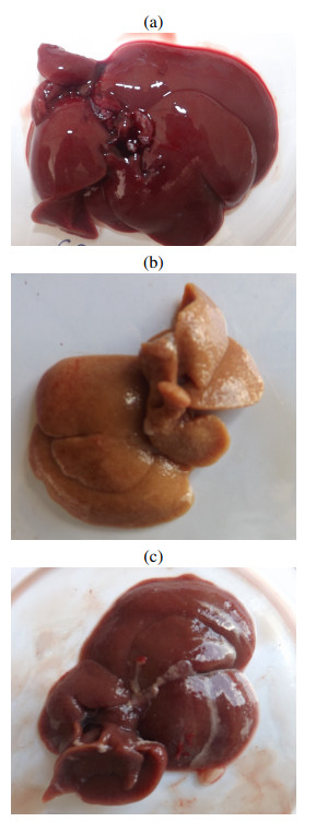
In this work, we propose a mathematical model that describes liver evolution and concentrations of alanine aminotransferase and aspartate aminotransferase in a group of rats damaged with carbon tetrachloride. Carbon tetrachloride was employed to induce cirrhosis. A second groups damaged with carbon tetrachloride was exposed simultaneously a plant extract as hepatoprotective agent. The model reproduces the data obtained in the experiment reported in [Rev. Cub. Plant. Med. 22(1), 2017], and predicts that using the plants extract helps to get a better natural recovery after the treatment. Computer simulations show that the extract reduces the damage velocity but does not avoid it entirely. The present paper is the first report in the literature in which a mathematical model reliably predicts the protective effect of a plant extract mixture in rats with cirrhosis disease. The results reported in this manuscript could be used in the future to help in fighting cirrhotic conditions in humans, though more experimental and mathematical work is required in that case.
Citation: Luis E. Ayala-Hernández, Gabriela Rosales-Muñoz, Armando Gallegos, María L. Miranda-Beltrán, Jorge E. Macías-Díaz. On a deterministic mathematical model which efficiently predicts the protective effect of a plant extract mixture in cirrhotic rats[J]. Mathematical Biosciences and Engineering, 2024, 21(1): 237-252. doi: 10.3934/mbe.2024011
In this work, we propose a mathematical model that describes liver evolution and concentrations of alanine aminotransferase and aspartate aminotransferase in a group of rats damaged with carbon tetrachloride. Carbon tetrachloride was employed to induce cirrhosis. A second groups damaged with carbon tetrachloride was exposed simultaneously a plant extract as hepatoprotective agent. The model reproduces the data obtained in the experiment reported in [Rev. Cub. Plant. Med. 22(1), 2017], and predicts that using the plants extract helps to get a better natural recovery after the treatment. Computer simulations show that the extract reduces the damage velocity but does not avoid it entirely. The present paper is the first report in the literature in which a mathematical model reliably predicts the protective effect of a plant extract mixture in rats with cirrhosis disease. The results reported in this manuscript could be used in the future to help in fighting cirrhotic conditions in humans, though more experimental and mathematical work is required in that case.

| [1] | R. L. Carithers, C. J. Mcclain, Chapter 84 - alcoholic liver disease, in Sleisenger and Fordtran's Gastrointestinal and Liver Disease (eds. M. Feldman, L. S. Friedman, and Lawrence J. Brand), e4. W.B. Saunders, Philadelphia, ninth edition edition, (2010), 1383–1400. https://doi.org/10.1016/B978-1-4160-6189-2.00084-6 |
| [2] |
S. K. Asrani, H. Devarbhavi, J. Eaton, P. S. Kamath, Burden of liver diseases in the world, J. Hepatol., 70 (2019), 151–171. https://doi.org/10.1016/j.jhep.2018.09.014 doi: 10.1016/j.jhep.2018.09.014

|
| [3] |
J. F. Perz, G. L. Armstrong, L. A. Farrington, Y. J. F. Hutin, B. P. Bell, The contributions of hepatitis B virus and hepatitis C virus infections to cirrhosis and primary liver cancer worldwide, J. Hepatol., 450 (2006), 529–538. https://doi.org/10.1016/j.jhep.2006.05.013 doi: 10.1016/j.jhep.2006.05.013
|
| [4] | J. Loscalzo, A. S. Fauci, D. L. Kasper, S. Hauser, D. Longo, J. L. Jameson, Harrison's Principles of Internal Medicine, (Vol. 1 & Vol. 2), McGraw Hill Professional, New York, 2022. |
| [5] |
L. García, I. Hernández, A. Sandoval, A. Salazar, J. Garcia, J. Vera, et al., Pirfenidone effectively reverses experimental liver fibrosis, J. Hepatol., 37 (2002), 797–805. https://doi.org/10.1016/S0168-8278(02)00272-6 doi: 10.1016/S0168-8278(02)00272-6
|
| [6] |
E. Fogden, J. Neuberger, Alternative medicines and the liver, Liver Int., 23 (2003), 213–220. https://doi.org/10.1034/j.1600-0676.2003.00843.x doi: 10.1034/j.1600-0676.2003.00843.x
|
| [7] |
I. Shimizu, Sho-saiko-to: Japanese herbal medicine for protection against hepatic fibrosis and carcinoma, J. Gastroenterol. Hepatol., 15 (2000), 84–90. https://doi.org/10.1046/j.1440-1746.2000.02138.x doi: 10.1046/j.1440-1746.2000.02138.x
|
| [8] |
N. Ghosh, R. Ghosh, V. Mandal, S. C. Mandal, Recent advances in herbal medicine for treatment of liver diseases, Pharm. Biol., 49 (2011), 970–988. https://doi.org/10.3109/13880209.2011.558515 doi: 10.3109/13880209.2011.558515
|
| [9] |
F. A. Crocenzi, M. G. Roma, Silymarin as a new hepatoprotective agent in experimental cholestasis: New possibilities for an ancient medication, Current Med. Chem., 13 (2006), 1055–1074. https://doi.org/10.2174/092986706776360950 doi: 10.2174/092986706776360950
|
| [10] | C. S. Fregozo, M. de la L. Beltrán, M. E. F. Soto, M. I. P. Vega, R. Y. R. Rodríguez, A. L. L. Velázquez, et al., Protective effect of rosmarinus officinalis l. on the expression of the glutamate transporter (glt-1) and neuronal damage in the frontal cortex of ccl4-induced hepatic damage, J. Med. Plant Res., 6 (2012), 5886–5894. |
| [11] |
C. S. Fregozo, M. L. M. Beltrán, M. E. F. Soto, M. I. P. Vega, C. B. Zárate, L. H. Ruiz, Expression of nmda receptor subunits in rat prefrontal cortex with ccl4-induced hepatic damage after a treatment with rosmarinus officinalis l, Neurología (English Edition), 27 (2012), 261–267. https://doi.org/10.1016/j.nrleng.2011.10.002 doi: 10.1016/j.nrleng.2011.10.002
|
| [12] | M. de la L. Beltrán, L. H. Ruiz, A. L. L. Velásquez, A. P. Cerda, Molecular phytotherapy as part of a complementary and alternative medicine for liver diseases, Investigación en Salud, 7 (2005), 64–70. |
| [13] | C. G. R. Muñoz, C. S. Fregozo, M. I. Pérez Vega, L. Y. C. Cruz, L. Huacuja Ruiz, M. de la L. M. Beltrán, Efecto hepatoprotector de una mezcla de siete plantas en cirrosis inducida con tetracloruro de carbono, Revista Cubana de Plantas Medicinales, 22 (2017), 1. |
| [14] |
J. Chhatwal, E. B. Tapper, Nonalcoholic fatty liver disease natural history: Role of mathematical modeling, Clin. Gastroenterol. Hepatol., (2022). https://doi.org/10.1016/j.cgh.2022.01.041 doi: 10.1016/j.cgh.2022.01.041
|
| [15] |
E. R. Dickson, P. M. Grambsch, T. R. Fleming, L. D. Fisher, A. Langworthy, Prognosis in primary biliary cirrhosis: Model for decision making, Hepatology, 10 (1989), 1–7. https://doi.org/10.1002/hep.1840100102 doi: 10.1002/hep.1840100102
|
| [16] |
J. A. Talwalkar, K. D. Lindor, Primary biliary cirrhosis, The Lancet, 362 (2003), 53–61. https://doi.org/10.1016/S0140-6736(03)13808-1 doi: 10.1016/S0140-6736(03)13808-1
|
| [17] |
A. Friedman, N. Siewe, Chronic hepatitis B virus and liver fibrosis: A mathematical model, Plos One, 13 (2018), e0195037. https://doi.org/10.1371/journal.pone.0195037 doi: 10.1371/journal.pone.0195037
|
| [18] |
D. Drasdo, S. Hoehme, J. G. Hengstler, How predictive quantitative modelling of tissue organisation can inform liver disease pathogenesis, J. Hepatol., 61 (2014), 951–956. https://doi.org/10.1016/j.jhep.2014.06.013 doi: 10.1016/j.jhep.2014.06.013
|
| [19] |
S. Höhme, J. G. Hengstler, M. Brulport, M. Schäfer, A. Bauer, R. Gebhardt, et al., Mathematical modelling of liver regeneration after intoxication with CCl4, Chemico-Biol. Interact., 168 (2007), 74–93. https://doi.org/10.1016/j.cbi.2007.01.010 doi: 10.1016/j.cbi.2007.01.010
|
| [20] |
J. A. Leedale, C. L. Mason, N. Brillant, S. D. Webb, J. W. Dear, Mathematical modelling and statistical analysis of indocyanine green and other biomarkers of hepatic function and drug-induced liver injury, Comput. Toxicol., 16 (2020), 100134. https://doi.org/10.1016/j.comtox.2020.100134 doi: 10.1016/j.comtox.2020.100134
|
| [21] |
A. Ghosh, C. Onsager, A. Mason, L. Arriola, W. Lee, A. Mubayi, The role of oxygen intake and liver enzyme on the dynamics of damaged hepatocytes: Implications to ischaemic liver injury via a mathematical model, PloS One, 16 (2021), e0230833. https://doi.org/10.1371/journal.pone.0230833 doi: 10.1371/journal.pone.0230833
|
| [22] |
A. Ghosh, I. Berger, C. H. Remien, A. Mubayi, The role of alcohol consumption on acetaminophen induced liver injury: Implications from a mathematical model, J. Theor. Biol., 519 (2021), 110559. https://doi.org/10.1016/j.jtbi.2020.110559 doi: 10.1016/j.jtbi.2020.110559
|
| [23] |
C. H. Remien, F. R. Adler, L. Waddoups, T. D. Box, N. L. Sussman, Mathematical modeling of liver injury and dysfunction after acetaminophen overdose: Early discrimination between survival and death, Hepatology, 56 (2012), 727–734. https://doi.org/10.1002/hep.25656 doi: 10.1002/hep.25656
|
| [24] |
C. H. Remien, N. L. Sussman, F. R. Adler, Mathematical modelling of chronic acetaminophen metabolism and liver injury, Math. Med. Biol. J. IMA, 31 (2014), 302–317. https://doi.org/10.1093/imammb/dqt010 doi: 10.1093/imammb/dqt010
|
| [25] |
M. S. Khatun, M. H. A. Biswas, Optimal control strategies for preventing hepatitis b infection and reducing chronic liver cirrhosis incidence, Infect. Disease Model., 5 (2020), 91–110. https://doi.org/10.1016/j.idm.2019.12.006 doi: 10.1016/j.idm.2019.12.006
|
| [26] | P. M. FRAsER, D. A. Franklin, Mathematical models for the diagnosis of liver disease: Problems arising in the use of conditional probability theory, QJM Int. J. Med., 43 (1974), 73–88. |
| [27] |
A. Parés, J. Rodés, Natural history of primary biliary cirrhosis, Clin. Liver Disease, 7 (2003), 779–794. https://doi.org/10.1016/S1089-3261(03)00100-4 doi: 10.1016/S1089-3261(03)00100-4
|
| [28] | P. M. Grambsch, E. R. Dickson, R. H. Wiesner, A. Langworthy, Application of the mayo primary biliary cirrhosis survival model to mayo liver transplant patients, In Mayo Clinic Proceed., 64 (1989), 699–704. https://doi.org/10.1016/S0025-6196(12)65350-6 |
| [29] |
V. P. Stadlbauer, G. A. K. Wright, M. Banaji, A. Mukhopadhya, R. Mookerjee, K. Moore, et al., Relationship between activation of the sympathetic nervous system and renal blood flow autoregulation in cirrhosis, Gastroenterology, 134 (2008), 111–119. https://doi.org/10.1053/j.gastro.2007.10.055 doi: 10.1053/j.gastro.2007.10.055
|
| [30] |
G. Peeters, C. Debbaut, P. Cornillie, T. De Schryver, D. Monbaliu, W. Laleman, et al., A multilevel modeling framework to study hepatic perfusion characteristics in case of liver cirrhosis, J. Biomechan. Eng., 137 (2015), 051007. https://doi.org/10.1115/1.4029280 doi: 10.1115/1.4029280
|
| [31] |
R. Veteläinen, A. K.van Vliet, T. M. van Gulik, Severe steatosis increases hepatocellular injury and impairs liver regeneration in a rat model of partial hepatectomy, Ann. Surgery, 245 (2007), 44. https://doi.org/10.1097/01.sla.0000225253.84501.0e doi: 10.1097/01.sla.0000225253.84501.0e
|
| [32] |
G. A. M. Tiberio, L. Tiberio, A. Benetti, E. Cervi, N. Montani, M. Dreano, et al., IL-6 Promotes compensatory liver regeneration in cirrhotic rat after partial hepatectomy, Cytokine, 42 (2008), 372–378. https://doi.org/10.1016/j.cyto.2008.03.012 doi: 10.1016/j.cyto.2008.03.012
|
| [33] |
U. Y. Sanzgiri, V. Srivatsan, S. Muralidhara, C. E. Dallas, J. V. Bruckner, Uptake, distribution, and elimination of carbon tetrachloride in rat tissues following inhalation and ingestion exposures, Toxicol. Appl. Pharmacol., 143 (1997), 120–129. https://doi.org/10.1006/taap.1996.8079 doi: 10.1006/taap.1996.8079
|
| [34] |
J. F. Zhao, R. Agarwal, Tissue distribution of silibinin, the major active constituent of silymarin, in mice and its association with enhancement of phase Ⅱ enzymes: Implications in cancer chemoprevention, Carcinogenesis, 20 (1999), 2101–2108. https://doi.org/10.1093/carcin/20.11.2101 doi: 10.1093/carcin/20.11.2101
|
| [35] |
C. Balzotti, M. Briani, B. De Filippo, B. Piccoli, A computational modular approach to evaluate no$\_x$ emissions and ozone production due to vehicular traffic, Discrete Continuous Dynam. Systems-Series B, 27 (2022). https://doi.org/10.3934/dcdsb.2021192 doi: 10.3934/dcdsb.2021192
|
| [36] | L. Rarità, A genetic algorithm to optimize dynamics of supply chains, In Optimization in Artificial Intelligence and Data Sciences: ODS, First Hybrid Conference, Rome, Italy, (2022), Springer, 107–115. https://doi.org/10.1007/978-3-030-95380-5_10 |
| [37] |
J. E. Macías-Díaz, N. Ahmed, M. Rafiq, Analysis and nonstandard numerical design of a discrete three-dimensional hepatitis b epidemic model, Mathematics, 7 (2019), 1157. https://doi.org/10.3390/math7121157 doi: 10.3390/math7121157
|
| [38] |
C. Balzotti, M. Briani, B. Piccoli, Emissions minimization on road networks via generic second order models, Networks Heterogen. Media, 18 (2023), 694–722. https://doi.org/10.3934/nhm.2023030 doi: 10.3934/nhm.2023030
|
| [39] | M. P. D'Arienzo, L. Rarità, Growth effects on the network dynamics with applications to the cardiovascular system, In AIP Conference Proceedings, AIP Publishing, 2293 (2020). https://doi.org/10.1063/5.0026464 |
| [40] | M. P. D'Arienzo, L. Rarità, Management of supply chains for the wine production, In AIP conference proceedings, AIP Publishing, 2293 (2020). https://doi.org/10.1063/5.0026462 |
| [41] |
S. Azam, J. E. Macías-Díaz, N. Ahmed, I. Khan, M. S. Iqbal, M. Rafiq, et al., Numerical modeling and theoretical analysis of a nonlinear advection-reaction epidemic system, Computer Methods Programs Biomed., 193 (2020), 105429. https://doi.org/10.1016/j.cmpb.2020.105429 doi: 10.1016/j.cmpb.2020.105429
|
Figures(4) / Tables(4)

Luis E. Ayala-Hernández, Gabriela Rosales-Muñoz, Armando Gallegos, María L. Miranda-Beltrán, Jorge E. Macías-Díaz. On a deterministic mathematical model which efficiently predicts the protective effect of a plant extract mixture in cirrhotic rats[J]. Mathematical Biosciences and Engineering, 2024, 21(1): 237-252. doi: 10.3934/mbe.2024011


 DownLoad: