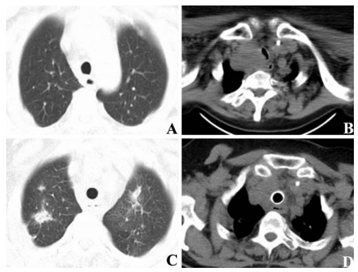
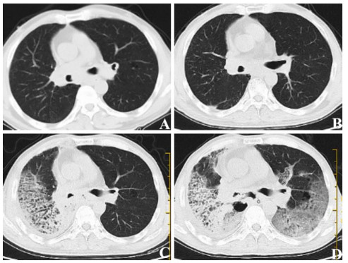
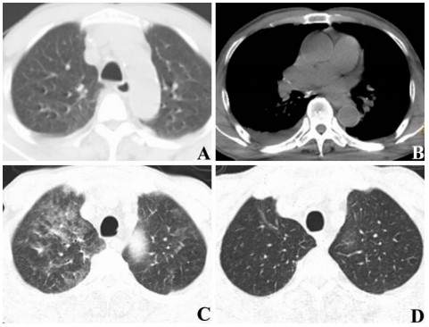
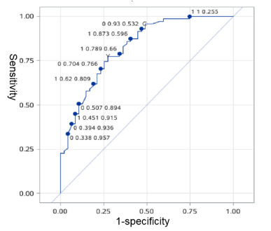
The precise radiotherapy of esophageal cancer may cause different degrees of radiation damage for lung tissues and cause radioactive pneumonia. However, the occurrence of radioactive pneumonia is related to many factors. To further clarify the correlation between the occurrence of radioactive pneumonia and related factors, a random forest model was used to build a risk prediction model for patients with esophageal cancer undergoing radiotherapy. In this study, we retrospectively reviewed 118 patients with esophageal cancer confirmed by pathology in our hospital. The health characteristics and related parameters of all patients were analyzed, and the predictive effect of radiation pneumonia was discussed using the random forest algorithm. After treatment, 71 patients developed radioactive pneumonia (60.17%). In univariate analyses, age, planning target volume length, Karnofsky performance score (KPS), pulmonary emphysema, with or without chemotherapy, and the ratio of planning target volume to planning gross tumor volume (PTV/PGTV) in mediastinum were significantly associated with radioactive pneumonia (P < 0.05 for each comparison). Multivariate analysis revealed that with or without pulmonary emphysema (OR = 7.491, P = 0.001), PTV/PGTV (OR = 0.205, P = 0.007), and KPS (OR = 0.251, P = 0.011) were independent predictors for radiation pneumonia. The results concluded that the analysis of radiation pneumonia-related factors based on the random forest algorithm could build a mathematical prediction model for the easily obtained data. This algorithm also could effectively analyze the risk factors of radiation pneumonia and formulate the appropriate treatment plan for esophageal cancer.
Citation: Na Li, Peng Luo, Chunyang Li, Yanyan Hong, Mingjun Zhang, Zhendong Chen. Analysis of related factors of radiation pneumonia caused by precise radiotherapy of esophageal cancer based on random forest algorithm[J]. Mathematical Biosciences and Engineering, 2021, 18(4): 4477-4490. doi: 10.3934/mbe.2021227
The precise radiotherapy of esophageal cancer may cause different degrees of radiation damage for lung tissues and cause radioactive pneumonia. However, the occurrence of radioactive pneumonia is related to many factors. To further clarify the correlation between the occurrence of radioactive pneumonia and related factors, a random forest model was used to build a risk prediction model for patients with esophageal cancer undergoing radiotherapy. In this study, we retrospectively reviewed 118 patients with esophageal cancer confirmed by pathology in our hospital. The health characteristics and related parameters of all patients were analyzed, and the predictive effect of radiation pneumonia was discussed using the random forest algorithm. After treatment, 71 patients developed radioactive pneumonia (60.17%). In univariate analyses, age, planning target volume length, Karnofsky performance score (KPS), pulmonary emphysema, with or without chemotherapy, and the ratio of planning target volume to planning gross tumor volume (PTV/PGTV) in mediastinum were significantly associated with radioactive pneumonia (P < 0.05 for each comparison). Multivariate analysis revealed that with or without pulmonary emphysema (OR = 7.491, P = 0.001), PTV/PGTV (OR = 0.205, P = 0.007), and KPS (OR = 0.251, P = 0.011) were independent predictors for radiation pneumonia. The results concluded that the analysis of radiation pneumonia-related factors based on the random forest algorithm could build a mathematical prediction model for the easily obtained data. This algorithm also could effectively analyze the risk factors of radiation pneumonia and formulate the appropriate treatment plan for esophageal cancer.

| [1] | Y. Zhao, L. Chen, S. Zhang, Q. Wu, X. Jiang, H. Zhu, et al., Predictive factors for acute radiation pneumonitis in postoperative intensity modulated radiation therapy and volumetric modulated arc therapy of esophageal cancer, Thorac. Cancer, 6 (2015), 49-57. |
| [2] | R. Wang, Q. Zeng, S. Luo, G. Shen, P. Li, S. Zhang, Dosimetric comparison of static intensity modulated radiotherapy, dynamic intensity modulated radiotherapy and volumetric modulated arc therapy for thoracic esophageal cancer: A single institutional experience, J. Med. Imaging Health Inf., 10 (2020), 628-632. |
| [3] | V. Verma, A. C. Moreno, S. H. Lin, Advances in radiotherapy management of esophageal cancer, J. Clin. Med., 5 (2016). |
| [4] | G. Suzuki, H. Yamazaki, E. Ogo, T. Abe, H. Eto, K. Muraki, et al., Multimodal approach for cervical esophageal carcinoma: Role of neoadjuvant chemotherapy, Anticancer Res., 34 (2014), 1989-1992. |
| [5] | Y. G. Suh, I. J. Lee, W. S. Koom, J. Cha, J. Y. Lee, S. K. Kim, et al., High-dose versus standard-dose radiotherapy with concurrent chemotherapy in stages ii-iii esophageal cancer, Jpn. J. Clin. Oncol., 44 (2014), 534-540. |
| [6] |
J. Kharofa, E. Gore, Symptomatic radiation pneumonitis in elderly patients receiving thoracic irradiation, Clin. Lung Cancer, 14 (2013), 283-287. doi: 10.1016/j.cllc.2012.10.005

|
| [7] | T. Ullah, H. Patel, G. M. Pena, R. Shah, A. M. Fein, A contemporary review of radiation pneumonitis, Curr. Opin. Pulm. Med., 26 (2020), 321-325. |
| [8] | L. Giuranno, J. Ient, D. De Ruysscher, M. A. Vooijs, Radiation-induced lung injury (rili), Front. Oncol., 9 (2019), 877. |
| [9] | X. Li, Y. Gong, D. Li, L. Xiang, Y. Ou, L. Jiang, et al., Low-dose radiation therapy promotes radiation pneumonitis by activating nlrp3 inflammasome, Int. J. Radiat. Oncol. Biol. Phys., 107 (2020), 804-814. |
| [10] |
S. Senthi, G. H. Griffioen, J. R. van S. de Koste, B. J. Slotman, S. Senan, Comparing rigid and deformable dose registration for high dose thoracic re-irradiation, Radiother. Oncol., 106 (2013), 323-326. doi: 10.1016/j.radonc.2013.01.018
|
| [11] |
M. R. Kaus, K. K. Brock, V. Pekar, L. A. Dawson, A. M. Nichol, D. A. Jaffray, Assessment of a model-based deformable image registration approach for radiation therapy planning, Int. J. Radiat. Oncol. Biol. Phys., 68 (2007), 572-580. doi: 10.1016/j.ijrobp.2007.01.056
|
| [12] | H. Yamashita, K. Nakagawa, N. Nakamura, H. Koyanagi, M. Tago, H. Igaki, et al., Exceptionally high incidence of symptomatic grade 2-5 radiation pneumonitis after stereotactic radiation therapy for lung tumors, Radiat. Oncol., 2 (2007), 21. |
| [13] |
Z. R. Zhou, Q. Han, S. X. Liang, X. D. He, N. Y. Cao, Y. J. Zi, Dosimetric factors and lyman normal-tissue complication modelling analysis for predicting radiation-induced lung injury in postoperative breast cancer radiotherapy: A prospective study, Oncotarget, 8 (2017), 33855-33863. doi: 10.18632/oncotarget.12979
|
| [14] | K. Hayashi, Y. Fujiwara, M. Nomura, M. Kamata, H. Kojima, M. Kohzai, et al., Predictive factors for pericardial effusion identified by heart dose-volume histogram analysis in oesophageal cancer patients treated with chemoradiotherapy, Br. J. Radiol., 88 (2015), 20140168. |
| [15] |
R. Awad, L. Nott, Radiation recall pneumonitis induced by erlotinib after palliative thoracic radiotherapy for lung cancer: Case report and literature review, Asia. J. Clin. Oncol., 12 (2016), 91-95. doi: 10.1111/ajco.12447
|
| [16] | K. Hayashi, N. Yamamoto, M. Karube, M. Nakajima, N. Matsufuji, H. Tsuji, et al., Prognostic analysis of radiation pneumonitis: Carbon-ion radiotherapy in patients with locally advanced lung cancer, Radiat. Oncol., 12 (2017), 91. |
| [17] | C. Henkenberens, S. Janssen, M. Lavae-Mokhtari, K. Leni, A. Meyer, H. Christiansen, et al., Inhalative steroids as an individual treatment in symptomatic lung cancer patients with radiation pneumonitis grade ii after radiotherapy-a single-centre experience, Radiat. Oncol., 11 (2016), 12. |
| [18] | I. R. Vogelius, D. C. Westerly, M. C. Aznar, G. M. Cannon, S. S. Korreman, T. R. Mackie, et al., Estimated radiation pneumonitis risk after photon versus proton therapy alone or combined with chemotherapy for lung cancer, Acta Oncol., 50 (2011), 772-776. |
| [19] | L. B. Marks, S. M. Bentzen, J. O. Deasy, F. M. Kong, J. D. Bradley, I. S. Vogelius, et al., Radiation dose-volume effects in the lung, Int. J. Radiat. Oncol. Biol. Phys., 76 (2010), S70-S76. |
| [20] | S. E. Schild, P. J. Stella, S. M. Geyer, J. A. Bonner, W. L. McGinnis, J. A. Mailliard, et al., The outcome of combined-modality therapy for stage iii non-small-cell lung cancer in the elderly, J. Clin. Oncol., 21 (2003), 3201-3206. |
| [21] | T. J. Robnett, M. Machtay, E. F. Vines, M. G. McKenna, K. M. Algazy, W. G. McKenna, Factors predicting severe radiation pneumonitis in patients receiving definitive chemoradiation for lung cancer, Int. J. Radiat. Oncol. Biol. Phys., 48 (2000), 89-94. |
| [22] | H. Jin, S. L. Tucker, H. H. Liu, X. Wei, S. S. Yom, S. Wang, et al., Dose-volume thresholds and smoking status for the risk of treatment-related pneumonitis in inoperable non-small cell lung cancer treated with definitive radiotherapy, Radiother. Oncol., 91 (2009), 427-432. |
| [23] | J. M. Monson, P. Stark, J. J. Reilly, D. J. Sugarbaker, G. M. Strauss, S. J. Swanson, et al., Clinical radiation pneumonitis and radiographic changes after thoracic radiation therapy for lung carcinoma, Cancer, 82 (1998), 842-850. |
| [24] |
G. B. Rodrigues, A prospective study on radiation pneumonitis following conformal radiation therapy in non-small-cell lung cancer: Clinical and dosimetric factors analysis, Radiother. Oncol., 75 (2005), 120-121. doi: 10.1016/j.radonc.2005.01.002
|
Figures(4) / Tables(4)

Na Li, Peng Luo, Chunyang Li, Yanyan Hong, Mingjun Zhang, Zhendong Chen. Analysis of related factors of radiation pneumonia caused by precise radiotherapy of esophageal cancer based on random forest algorithm[J]. Mathematical Biosciences and Engineering, 2021, 18(4): 4477-4490. doi: 10.3934/mbe.2021227


 DownLoad: