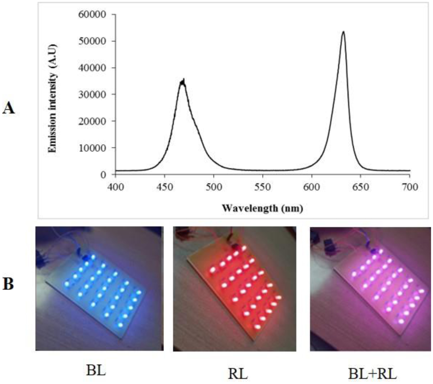
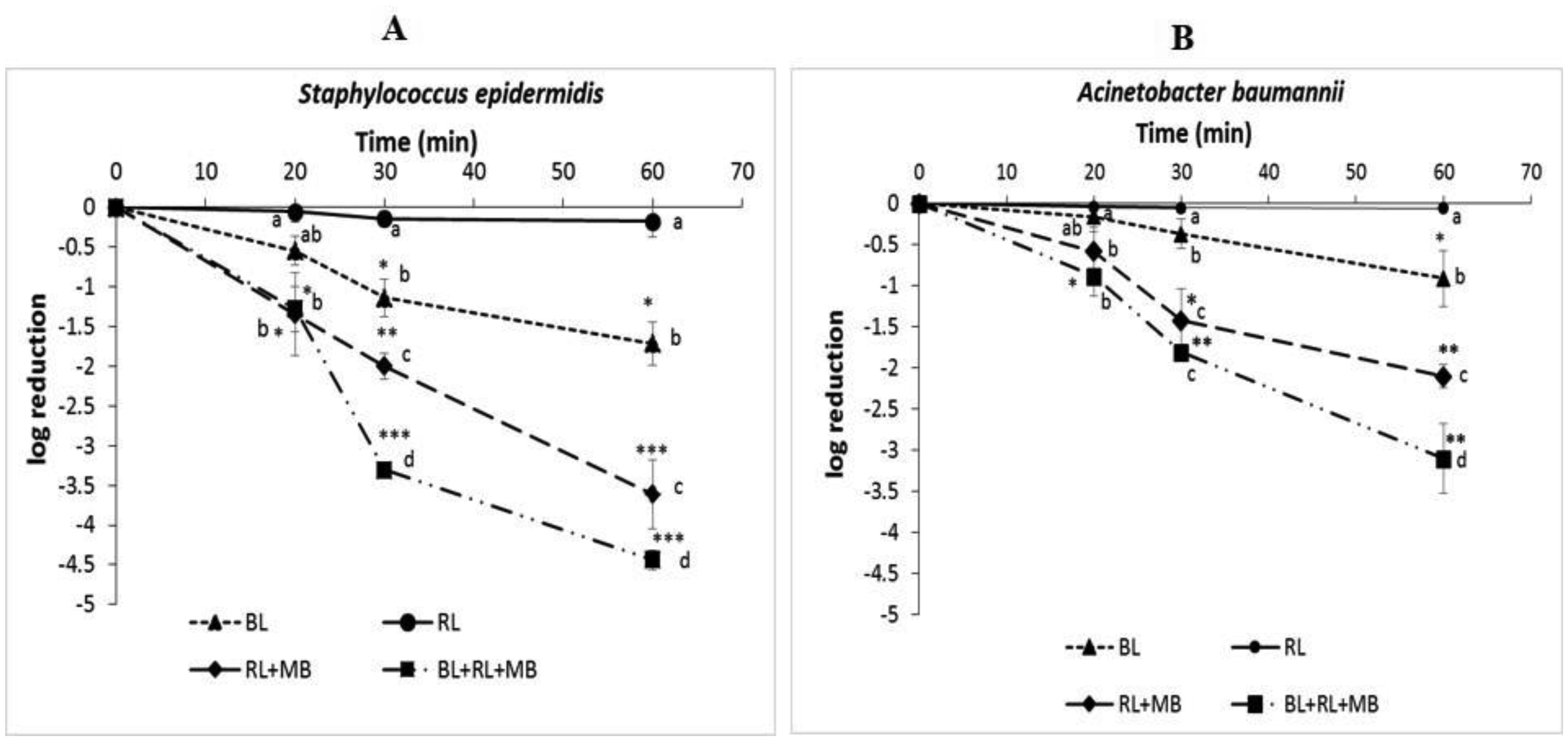
Due to increasing antibiotic resistance and a lack of new antibiotics, alternative treatments are urgently needed. This study investigates photodynamic therapy (PDT), which uses light-activated photosensitizers to produce reactive oxygen species that effectively inactivate bacteria. We evaluated the antibacterial efficacy of PDT against two pathogens that are resistant to current antibiotics, namely Staphylococcus epidermidis (Gram-positive) and Acinetobacter baumannii (Gram-negative), by testing various illumination protocols. The results showed that combining blue light (468 nm) and red light (632 nm) with methylene blue (MB) produced a synergistic effect in bacterial inactivation compared with protocols using either blue or red light individually in combination with methylene blue (MB). Specifically, after just 30 minutes of exposure, S. epidermidis showed a 3.3 log reduction (99.95%), while A. baumannii showed a 3.1 log reduction (99.92%) after 60 minutes. Overall, S. epidermidis was more sensitive to all tested protocols than A. baumannii. We also examined the effects of this protocol on antibiotic susceptibility. For most antibiotics tested, there was no change in the size of the inhibition zones. However, for linezolid, we observed a significant increase in the inhibition zone's diameter, indicating a possible enhanced susceptibility to this antibiotic.
Citation: Jaouhra Cherif, Anis Raddaoui, Nada Souissi, Nakkach Mohamed. Synergistic antibacterial activity of combined blue and red light photodynamic therapy[J]. AIMS Biophysics, 2025, 12(4): 572-585. doi: 10.3934/biophy.2025028
Due to increasing antibiotic resistance and a lack of new antibiotics, alternative treatments are urgently needed. This study investigates photodynamic therapy (PDT), which uses light-activated photosensitizers to produce reactive oxygen species that effectively inactivate bacteria. We evaluated the antibacterial efficacy of PDT against two pathogens that are resistant to current antibiotics, namely Staphylococcus epidermidis (Gram-positive) and Acinetobacter baumannii (Gram-negative), by testing various illumination protocols. The results showed that combining blue light (468 nm) and red light (632 nm) with methylene blue (MB) produced a synergistic effect in bacterial inactivation compared with protocols using either blue or red light individually in combination with methylene blue (MB). Specifically, after just 30 minutes of exposure, S. epidermidis showed a 3.3 log reduction (99.95%), while A. baumannii showed a 3.1 log reduction (99.92%) after 60 minutes. Overall, S. epidermidis was more sensitive to all tested protocols than A. baumannii. We also examined the effects of this protocol on antibiotic susceptibility. For most antibiotics tested, there was no change in the size of the inhibition zones. However, for linezolid, we observed a significant increase in the inhibition zone's diameter, indicating a possible enhanced susceptibility to this antibiotic.

| [1] |
Cassini A, Högberg LD, Plachouras D, et al. (2019) Attributable deaths and disability-adjusted life-years caused by infections with antibiotic-resistant bacteria in the EU and the European Economic Area in 2015: a population-level modelling analysis. Lancet Infect Dis 19: 56-66. https://doi.org/10.1016/S1473-3099(18)30605-4 
|
| [2] |
van Duin D, Paterson DL (2020) Multidrug-resistant bacteria in the community: an update. Infect Dis Clin North Am 34: 709-722. https://doi.org/10.1016/j.idc.2020.08.002
|
| [3] | Medina E, Pieper DH (2016) Tackling threats and future problems of multidrug-resistant bacteria. Curr Top Microbiol Immunol 398: 3-33. https://doi.org/10.1007/82_2016_492 |
| [4] |
Hoenes K, Bauer R, Meurle T, et al. (2021) Inactivation effect of violet and blue light on ESKAPE pathogens and closely related non-pathogenic bacterial species – A promising tool against antibiotic-sensitive and antibiotic-resistant microorganisms. Front Microbiol 11: 612367. https://doi.org/10.3389/fmicb.2020.612367
|
| [5] |
Willis JA, Cheburkanov V, Chen S, et al. (2022) Breaking down antibiotic resistance in methicillin-resistant Staphylococcus aureus: combining antimicrobial photodynamic and antibiotic treatments. Proc Natl Acad Sci USA 119: e2208378119. https://doi.org/10.1073/pnas.2208378119
|
| [6] |
Hessling M, Spellerberg B, Hoenes K (2017) Photoinactivation of bacteria by endogenous photosensitizers and exposure to visible light of different wavelengths – a review on existing data. FEMS Microbiol Lett 364: fnw270. https://doi.org/10.1093/femsle/fnw270
|
| [7] |
Huang S, Lin S, Qin H, et al. (2023) The parameters affecting antimicrobial efficiency of antimicrobial blue light therapy: a review and prospect. Biomedicines 11: 1197. https://doi.org/10.3390/biomedicines11041197
|
| [8] |
Wang Y, Wang Y, Wang Y, et al. (2017) Antimicrobial blue light inactivation of pathogenic microbes: state of the art. Drug Resist Updat 33–35: 1-22. https://doi.org/10.1016/j.drup.2017.10.002
|
| [9] |
Hoenes K, Wenzel U, Spellerberg B, et al. (2020) Photoinactivation sensitivity of Staphylococcus carnosus to visible-light irradiation as a function of wavelength. Photochem Photobiol 96: 156-169. https://doi.org/10.1111/php.13168
|
| [10] |
Galo IDC, Prado RP, Santos WGD (2021) Blue and red light photoemitters as approach to inhibit Staphylococcus aureus and Pseudomonas aeruginosa growth. Braz J Biol 82: e231742. https://doi.org/10.1590/1519-6984.231742
|
| [11] |
Leanse LG, Harrington OD, Fang Y, et al. (2018) Evaluating the potential for resistance development to antimicrobial blue light (at 405 nm) in Gram-negative bacteria: in vitro and in vivo studies. Front Microbiol 9: 2403. https://doi.org/10.3389/fmicb.2018.02403
|
| [12] |
Rapacka-Zdonczyk A, Wozniak A, Nakonieczna J, et al. (2021) Development of antimicrobial phototreatment tolerance: Why the methodology matters. Int J Mol Sci 22: 2224. https://doi.org/10.3390/ijms22042224
|
| [13] |
Hamblin MR (2017) Mechanisms and applications of the anti-inflammatory effects of photobiomodulation. AIMS Biophys 4: 337-361. https://doi.org/10.3934/biophy.2017.3.337
|
| [14] |
Salman S, Guermonprez C, Peno-Mazzarino L, et al. (2023) Photobiomodulation controls keratinocytes inflammatory response through Nrf2 and reduces Langerhans cells activation. Antioxidants 12: 766. https://doi.org/10.3390/antiox12030766
|
| [15] |
Piksa M, Fortuna W, Lian C, et al. (2023) Treatment of antibiotic-resistant bacteria colonizing diabetic foot ulcers by OLED induced antimicrobial photodynamic therapy. Sci Rep 13: 14087. https://doi.org/10.1038/s41598-023-39363-4
|
| [16] |
Pérez-Laguna V, García-Luque I, Ballesta S, et al. (2021) Photodynamic therapy combined with antibiotics or antifungals against microorganisms that cause skin and soft tissue infections: a planktonic and biofilm approach to overcome resistances. Pharmaceuticals (Basel) 14: 603. https://doi.org/10.3390/ph14070603
|
| [17] |
da Fonseca AS, Mencalha AL, de Paoli F (2021) Antimicrobial photodynamic therapy against Acinetobacter baumannii. Photodiagnosis Photodyn Ther 35: 102430. https://doi.org/10.1016/j.pdpdt.2021.102430
|
| [18] | Siciliano V, Passerotto RA, Chiuchiarelli M, et al. (2023) Difficult-to-treat pathogens: a review on the management of multidrug-resistant Staphylococcus epidermidis. Life (Basel) 13: 1126. https://doi.org/10.3390/life13051126 |
| [19] | CA-SFM, Comité de l'Antibiogramme de la Société Française de Microbiologie, Paris, France, 2024. Available from: https://www.sfm-microbiologie.org/boutique/_comite-de-lantibiogramme-de-la-sfm-ca-sfm-v1-0-juin-2024/ |
| [20] |
Bliss CI (1939) The toxicity of poisons applied jointly. Ann Appl Biol 26: 585-615. https://doi.org/10.1111/j.1744-7348.1939.tb06990.x
|
| [21] |
Goldoni M, Johansson C (2007) A mathematical approach to study combined effects of toxicants in vitro: evaluation of the Bliss independence criterion and the Loewe additivity model. Toxicol In Vitro 21: 759-769. https://doi.org/10.1016/j.tiv.2007.03.003
|
| [22] |
Maclean M, MacGregor SJ, Anderson JG, et al. (2009) Inactivation of bacterial pathogens following exposure to light from a 405-nanometer light-emitting diode array. Appl Environ Microbiol 75: 1932-1937. https://doi.org/10.1128/AEM.01892-08
|
| [23] |
Murdoch LE, McKenzie K, Maclean M, et al. (2013) Lethal effects of high-intensity violet 405-nm light on Saccharomyces cerevisiae, Candida albicans, and on dormant and germinating spores of Aspergillus niger. Fungal Biol 117: 519-527. https://doi.org/10.1016/j.funbio.2013.05.004
|
| [24] |
Choby JE, Skaar EP (2016) Heme synthesis and acquisition in bacterial pathogens. J Mol Biol 428: 3408-3428. https://doi.org/10.1016/j.jmb.2016.03.018
|
| [25] |
Dailey HA, Dailey TA, Gerdes S, et al. (2017) Prokaryotic heme biosynthesis: multiple pathways to a common essential product. Microbiol Mol Biol Rev 81: e00048-16. https://doi.org/10.1128/MMBR.00048-16
|
| [26] |
Hyun JE, Moon SK, Lee SY (2021) Antibacterial activity and mechanism of 460–470 nm light-emitting diodes against pathogenic bacteria and spoilage bacteria at different temperatures. Food Control 123: 107721. https://doi.org/10.1016/j.foodcont.2020.107721
|
| [27] |
Nitzan Y, Salmon-Divon M, Shporen E, et al. (2004) ALA induced photodynamic effects on Gram positive and negative bacteria. Photochem Photobiol Sci 3: 430-435. https://doi.org/10.1039/b315633h
|
| [28] |
Hadi J, Wu S, Brightwell G (2020) Antimicrobial blue light versus pathogenic bacteria: Mechanism, application in the food industry, hurdle technologies and potential resistance. Foods 9: 1895. https://doi.org/10.3390/foods9121895
|
| [29] |
Kim S, Kim J, Lim W, et al. (2013) In vitro bactericidal effects of 625, 525, and 425 nm wavelength (red, green, and blue) light-emitting diode irradiation. Photomed Laser Surg 31: 554-562. https://doi.org/10.1089/pho.2012.3343
|
| [30] |
Pereira PR, de Paula JB, Cielinski J, et al. (2014) Effects of low intensity laser in in vitro bacterial culture and in vivo infected wounds. Rev Col Bras Cir 41: 49-55. https://doi.org/10.1590/s0100-69912014000100010
|
| [31] | Angarano V, Akkermans S, Smet C, et al. (2020) The potential of violet, blue, green and red light for the inactivation of P. fluorescens as planktonic cells, individual cells on a surface and biofilms. Food Bioprocess Technol 124: 184-195. https://doi.org/10.1016/j.fbp.2020.07.019 |
| [32] |
Martins Antunes de Melo WC, Celiešiūtė-Germanienė R, Šimonis P, et al. (2021) Antimicrobial photodynamic therapy (aPDT) for biofilm treatments. Possible synergy between aPDT and pulsed electric fields. Virulence 12: 2247-2272. https://doi.org/10.1080/21505594.2021.1960105
|
| [33] |
Vieira C, Gomes ATPC, Mesquita MQ, et al. (2018) An insight into the potentiation effect of potassium iodide on aPDT efficacy. Front Microbiol 9: 2665. https://doi.org/10.3389/fmicb.2018.02665
|
| [34] |
Minnock A, Vernon DI, Schofield J, et al. (2000) Mechanism of uptake of a cationic water-soluble pyridinium zinc phthalocyanine across the outer membrane of Escherichia coli. Antimicrob Agents Chemother 44: 522-527. https://doi.org/10.1128/AAC.44.3.522-527.2000
|
| [35] |
Kim MJ, Tang CH, Bang WS, et al. (2017) Antibacterial effect of 405 ± 5 nm light emitting diode illumination against Escherichia coli O157:H7, Listeria monocytogenes, and Salmonella on the surface of fresh-cut mango and its influence on fruit quality. Int J Food Microbiol 244: 82-89. https://doi.org/10.1016/j.ijfoodmicro.2016.12.023
|
| [36] |
Wu J, Chu Z, Ruan Z, et al. (2018) Changes of intracellular porphyrin, reactive oxygen species, and fatty acids profiles during inactivation of methicillin-resistant Staphylococcus aureus by antimicrobial blue light. Front Physiol 9: 1658. https://doi.org/10.3389/fphys.2018.01658
|
| [37] |
Chu Z, Hu X, Wang X, et al. (2019) Inactivation of Cronobacter sakazakii by blue light illumination and the resulting oxidative damage to fatty acids. Can J Microbiol 65: 922-929. https://doi.org/10.1139/cjm-2019-0054
|
| [38] |
Kruszewska-Naczk B, Grinholc M, Rapacka-Zdonczyk A (2024) Mimicking the effects of antimicrobial blue light: Exploring single stressors and their impact on microbial growth. Antioxidants 13: 1583. https://doi.org/10.3390/antiox13121583
|
| [39] |
Bowman C, Bumah VV, Niesman IR, et al. (2021) Structural membrane changes induced by pulsed blue light on methicillin-resistant Staphylococcus aureus (MRSA). J Photochem Photobiol B 216: 112150. https://doi.org/10.1016/j.jphotobiol.2021.112150
|
| [40] |
Leanse LG, Anjos CD, Kaler KR, et al. (2023) Blue light potentiates antibiotics in bacteria via parallel pathways of hydroxyl radical production and enhanced antibiotic uptake. Adv Sci 10: e2303731. https://doi.org/10.1002/advs.202303731
|
Figures(2) / Tables(2)

Jaouhra Cherif, Anis Raddaoui, Nada Souissi, Nakkach Mohamed. Synergistic antibacterial activity of combined blue and red light photodynamic therapy[J]. AIMS Biophysics, 2025, 12(4): 572-585. doi: 10.3934/biophy.2025028


 DownLoad: