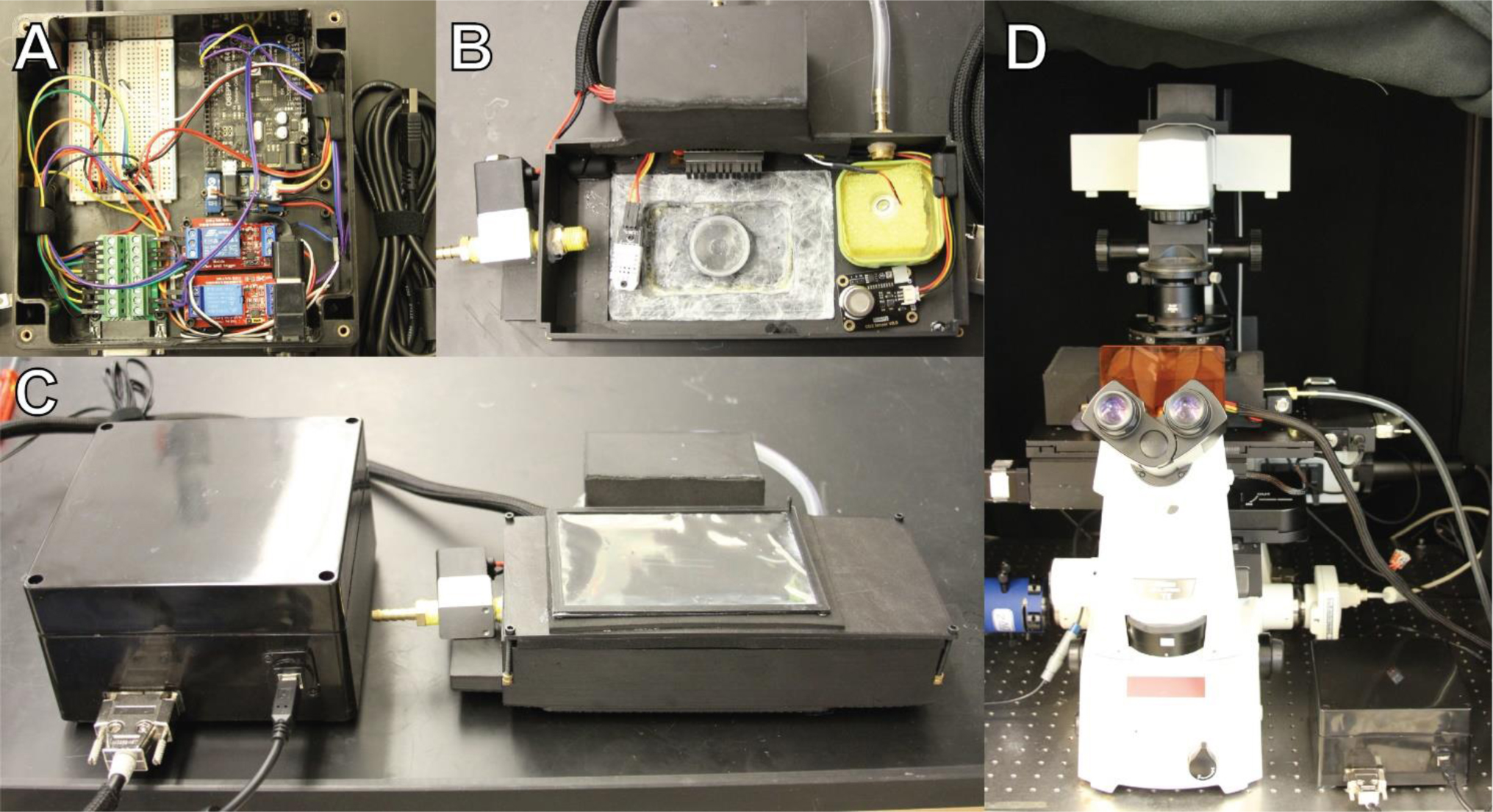
Live imaging of human or other mammalian cells at multi-hour time scales with minimal perturbation to their growth state requires that the specimen's optimal growth conditions are met while fixed to a microscope stage. In general, the ideal conditions include culturing in complete growth media, an ambient temperature of 36–37 °C, and a humidity-controlled atmosphere typically comprised of 5–7% CO2. Commercially available devices that achieve these conditions are not a financially viable option for many labs, with the price ranging anywhere from $12,000 to $40,000. The advent of 3D printing technologies have allowed for low-cost rapid prototyping with precision comparable to traditional fabrication methods, thus opening the possibility for the in-lab design and production of otherwise prohibitively expensive equipment such as stage-top incubation devices. Additionally, the continued usefulness and widespread availability of single-board computers (SBC) such as Arduino and Raspberry Pi simplify the process by which these devices can be controlled. Here, we report the production of a do-it-yourself (DIY) device for stage-top incubation with temperature and atmospheric control with a cost reduction of approximately 100x.
Citation: Michael Worcester, Shayan Nejad, Pratyasha Mishra, Quintin Meyers, Melissa Gomez, Thomas Kuhlman. A low-cost stage-top incubation device for live human cell imaging using rapid prototyping methods[J]. AIMS Biophysics, 2025, 12(2): 164-173. doi: 10.3934/biophy.2025010
Live imaging of human or other mammalian cells at multi-hour time scales with minimal perturbation to their growth state requires that the specimen's optimal growth conditions are met while fixed to a microscope stage. In general, the ideal conditions include culturing in complete growth media, an ambient temperature of 36–37 °C, and a humidity-controlled atmosphere typically comprised of 5–7% CO2. Commercially available devices that achieve these conditions are not a financially viable option for many labs, with the price ranging anywhere from $12,000 to $40,000. The advent of 3D printing technologies have allowed for low-cost rapid prototyping with precision comparable to traditional fabrication methods, thus opening the possibility for the in-lab design and production of otherwise prohibitively expensive equipment such as stage-top incubation devices. Additionally, the continued usefulness and widespread availability of single-board computers (SBC) such as Arduino and Raspberry Pi simplify the process by which these devices can be controlled. Here, we report the production of a do-it-yourself (DIY) device for stage-top incubation with temperature and atmospheric control with a cost reduction of approximately 100x.

| [1] |
Segeritz CP, Vallier L (2017) Cell culture: growing cells as model systems in vitro. Basic Science Methods for Clinical Researchers . Academic Press 151-172. https://doi.org/10.1016/B978-0-12-803077-6.00009-6 
|
| [2] | Heating System Slide/Dish–Silver Line. Available from: https://ibidi.com/heating-systems/292-ibidi-heating-system-slidedish-silver-line.html |
| [3] |
Rossi VM, Davidson KC, Moore LE (2022) Arduino-based, low-cost imaging incubator for extended live cell imaging. Appl Optics 61: 5282. https://doi.org/10.1364/AO.460443
|
| [4] | Ay FC, Bilici N, Varol R, et al. Study on the concept and development of a mobile incubator (2022) arXiv:2208.09697. |
| [5] | 293T Cells. Available from: https://www.thermofisher.com/us/en/home/technical-resources/cell-lines/2/cell-lines-detail-153.html |
| [6] |
Schindelin J, Arganda-Carreras I, Frise E, et al. (2012) Fiji: an open-source platform for biological-image analysis. Nat Methods 9: 676-682. https://doi.org/10.1038/nmeth.2019
|
| [7] |
Stirling DR, Swain-Bowden MJ, Lucas AM, et al. (2021) CellProfiler 4: improvements in speed, utility and usability. BMC Bioinformatics 22: 433. https://doi.org/10.1186/s12859-021-04344-9
|
| [8] |
Geva-Zatorsky N, Dekel E, Batchelor E, et al. (2010) Fourier analysis and systems identification of the p53 feedback loop. Proc Natl Acad Sci USA 107: 13550-13555. https://doi.org/10.1073/pnas.1001107107
|
| [9] |
Fobian SF, Amin M, Sacchetti A, et al. (2025) Investigating the delivery of PD-L1-targeted immunoliposomes in a dynamic cervical cancer-on-a-chip model. J Control Release 379: 236-250. https://doi.org/10.1016/j.jconrel.2025.01.014
|
| [10] |
Mannell H, Kameritsch P, Beck H, et al. (2021) Cx43 promotes endothelial cell migration and angiogenesis via the tyrosine phosphatase SHP-2. Int J Mol Sci 23: 294. https://doi.org/10.3390/ijms23010294
|


Figures(5)

Michael Worcester, Shayan Nejad, Pratyasha Mishra, Quintin Meyers, Melissa Gomez, Thomas Kuhlman. A low-cost stage-top incubation device for live human cell imaging using rapid prototyping methods[J]. AIMS Biophysics, 2025, 12(2): 164-173. doi: 10.3934/biophy.2025010


 DownLoad: