Citation: Jay L. Brewster. Signaling hubs at ER/mitochondrial membrane associations[J]. AIMS Biophysics, 2017, 4(2): 222-239. doi: 10.3934/biophy.2017.2.222
[1] | Mahmoud Abdallat, Abdallah Barjas Qaswal, Majed Eftaiha, Abdel Rahman Qamar, Qusai Alnajjar, Rawand Sallam, Lara Kollab, Mohammad Masa'deh, Anas Amayreh, Hiba Mihyar, Hesham Aboushakra, Bayan Alkelani, Rawan Owaimer, Mohannad Abd-Alhadi, Salwa Ireiqat, Fahed Turk, Ahmad Daoud, Bashar Darawsheh, Ahmad Hiasat, Majd Alhalaki, Shahem Abdallat, Salsabiela Bani Hamad, Rand Murshidi . A mathematical modeling of the mitochondrial proton leak via quantum tunneling. AIMS Biophysics, 2024, 11(2): 189-233. doi: 10.3934/biophy.2024012 |
[2] | Jacques Fantini, Francisco J. Barrantes . How membrane lipids control the 3D structure and function of receptors. AIMS Biophysics, 2018, 5(1): 22-35. doi: 10.3934/biophy.2018.1.22 |
[3] | Satoru Ito, Kishio Furuya, Masahiro Sokabe, Yoshinori Hasegawa . Cellular ATP release in the lung and airway. AIMS Biophysics, 2016, 3(4): 571-584. doi: 10.3934/biophy.2016.4.571 |
[4] | Daniela Meleleo, Cesare Sblano . Influence of cholesterol on human calcitonin channel formation. Possible role of sterol as molecular chaperone. AIMS Biophysics, 2019, 6(1): 23-38. doi: 10.3934/biophy.2019.1.23 |
[5] | Iva Valkova, Petar Todorov, Victoria Vitkova . VV-hemorphin-5 association to lipid bilayers and alterations of membrane bending rigidity. AIMS Biophysics, 2022, 9(4): 294-307. doi: 10.3934/biophy.2022025 |
[6] | István P. Sugár, Parkson Lee-Gau Chong . Monte Carlo simulations of the distributions of intra- and extra-vesicular ions and membrane associated charges in hybrid liposomes composed of negatively charged tetraether and zwitterionic diester phospholipids. AIMS Biophysics, 2017, 4(2): 316-336. doi: 10.3934/biophy.2017.2.316 |
[7] | Marek K. Korzeniowski, Barbara Baird, David Holowka . STIM1 activation is regulated by a 14 amino acid sequence adjacent to the CRAC activation domain. AIMS Biophysics, 2016, 3(1): 99-118. doi: 10.3934/biophy.2016.1.99 |
[8] | Oleg A. Karpov, Gareth W. Fearnley, Gina A. Smith, Jayakanth Kankanala, Michael J. McPherson, Darren C. Tomlinson, Michael A. Harrison, Sreenivasan Ponnambalam . Receptor tyrosine kinase structure and function in health and disease. AIMS Biophysics, 2015, 2(4): 476-502. doi: 10.3934/biophy.2015.4.476 |
[9] | Matteo Corradini, Marta Checchi, Marzia Ferretti, Francesco Cavani, Carla Palumbo, Vincenza Rita Lo Vasco . Endoplasmic reticulum localization of phosphoinositide specific phospholipase C enzymes in U73122 cultured human osteoblasts. AIMS Biophysics, 2023, 10(1): 25-49. doi: 10.3934/biophy.2023004 |
[10] | Emmanuel Reyes-Uribe, Nathalia Serna-Marquez, Eduardo Perez Salazar . DDRs: receptors that mediate adhesion, migration and invasion in breast cancer cells. AIMS Biophysics, 2015, 2(3): 303-317. doi: 10.3934/biophy.2015.3.303 |
Organelle systems comprise a dynamic network of critical cellular functions and are managed by signaling networks foundational to living systems. Studies of stress signaling from the endoplasmic reticulum (ER) identified critical influences upon the nucleus, mitochondria, and the cytoplasm. As ER signal mediators have been characterized, the continual exchange of regulatory information between the ER and other organelles has become clear. Disruption of these signaling processes creates complex phenotypes; including altered biochemical pathways, loss of Ca2+ homeostasis in the cell, physical changes to the mitochondria and ER, and inappropriate activation of cellular stress response programming. Studies of ER/mitochondrial communication have revealed that dysfunction at this signaling hub generates profound consequences. Genetic analysis of heritable neurodegenerative disease in humans has identified several MAM components to be responsible for disease initiation.
The ER and mitochondria comprise a major proportion of cellular membranes. These membranes display complex functional organization; mediating function of the endomembrane system (vesicle mediated transport), lipid biosynthetic processes, and exchange of critical cellular molecules. The ER membrane network is a dynamic labyrinth of tubes connected with larger sacs, and this membrane is continuous with the outer nuclear membrane. The lumen of the ER and the nuclear inter-membrane space are contiguous and surrounded by a shared sheet of membrane. Domains within this single membrane are functionally distinct forming sites of lipid biogenesis (smooth ER), ribosome-associated regions involved in protein production (rough ER), and the regulation of nuclear import/export at the nucleus. This lumen space is central for accumulation of cellular Ca2+ at concentrations of up to 500 μM, being imported via the sarco-endoplasmic reticulum Ca2+ ATPase transporter (SERCA) [1]. Calcium release from the ER is primarily mediated by two gene families, the inositol 1, 4, 5-trisphosphate receptor family (ITPR1-3), and the ryanodine receptor family (RyR1-3) [2,3]. The ER also serves in lipid and phospholipid biosynthesis, and in the processing of proteins destined for the endomembrane transport system. The overall composition of ER enzymes and architecture varies according to differentiated cell type.
Mitochondria contain their own genome and are structurally organized in a similar manner to Gram-negative bacterium, with outer and inner membranes that are functionally and biochemically distinct. The outer mitochondrial membrane (OMM) is porous relative to the inner membrane and contains pore-forming membrane proteins such as the voltage-dependent anion channel (VDAC), which facilitates movement of diverse small molecules, including Ca2+ and ATP. The inner mitochondrial membrane (IMM) provides a significant barrier to biomolecule passage. It has extensive involutions generating isolated structural pockets known as cristae, and contains a high-density of membrane proteins including electron transport components and channels facilitating import and export of ETS substrates and products. Mitochondria have their own ribosomes and tRNAs, producing some key proteins for mitochondrial function from within the matrix, though most mitochondrial proteins are produced from the nuclear genome and translocated to the organelle [4]. Similar to the ER, mitochondria are dynamic in structure, displaying the ability to divide via fission to generate multiple small mitochondria, or to fuse to generate larger mitochondrial structures [5,6]. Changes in mitochondrial number and morphology are an indicator of cellular stress and an established pathology of human disease [7]. Aging studies have shown that mitochondrial organization and structure; in particular membrane cristae, are distinct in older mitochondria [4,8].
The labyrinth of ER tubules and sacs is infiltrated with mitochondria, placing these organelles in close proximity. As measured with live cell imaging techniques, physical interaction between the organelles is extensive, and is critical in the regulation of mitochondrial behavior. The ER is the primary Ca2+ storage site in metazoan cells, and Ca2+ efflux plays a role in communication from the ER to mitochondria and the cytosol. Mitochondrial Ca2+ uptake regulates specific enzymes of the TCA cycle in the matrix, elevates levels of reactive oxygen species (ROS) generated by the mitochondria, induces mitochondrial division, and can generate apoptosis-inducing mitochondrial dynamics. Close mitochondrial interaction with the ER was first imaged over 50 years ago using electron microscopy [9]. Time-lapse 3D imaging of the interwoven ER/mitochondrial membrane network began with the remarkable work of Rizzuto and colleagues [10]. In order to label the ER and mitochondria, cultured cells were transfected with recombinant fluorescent proteins localized to each structure. Membrane domains of close apposition (10-25 nm apart) were observed to form between the ER and mitochondrial membranes, and to involve approximately 5-20% of the total mitochondrial surface area [10]. These regions are now referred to as Mitochondria Associated Membranes (MAM): raft domains that are enriched with transmembrane proteins. ITPR channel proteins localize to MAM domains. These channels normally signal via either oscillating or wave pulses of Ca2+ current. Severe organelle stress generates waves of Ca2+ current from the ITPR, resulting in an overload of mitochondrial Ca2+ in the intermembrane space (IMS) and matrix [11]. The Bcl-XL protein, a Bcl-2 family member, is partially localized to MAM domains, and has been shown to bind to the ITPR to influence Ca2+ release dynamics into the mitochondria [12]. The p53 tumor suppressor, a protein that regulates cellular responses to mutation and stress, has been shown to bind SERCA on the ER membrane to induce an overloading of Ca2+ in the ER lumen [13,14]. This is sufficient to activate apoptosis via ITPR mediated Ca2+ overloading of mitochondria, and it notably lost in oncogenic mutation of p53 and SERCA genes [14]. In original studies of MAM structures, elevation of Ca2+ in mitochondrial IMS was adjacent to MAM structures. Since that initial description, MAM domains have gained intense attention as a key site of communication between the ER and mitochondria. Examples of ITPR-mediated activation of intrinsic apoptosis are accumulating in disease studies; and include poliovirus infection [15], diabetes progression [16], and Alzheimer's Disease [17].
The ITPR is not only a site of Ca2+ release from the ER, it also is one contact point for building physical connections to the mitochondria. A yeast two-hybrid screen for proteins that bind to VDAC revealed the cytosolic GRP75 chaperone as a binding partner [18]. In evaluating this interaction in vivo, GRP75 was shown to simultaneously bind VDAC at the mitochondria and the ITPR at the ER; physically linking these two channels and enabling Ca2+ transfer into the mitochondria (Figure 1). This mechanism benefitted the study of Ca2+ entry into the matrix of the mitochondria from the IMS. The central Ca2+ entry point into the matrix, through a Ca2+ impermeable membrane, is the mitochondrial Ca2+ uniporter channel (MCU). This channel exhibits very low affinity for Ca2+, with a Kd in the range of 20-50 μM, making it an effective transporter only when [Ca2+] in the IMS is severely elevated [3]. Localized influx of Ca2+ from the ITPR through the VDAC sufficiently elevates IMS Ca2+ to provide influx via MCU into the matrix. Though GRP75 serves to crosslink VDAC and ITPR, this interaction does not qualify as an organelle tether as deletion of all ITPR genes in cultured B cells (DT40) does not perturb the generation and stabilization of MAM associations [19]. Thus responsibility for stabilizing these close interactions is likely due to other proteins.
The impacts of Ca2+ signals transmitted from the ER or the cytosol to the mitochondria are remarkably diverse. Elevation of matrix Ca2+ stores stimulates key enzymes of the TCA cycle such as pyruvate dehydrogenase, and subsequently elevates production of ATP [20,21]. The Na+/Ca2+/Li+ exchange channel (NCLX) in the inner mitochondrial membrane regulates calcium levels in the mitochondrial matrix [22]. Calcium overload in the mitochondria has been linked to hyper-production of reactive oxygen species (ROS), a causative agent in activation mitochondria-initiated intrinsic apoptosis [23,24]. The initiating factors in activating mitochondrial outer membrane permeabilization (MOMP) are not well understood, though this permeabilization is a key factor in intrinsic apoptosis. Activated monomers of Bax/Bak assemble in the mitochondria membrane, overwhelming the inhibitory capacity of anti-apoptotic Bcl-2 family members, and ultimately a large "Bax pore" or permeability transition pore (PTP) is formed. This structure releases a number of pro-apoptotic factors from the mitochondria, most famously cytochrome c. The inner mitochondrial membrane is an extensively involuted structure, displaying cristae that are isolated pockets of IMS components. Electrostatic interactions between soluble electron carrier cytochrome c and reduced lipids in the inner membrane result in approximately 85% localization of cytochrome c to cristae. Following formation of the PTP, access of cytosolic components to the IMS facilitates reorganization of the mitochondrial cristae, releasing cytochrome c from cristae, and subsequently to the cytosol [25]. Cytosolic cytochrome c activates caspase-9 via formation of the apoptosome structure, and a subsequent induction of executioner caspases.
Lipid rafts are defined as regions of lateral heterogeneity within membranes, and are typically enriched with sphingolipids, cholesterol, and proteins [26]. The first biochemical characterization of lipid rafts was published in 1990, defining a method of isolating distinct membrane components from rat liver mitochondria [27]. Sub-fractions were generated via centrifugation techniques, and a raft-containing fraction with both mitochondrial and ER membranes was identified with unique enzymatic and biochemical characteristics [27,28]. The physical processes of generation and maintenance of these raft domains remains technically challenging to experimentally evaluate. However, it is clear that these domains offer critical organization to biological membranes, allowing regional concentration of functionally related membrane components.
Sphingolipids are enriched in lipid raft domains, and disruption of their accumulation has been linked to abnormalities in MAM function. Sphingolipids are amino alcohols with a long hydrocarbon chain that comprise a complex group of membrane lipid components. Gangliosides are one class of sphingolipid, containing bulky head groups with sialic acids and sugars. Gangliosides are enriched in plasma membranes, and comprise~5% of the lipids in the membranes of cells in the vertebrate nervous system. Characterization of GM1-ganglioside accumulation in neuronal lysosomal storage disease models provides an additional insight into MAM domains and lipid dynamics. GM1-gangliosidosis in cultured neurons was shown to induce ER stress and apoptosis via the mitochondria [29]. Analysis of this phenomenon revealed GM1-gangliosides to accumulate in glycophospholipid-enriched microdomains (GEM) of MAM fractions. These accumulated GM1-gangliosides bound the ITPR, elevating Ca2+ flux from the ER to generate mitochondrial Ca2+ overload, and apoptosis [30,31]. GD3-gangliosides are enriched in autophagosome vesicle membranes. As autophagy initiation is primarily regulated via the mitochondria, GD3-enriched MAM domains are suspected to play a role in autophagy. Inhibition of GD3 biosynthesis via ablation of biosynthetic genes was shown to perturb MAM function in signaling, and to disrupt autophagy activation [32].
The movement and maintenance of organelle-specific lipid composition is a critical consideration for understanding cell function. Shuttling of membrane components between leaflets of a bilayer is mediated by "flippases" (inward), "floppases" (outward), and "scramblases" (bidirectional) [33]. Vesicle movement from the ER and Golgi enables distribution of membrane components about the cell. Close apposition of membranes at ER-mitochondrial MAM sites and ER-endosomal sites, enables non-vesicle mediated lipid exchange [34,35]. Lipid-transfer proteins (LTP) mediate non-vesicular lipid transport between juxtaposed membranes, though the mechanism of this transfer is poorly understood [35,36]. Characterization of MAM interactions between ER and mitochondria has provided opportunities to examine non-vesicle mediated lipid exchange (for review, [37]). Cholesterol biosynthesis occurs primarily via enzymes in the ER membrane, and provision of cholesterol to the mitochondria via MAM domains is required to feed steroid biosynthetic pathways [38]. The Caveolin-1 membrane scaffolding protein is enriched in MAM domains and plays a central role in the recruitment and regulation of cholesterol and steroid levels in these membrane regions [39].
The ER also serves as a central biosynthetic site for membrane phospholipids. Phosphatidylserine (PS) produced in the ER is translocated to the mitochondrial inner membrane, and then converted to phosphatidylethanolamine (PE) via activity of phosphatidylserine decarboxylase (Psd1). PE is distributed throughout the cell from the mitochondria, with some being transported back to the ER membrane in order to be converted to phosphatidylcholine (PC). These exchanges have been localized to transport processes in MAM domains [40]. During mitochondrial stress, conversion of PS to PE is inhibited, and PS arriving from the ER accumulates in the mitochondrial membranes. This accumulation contributes to a distribution and enrichment of PS in cellular membranes during stress (organelle scrambling), a key characteristic of apoptotic activation.
Tethering the close association between the ER and mitochondria is accomplished by protease sensitive structures that are structurally diverse. In Saccharomyces cerevisiae, an elegant strategy was developed to identify genes involved in tethering [41]. Mutagenized yeast cells were screened for mutations that generate an inability of cells to grow/survive on non-fermentable carbon sources that require mitochondrial function for sufficient metabolic support. An artificial ER-mitochondrial tether was used to screen mutant cell lines, and to identifying those with defects in tethering (Figure 2A). This artificial tether included ER and mitochondrial localization domains fused to opposite ends of green fluorescent protein (GFP). Expression of the artificial tether in yeast cells results in GFP fluorescence localization to MAM sites. The mutant cell lines rescued by this artificial tether identified four genes: MDM10, MDM34, MMM1, and MDM12. Each of the four genes is required for normal MAM structure and Ca2+ signaling dynamics. Mdm10 and Mdm34 were shown to localize to the outer mitochondrial membrane, Mmm1 to the ER membrane, and Mdm12 to the cytosol. All four proteins assemble into a single MAM complex known as the ER-mitochondria encounter structure (ERMES) [40,42]. No homologous set of genes has yet been identified in higher eukaryotes. Disruption of the ERMES complex also generated lipid-distribution defects to identical to genetic ablation of PSD1, encoding the enzyme that mediates conversion of phosphatidylserine PS to PE in the mitochondrial inner membrane [41]. ERMES-deficient cells did not display a complete loss of lipid trafficking between the ER and mitochondria, indicating additional mechanisms must exist in yeast. A second structure, known as the EMC complex (ER membrane protein complex), has subsequently been identified, and appears to have redundant lipid transfer characteristics [43].
VAPB is a member of the VAMP-associated protein (VAP) family, which are ER resident proteins involved in diverse functions that include lipid transport, the unfolded protein response, membrane trafficking (for review, [44]). Analysis of VAPB function revealed binding to a mitochondrial outer membrane protein, protein tyrosine phosphatase interacting protein 51 (PTPIP51) [45,46]. When expression of either PTPIP51 or VAPB was inhibited with siRNA, ER-mitochondrial associations were lost, and cells displayed abnormal mitochondrial uptake of Ca2+ during ER stress conditions [46].
Mitofusin-1/2 (Mfn1/2) are GTPase enzymes that predominantly locate to the mitochondrial outer membrane and were initially discovered as mediators of mitochondrial fusion events [47]. These proteins localize to MAM domains, with Mfn2 being shown to also localize to the ER side of MAM domains and forming homo/heterodimers with Mfn1/2 [48]. There are conflicting publications regarding the role of mitofusin proteins as tethers or as tethering antagonists that separate membranes at appropriate distance [49]. However, a recent study carefully examined the impact of MFN2 ablation using electron microscopy and fluorescence energy transfer (FRET) imaging, revealing an increase in MAM membrane distance and disruption of Ca2+ signaling [47]. Mutations in MFN2 are the most common cause of type 2 Charcot-Marie-Tooth (CMT) disease, a form of hereditary peripheral neuropathy [51].
An unusual example of signaling from the mitochondrial to the ER is via the Bap31 polytopic transmembrane protein. Bap31 function was initially established as a sorting factor, interacting with nascent membrane proteins at the Sec61 complex in the ER membrane and sorting those proteins to additional enzymes for appropriate processing [52]. Mitochondrial outer membrane protein Fis1 binds ER Bap31 in MAMs and facilitates cleavage of Bap31 by active caspase-8 following treatment with cellular stressors known to activate intrinsic apoptosis (Actinomycin C, etoposide) [53]. Caspase-8 protease induction cleaves the cytosolic tail of Bap31 to generate a 20-kDa form of Bap31, p20Bap31 [54,55]. This truncated p20Bap31 associates with the ITPR to stimulate Ca2+ release at MAM domains, generating mitochondrial Ca2+ overload, and a subsequent activation of the PTP complex [53,54]. Ectopic expression of an ER-restricted Bcl-2 was sufficient to block the ability of p20Bap31 to induce Ca2+ release sufficient to activate cell death [58]. Inhibiting this process by expressing an engineered form of Bap31, missing the caspase-8 cleavage site, was protective against activators of intrinsic apoptosis [59].
An additional ER/mitochondrial point of contact is revealed in the regulation of mitochondrial division. Stressors of the ER and mitochondria can induce mitochondrial fission to precede mitophagy, the selective destruction of mitochondria via autophagy. The mitochondrial outer membrane protein, FUNDC1, is known as a mitophagy receptor, capable of stimulating fission. FUNDC1 is localized to the MAM via interaction of a cytosolic loop domain with calnexin (CANX), an ER MAM component [60]. Induction of mitophagy with hypoxic conditions causes FUNDC1 to disassociate from CANX, and to subsequently bind/recruit dynamin-related protein (Drp1) dimers from the cytosol. Drp1 is a GTPase that plays a central role in mitochondrial division, using self-assembly to form a polymeric ring structure about the mitochondria to induce fission [61,62]. Loss of any of these three proteins generates defects in mitochondrial fission during hypoxia. These examples of ER-mitochondrial contact points (Figure 2B) reveal diverse mechanisms of organelle contact and regulation at the MAM domain.
Disruption of normal synthesis/processing of lipids or proteins synthesized by the ER generates stress signaling that can lead to adaptation or apoptosis. With regard to abnormalities in protein synthesis, stress signaling mediators include Ca2+-binding chaperones such as BiP/Grp78, a key regulatory chaperone in the ER lumen [63]. Accumulation of misfolded proteins saturates the chaperone activity of BiP/GRP78, activating stress signaling known as the unfolded protein response (UPR). Signaling activators of the UPR are located in the ER membrane, and have inhibitory regulatory interactions with BiP/GRP78 that are released during unfolded protein accumulation. These activators include the PKR-like ER kinase (PERK), the inositol-requiring enzyme 1 (IRE1), and the activating transcription factor 6 (ATF6). The UPR activates a pause in general cellular translation, adaptive changes in gene expression to restore ER function, autophagy to remove damaged organelles, and eventually apoptosis [64]. Both PERK and IRE1 have been shown to localize to MAM domains, and that localization plays a role in ER stress signaling. Several pathways downstream of ER stress are proapoptotic, including Ca2+ release via the ITPR, activation of JNK and p38 stress activated kinases, and direct activation of caspases [64,65]. GRP78 dissociation from IRE1 induces endoribonuclease activity and the splicing of Xbp1 pre-mRNA to enable translation of the encoded transcription factor, and activated expression of stress-response genes in the nucleus. Active IRE1 can also stimulate JNK signaling in the cytosol [64]. Some components of ER stress signaling are localized to MAMs, most notably PERK and IRE1, whose MAM localization enables interactions with regulatory partners. Physically uncoupling the mitochondria and ER generates ER stress signaling [66]. The Sigma-1 Receptor (Sig1R) is a protective chaperone protein located in the ER membrane at MAMs, and when activated, binds to IRE1 and the ITPR [67,68]. Similar to the regulation of other ER stress signaling proteins in the ER membrane, the BiP/GRP78 chaperone binds to Sig1R to inhibit its activation (Figure 3). During ER stress, this interaction is lost as unfolded proteins accumulate and saturate BiP/GRP78 activity. Release of Sig1R enables its diffusion within the membrane to ITPR, resulting in regulation of Ca2+ flux to benefit cell survival [68,69]. Sig1R binding of IRE1 during ER stress results in a stabilization of an active IRE1 protein complex, sustaining protective endoribonuclease activity [69]. The protective function of Sig1R is noted in the nervous system, in models of hereditary blindness and neurodegeneration. The simple activation of Sig1R, using a chemical agonist, has been shown to preserve rod and cone photoreceptor cells survival and function in a mouse model of heritable retinal degeneration [70]. The impacts of Sig1R signaling related to Ca2+ homeostasis and cellular responses to ROS are quite diverse, but its role in several neurodegenerative diseases is clear and the potential for pharmacological modulation of this mechanism is promising [71].
PERK (PKR-like ER kinase) is an ER transmembrane protein kinase regulated by BiP/GRP78 interaction and whose kinase active form phosphorylates eIF2α to suppress translation initiation during ER stress [64]. PERK localizes to the MAM, and is required for stabilization of MAM associations and ER morphology. Apoptosis activation during elevation of ROS (photo-oxidative treatment) was shown to be dependent upon the PERK protein [72]. Perhaps most surprising in this study was that a kinase dead PERK mutant was sufficient to support MAM structure and apoptotic signaling following ROS elevation. This suggests a tethering function for PERK that facilitates signal transfer from the ER to mitochondria, though no mitochondrial contact point has been identified.
The list of neuronal disorders associated with MAM dysfunction has grown rapidly over the past few years. It is clear that communication between the ER and mitochondria, especially communication via Ca2+ regulation is a particularly vulnerability of neuronal cells. Highlighted here are two disorders whose heritable forms have identified MAM proteins as potential sites of disease origin.
Alzheimer's Disease (AD) is a complex neurodegenerative disorder whose primary characteristics in the brain include neuronal loss associated with accumulation of intra/extra-cellular proteinacious deposits (plaques and tangles). Familial forms of AD result from mutations in presenilin genes (PSEN1, PSEN2), which encode subunits of the γ-secretase intramembrane protease complex, and amyloid precursor protein (APP), a substrate of this protease complex. The "amyloid cascade hypothesis" defines dysfunction of APP processing and accumulation of toxic Ab proteins as the primary origin of AD pathology via neuronal apoptosis [73,74]. However, an AD phenotype includes abnormal ER stress signaling, loss of Ca2+ homeostasis, abnormal phospholipid distribution, and abnormal cholesterol production [75,76,77]. Studies of mitochondria in AD have revealed a dramatic increase in MAM associations, and an increased ER-mitochondrial exchange of cholesterol and phospholipids. It has been noted that AD sufferers also display abnormal serum cholesterol levels [78].
Membrane-localized γ-secretase activity is found in lipid rafts, most predominantly in MAM domains [79]. Presenilin mutations results in abnormal γ-secretase processing of the stromal interaction module 1 (STIM1) transmembrane protein. This protein regulates Ca2+ and K+ ion channels both at the ER and the plasma membrane [80]. Loss of STIM1 causes dysregulation of Ca2+levels in the ER and abnormal capacitative Ca2+ entry (CCE) at the cell membrane [78,79]. Overexpression of STIM1 in presenilin mutant hippocampal cell lines was sufficient to restore Ca2+ homeostasis and arrest loss of dendritic spines [81].
ER stress signaling is also induced in neurons during AD progression. PERK mediated phosphorylation of eIF2α inhibits the activation of translation of most transcripts, though some translation is induced. PERK activity induces expression of secretase b components, whose processing activity increases the production and accumulation of Ab peptides [83]. The affiliation of abnormalities in AD with ER stress and dysfunction of the MAM has brought attention to ER-mitochondrial association as a potential origin of disease. Potentially unifying characteristics of AD-based neuronal dysfunction are increased MAM associations, ER stress signaling, and abnormalities in lipid transport.
Amyotrophic lateral sclerosis (ALS) is a progressive neurological deterioration of nerves that control voluntary muscles. Though only 5-10% of ALS cases are hereditary in nature, analysis of the genetic basis of disease in these cases has offered important insight into the cellular basis of ALS progression. Approximately 20% of heritable forms of ALS are due to mutation of Cu-Zn superoxide dismutase (SOD1), a key enzyme in the destruction of ROS. Both autosomal recessive loss of function and autosomal dominant gain of function mutations in SOD1 have been shown to cause ROS elevation, neuronal apoptosis, and ALS progression [84]. A number of other genes have been implicated in hereditary ALS, and two of these genes play a direct role in MAM functionality, the previously described SigR1 and VAPB proteins. Analysis of Chinese and Italian families displaying cases of distal hereditary motor neuropathy (dHMN) revealed novel mutations in Sig1R gene [85,86]. Also, heritable juvenile ALS (ALS16) results from mutation of Sig1R. In one remarkable study, mutation in either SIGMAR1 or SOD1 (G85R) were shown to cause similar disruptions of MAM associations in motor neurons and to decrease ITPR3 co-purification with MAM [87]. Regulation of Ca2+ flux from the ER was also abnormal in cells expressing this mutant SIGMAR1. Mice carrying either mutation displayed loss of motor neurons similar to progression of ALS. This study also identified direct interactions between SOD1 and SigR1, potentially revealing an additional mechanism of regulation. Sig1R activity can be induced using agonists, which have been shown to be effective in protecting neuronal cells in neurodegenerative disease models [88,89]. Agonist stimulation of SigR1 in cells expressing the dominant SOD1 (G85R) mutant generated a normal SigR1-ITPR3 interaction and restored Ca2+ flux response [87].
An additional example of MAM involvement in ALS progression involves the regulation of VAPB-PTPIP51 interaction by the transactive response DNA binding protein 43 (TDP-43) [45]. TDP-43 accumulation is a central feature of ALS and fronto-temporal dementia (FTD). Mutation of the TDP43 gene is established as causative for some heritable forms of these diseases [90]. Ectopic expression of either wild-type or ALS-associated mutant TDP-43 induced diminished VAPB-PRPIP51 interaction, MAM deterioration, and a disruption of Ca2+ homeostasis. TDP-43 had previously been shown to cause activation of glycogen synthase kinase-3 GSK-3β [91]. In cells expressing wild-type or mutant TDP-43, inhibition of GSK-3β was sufficient to block TDP-43-induced dissociation of VAPB-PTPIP51 and loss of Ca2+ homeostasis [45]. These recent analyses of ALS highlight a clear role for MAM dysfunction in cell damage and the loss over time of motor neurons.
Characterization of signaling between the ER and mitochondria has dramatically intensified over the past 10 years, revealing membrane associations between these organelles to be complex signaling hubs. These domains are organized as lipid rafts with a broad array of responsibilities for biomolecule exchange, enzymatic activity, regulation of Ca2+ homeostasis, and signaling with profound cellular implications. There are many open questions regarding the mechanistic details of MAM domain function, some of which are especially difficult to experimentally address. The relevance of MAM domain function to disease is already quite clear, offering critical insight to neurodegenerative disorders in particular. As regulators of MAM domains are identified, targets for pharmacological intervention, such as Sig1R, will increase in number. The study of organelle communication, a field in infancy, holds tremendous promise for clarifying the fundamentals of cellular behavior.
The author declares no conflicts of interest in this paper.
[1] |
MacLennan DH, Rice WJ, Green NM (1997) The mechanism of Ca2+ transport by sarco (endo) plasmic reticulum Ca2+-ATPases. J Biol Chem 272: 28815–28818. doi: 10.1074/jbc.272.46.28815
![]() |
[2] |
Clapham DE (1995) Calcium signaling. Cell 80: 259–268. doi: 10.1016/0092-8674(95)90408-5
![]() |
[3] |
Clapham DE (2007) Calcium signaling. Cell 131: 1047–1058. doi: 10.1016/j.cell.2007.11.028
![]() |
[4] |
Kuhlbrandt W (2015) Structure and function of mitochondrial membrane protein complexes. BMC Biol 13: 89. doi: 10.1186/s12915-015-0201-x
![]() |
[5] |
Westermann B (2010) Mitochondrial fusion and fission in cell life and death. Nat Rev Mol Cell Biol 11: 872–884. doi: 10.1038/nrm3013
![]() |
[6] |
Westermann B (2010) Mitochondrial dynamics in model organisms: what yeasts, worms and flies have taught us about fusion and fission of mitochondria. Semin Cell Dev Biol 21: 542–549. doi: 10.1016/j.semcdb.2009.12.003
![]() |
[7] |
Archer SL (2013) Mitochondrial dynamics-mitochondrial fission and fusion in human diseases. N Engl J Med 369: 2236–2251. doi: 10.1056/NEJMra1215233
![]() |
[8] |
Daum B, Walter A, Horst A, et al. (2013) Age-dependent dissociation of ATP synthase dimers and loss of inner-membrane cristae in mitochondria. Proc Natl Acad Sci USA 110: 15301–15306. doi: 10.1073/pnas.1305462110
![]() |
[9] | Robertson JD (1960) The molecular structure and contact relationships of cell membranes. Prog Biophys Mol Biol 10: 343–418. |
[10] |
Rizzuto R, Pinton P, Carrington W, et al. (1998) Close contacts with the endoplasmic reticulum as determinants of mitochondrial Ca2+ responses. Science 280: 1763–1766. doi: 10.1126/science.280.5370.1763
![]() |
[11] |
Garcia-Perez C, Roy SS, Naghdi S, et al. (2012) Bid-induced mitochondrial membrane permeabilization waves propagated by local reactive oxygen species (ROS) signaling. Proc Natl Acad Sci USA 109: 4497–4502. doi: 10.1073/pnas.1118244109
![]() |
[12] |
Williams A, Hayashi T, Wolozny D, et al. (2016) The non-apoptotic action of Bcl-xL: regulating Ca(2+) signaling and bioenergetics at the ER-mitochondrion interface. J Bioenerg Biomembr 48: 211–225. doi: 10.1007/s10863-016-9664-x
![]() |
[13] |
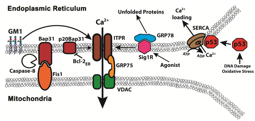
Giorgi C, Bonora M, Sorrentino G, et al. (2015) p53 at the endoplasmic reticulum regulates apoptosis in a Ca2+-dependent manner. Proc Natl Acad Sci USA 112: 1779–1784. doi: 10.1073/pnas.1410723112
![]() |
[14] | Haupt S, Raghu D, Haupt Y (2015) p53 Calls upon CIA (Calcium Induced Apoptosis) to Counter Stress. Front Oncol 5: 57. |
[15] | Brisac C, Teoule F, Autret A, et al. (2010) Calcium flux between the endoplasmic reticulum and mitochondrion contributes to poliovirus-induced apoptosis. J Virol 84: 12226–12235. |
[16] |
Luciani DS, Gwiazda KS, Yang TL, et al. (2009) Roles of IP3R and RyR Ca2+ channels in endoplasmic reticulum stress and beta-cell death. Diabetes 58: 422–432. doi: 10.2337/db07-1762
![]() |
[17] |
Toglia P, Ullah G (2016) The gain-of-function enhancement of IP3-receptor channel gating by familial Alzheimer's disease-linked presenilin mutants increases the open probability of mitochondrial permeability transition pore. Cell Calcium 60: 13–24. doi: 10.1016/j.ceca.2016.05.002
![]() |
[18] |
Szabadkai G, Bianchi K, Varnai P, et al. (2006) Chaperone-mediated coupling of endoplasmic reticulum and mitochondrial Ca2+ channels. J Cell Biol 175: 901–911. doi: 10.1083/jcb.200608073
![]() |
[19] |
Csordas G, Renken C, Varnai P, et al. (2006) Structural and functional features and significance of the physical linkage between ER and mitochondria. J Cell Biol 174: 915–921. doi: 10.1083/jcb.200604016
![]() |
[20] |
Hansford RG (1994) Physiological role of mitochondrial Ca2+ transport. J Bioenerg Biomembr 26: 495–508. doi: 10.1007/BF00762734
![]() |
[21] |
Rutter GA, Burnett P, Rizzuto R, et al. (1996) Subcellular imaging of intramitochondrial Ca2+ with recombinant targeted aequorin: significance for the regulation of pyruvate dehydrogenase activity. Proc Natl Acad Sci USA 93: 5489–5494. doi: 10.1073/pnas.93.11.5489
![]() |
[22] |
Palty R, Hershfinkel M, Sekler I (2012) Molecular identity and functional properties of the mitochondrial Na+/Ca2+ exchanger. J Biol Chem 287: 31650–31657. doi: 10.1074/jbc.R112.355867
![]() |
[23] | Adam-Vizi V, Starkov AA (2010) Calcium and mitochondrial reactive oxygen species generation: how to read the facts. J Alzheimers Dis 20 Suppl 2: S413–S426. |
[24] |
Hansson MJ, Mansson R, Morota S, et al. (2008) Calcium-induced generation of reactive oxygen species in brain mitochondria is mediated by permeability transition. Free Radic Biol Med 45: 284–294. doi: 10.1016/j.freeradbiomed.2008.04.021
![]() |
[25] |
Varanita T, Soriano ME, Romanello V, et al. (2015) The OPA1-dependent mitochondrial cristae remodeling pathway controls atrophic, apoptotic, and ischemic tissue damage. Cell Metab 21: 834–844. doi: 10.1016/j.cmet.2015.05.007
![]() |
[26] | Sonnino S, Prinetti A (2013) Membrane domains and the "lipid raft" concept. Curr Med Chem 20: 4–21. |
[27] | Vance JE (1990) Phospholipid synthesis in a membrane fraction associated with mitochondria. J Biol Chem 265: 7248–7256. |
[28] | Rusinol AE, Cui Z, Chen MH, et al. (1994) A unique mitochondria-associated membrane fraction from rat liver has a high capacity for lipid synthesis and contains pre-Golgi secretory proteins including nascent lipoproteins. J Biol Chem 269: 27494–27502. |
[29] |
Tessitore A, del P Martin M, Sano R, et al. (2004) GM1-ganglioside-mediated activation of the unfolded protein response causes neuronal death in a neurodegenerative gangliosidosis. Mol Cell 15: 753–766. doi: 10.1016/j.molcel.2004.08.029
![]() |
[30] |
Sano R, Annuziata I, Patterson A, et al. (2009) GM1-ganglioside accumulation at the mitochondria-associated ER membranes links ER stress to Ca(2+)-dependent mitochondrial apoptosis. Mol Cell 36: 500–511. doi: 10.1016/j.molcel.2009.10.021
![]() |
[31] | Annunziata I, Patterson A, D'Azzo A (2013) Mitochondria-associated ER membranes (MAMs) and glycosphingolipid enriched microdomains (GEMs): isolation from mouse brain. J Vis Exp 73: e50215. |
[32] |
Garofalo T, Matarrese P, Manganeli V, et al. (2016) Evidence for the involvement of lipid rafts localized at the ER-mitochondria associated membranes in autophagosome formation. Autophagy 12: 917–935. doi: 10.1080/15548627.2016.1160971
![]() |
[33] |
Pomorski TG, Menon AK (2016) Lipid somersaults: Uncovering the mechanisms of protein-mediated lipid flipping. Prog Lipid Res 64: 69–84. doi: 10.1016/j.plipres.2016.08.003
![]() |
[34] | Vance JE, Aasman EJ, Szarka R (1991) Brefeldin A does not inhibit the movement of phosphatidylethanolamine from its sites for synthesis to the cell surface. J Biol Chem 266: 8241–8247. |
[35] |
Lev S (2010) Non-vesicular lipid transport by lipid-transfer proteins and beyond. Nat Rev Mol Cell Biol 11: 739–750. doi: 10.1038/nrm2971
![]() |
[36] |
D'Angelo G, Vicinanza M, De Matteis MA (2008) Lipid-transfer proteins in biosynthetic pathways. Curr Opin Cell Biol 20: 360–370. doi: 10.1016/j.ceb.2008.03.013
![]() |
[37] |
Tatsuta T, Scharwey M, Langer T (2014) Mitochondrial lipid trafficking. Trends Cell Biol 24: 44–52. doi: 10.1016/j.tcb.2013.07.011
![]() |
[38] |
Miller WL (2013) Steroid hormone synthesis in mitochondria. Mol Cell Endocrinol 379: 62–73. doi: 10.1016/j.mce.2013.04.014
![]() |
[39] | Salavila A, Navarrolerida I, Sanchezalvarez M, et al. (2016) Interplay between hepatic mitochondria-associated membranes, lipid metabolism and caveolin-1 in mice. Sci Rep 6: 27351. |
[40] |
Kojima R, Endo T, Tamura Y (2016) A phospholipid transfer function of ER-mitochondria encounter structure revealed in vitro. Sci Rep 6: 30777. doi: 10.1038/srep30777
![]() |
[41] |
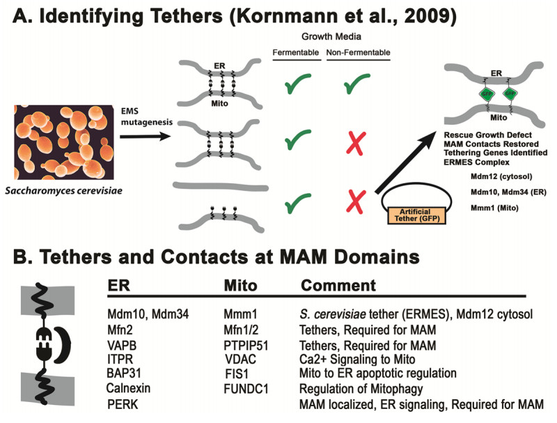
Kornmann B, Currie E, Collins SR, et al. (2009) An ER-mitochondria tethering complex revealed by a synthetic biology screen. Science 325: 477–481. doi: 10.1126/science.1175088
![]() |
[42] |
Kornmann B, Walter P (2010) ERMES-mediated ER-mitochondria contacts: molecular hubs for the regulation of mitochondrial biology. J Cell Sci 123: 1389–1393. doi: 10.1242/jcs.058636
![]() |
[43] |
Lahiri S, Chao JT, Tavassoli S, et al. (2014) A conserved endoplasmic reticulum membrane protein complex (EMC) facilitates phospholipid transfer from the ER to mitochondria. PLoS Biol 12: e1001969. doi: 10.1371/journal.pbio.1001969
![]() |
[44] |
Lev S, Ben Halaevy D, Peretti D, et al. (2008) The VAP protein family: from cellular functions to motor neuron disease. Trends Cell Biol 18: 282–290. doi: 10.1016/j.tcb.2008.03.006
![]() |
[45] | Stoica R, De Vos KJ, Paillusson S, et al. (2014) ER-mitochondria associations are regulated by the VAPB-PTPIP51 interaction and are disrupted by ALS/FTD-associated TDP-43. Nat Commun 5: 3996. |
[46] |
De Vos KJ, Morotz GM, Stoica R, et al. (2012) VAPB interacts with the mitochondrial protein PTPIP51 to regulate calcium homeostasis. Hum Mol Genet 21: 1299–1311. doi: 10.1093/hmg/ddr559
![]() |
[47] | Santel A, Fuller MT (2001) Control of mitochondrial morphology by a human mitofusin. J Cell Sci 114: 867–874. |
[48] |
de Brito OM, Scorrano L (2008) Mitofusin 2 tethers endoplasmic reticulum to mitochondria. Nature 456: 605–610. doi: 10.1038/nature07534
![]() |
[49] |
Filadi R, Greotti E, Turacchio G, et al. (2015) Mitofusin 2 ablation increases endoplasmic reticulum-mitochondria coupling. Proc Natl Acad Sci USA 112: E2174–E2181. doi: 10.1073/pnas.1504880112
![]() |
[50] |
Naon D, Zaninello M, Giacomello M, et al. (2016) Critical reappraisal confirms that Mitofusin 2 is an endoplasmic reticulum-mitochondria tether. Proc Natl Acad Sci USA 113: 11249–11254. doi: 10.1073/pnas.1606786113
![]() |
[51] |
Ouvrier R, Grew S (2010) Mechanisms of disease and clinical features of mutations of the gene for mitofusin 2: an important cause of hereditary peripheral neuropathy with striking clinical variability in children and adults. Dev Med Child Neurol 52: 328–330. doi: 10.1111/j.1469-8749.2010.03613.x
![]() |
[52] |
Wang B, Heath-Engel H, Zhang D, et al. (2008) BAP31 interacts with Sec61 translocons and promotes retrotranslocation of CFTRDeltaF508 via the derlin-1 complex. Cell 133: 1080–1092. doi: 10.1016/j.cell.2008.04.042
![]() |
[53] |
Iwasawa R, Mahul-Mellier AL, Datler C, et al. (2011) Fis1 and Bap31 bridge the mitochondria-ER interface to establish a platform for apoptosis induction. EMBO J 30: 556–568. doi: 10.1038/emboj.2010.346
![]() |
[54] |
Wang B, Nguyen M, Chang NC, et al. (2011) Fis1, Bap31 and the kiss of death between mitochondria and endoplasmic reticulum. EMBO J 30: 451–452. doi: 10.1038/emboj.2010.352
![]() |
[55] |
Ng FW, Nguyen M, Kwan T, et al. (1997) p28 Bap31, a Bcl-2/Bcl-XL- and procaspase-8-associated protein in the endoplasmic reticulum. J Cell Biol 139: 327–338. doi: 10.1083/jcb.139.2.327
![]() |
[56] | Rizzuto R, Marchi S, Bonora M, et al. (2009) Ca(2+) transfer from the ER to mitochondria: when, how and why. Biochim Biophys Acta 1787: 1342–1351. |
[57] | Breckenridge DG, Stojanovic M, Marcellus RC, et al. (2003) Caspase cleavage product of BAP31 induces mitochondrial fission through endoplasmic reticulum calcium signals, enhancing cytochrome c release to the cytosol. J Cell Biol 160: 1115–1127. |
[58] |
Heath-Engel HM, Wang B, Shore GC (2012) Bcl2 at the endoplasmic reticulum protects against a Bax/Bak-independent paraptosis-like cell death pathway initiated via p20Bap31. Biochim Biophys Acta 1823: 335–347. doi: 10.1016/j.bbamcr.2011.11.020
![]() |
[59] | Nguyen M, Breckenridge DG, Ducret A, et al. (2000) Caspase-resistant BAP31 inhibits fas-mediated apoptotic membrane fragmentation and release of cytochrome c from mitochondria. Mol Cell Biol 20: 6731–6740. |
[60] |
Wu W, Li W, Chen H, et al. (2016) FUNDC1 is a novel mitochondrial-associated-membrane (MAM) protein required for hypoxia-induced mitochondrial fission and mitophagy. Autophagy 12: 1675–1676. doi: 10.1080/15548627.2016.1193656
![]() |
[61] |
Smirnova E, Griparic L, Shurland DL, et al. (2001) Dynamin-related protein Drp1 is required for mitochondrial division in mammalian cells. Mol Biol Cell 12: 2245–2256. doi: 10.1091/mbc.12.8.2245
![]() |
[62] |
Friedman JR, Lackner LL, West M, et al. (2011) ER tubules mark sites of mitochondrial division. Science 334: 358–362. doi: 10.1126/science.1207385
![]() |
[63] | Wang M, Wey S, Zhang Y, et al. (2009) Role of the unfolded protein response regulator GRP78/BiP in development, cancer, and neurological disorders. Antioxid Redox Signal 11: 2307–2316. |
[64] |
Sano R, Reed JC (2013) ER stress-induced cell death mechanisms. Biochim Biophys Acta 1833: 3460–3470. doi: 10.1016/j.bbamcr.2013.06.028
![]() |
[65] |
Lumley EC, Osborn AR, Scott JC, et al. (2017) Moderate endoplasmic reticulum stress activates a PERK and p38-dependent apoptosis. Cell Stress Chaperones 22: 43–54.. doi: 10.1007/s12192-016-0740-2
![]() |
[66] |
Simmen T, Aslan JE, Blagoveshchenskaya AD, et al. (2005) PACS-2 controls endoplasmic reticulum-mitochondria communication and Bid-mediated apoptosis. EMBO J 24: 717–729. doi: 10.1038/sj.emboj.7600559
![]() |
[67] | Cormaci G, Mori T, Hayashi T, et al. (2007) Protein kinase A activation down-regulates, whereas extracellular signal-regulated kinase activation up-regulates sigma-1 receptors in B-104 cells: Implication for neuroplasticity. J Pharmacol Exp Ther 320: 202–210. |
[68] |
Hayashi T, Su TP (2007) Sigma-1 receptor chaperones at the ER-mitochondrion interface regulate Ca(2+) signaling and cell survival. Cell 131: 596–610. doi: 10.1016/j.cell.2007.08.036
![]() |
[69] |
Mori T, Hahashi T, Hayashi E, et al. (2013) Sigma-1 receptor chaperone at the ER-mitochondrion interface mediates the mitochondrion-ER-nucleus signaling for cellular survival. PLoS One 8: e76941. doi: 10.1371/journal.pone.0076941
![]() |
[70] |
Wang J, Saul A, Roon P, et al. (2016) Activation of the molecular chaperone, sigma 1 receptor, preserves cone function in a murine model of inherited retinal degeneration. Proc Natl Acad Sci USA 113: E3764–E3772. doi: 10.1073/pnas.1521749113
![]() |
[71] | Chu UB, Ruoho AE (2016) Biochemical pharmacology of the Sigma-1 receptor. Mol Pharmacol 89: 142–153. |
[72] | Verfaillie T, Rubio N, Garg AD, et al. (2012) PERK is required at the ER-mitochondrial contact sites to convey apoptosis after ROS-based ER stress. Cell Death Differ 19: 1880–1891. |
[73] |
Hardy JA, Higgins GA (1992) Alzheimer's disease: the amyloid cascade hypothesis. Science 256: 184–185. doi: 10.1126/science.1566067
![]() |
[74] | Karran E, De SB (2016) The amyloid cascade hypothesis: are we poised for success or failure? J Neurochem 139 Suppl 2: 237–252. |
[75] |
Herrup K (2015) The case for rejecting the amyloid cascade hypothesis. Nat Neurosci 18: 794–799. doi: 10.1038/nn.4017
![]() |
[76] |
Castrillo JI, Oliver SG (2016) Alzheimer's as a systems-level disease involving the interplay of multiple cellular networks. Methods Mol Biol 1303: 3–48. doi: 10.1007/978-1-4939-2627-5_1
![]() |
[77] | Bartley MG, Marquardt K, Kirchhof D, et al. (2012) Overexpression of amyloid-beta protein precursor induces mitochondrial oxidative stress and activates the intrinsic apoptotic cascade. J Alzheimers Dis 28: 855–868. |
[78] |
Benussi L, Ghidroni R, Dal Piaz F, et al. (2017) The level of 24-Hydroxycholesteryl Esters is an Early Marker of Alzheimer's Disease. J Alzheimers Dis 56: 825–833. doi: 10.3233/JAD-160930
![]() |
[79] |
Area-Gomez E, de Groof AJ, Boldogh I, et al. (2009) Presenilins are enriched in endoplasmic reticulum membranes associated with mitochondria. Am J Pathol 175: 1810–1816. doi: 10.2353/ajpath.2009.090219
![]() |
[80] | Leech CA, Kopp RF, Nelson HA, et al. (2016) Stromal Interaction Molecule 1 (STIM1) Regulates ATP-Sensitive Potassium (KATP) and Store-Operated Ca2+ Channels in MIN6 beta-Cells. J Biol Chem 292: 2266–2277. |
[81] |
Tong BC, Lee CS, Cheng WH, et al. (2016) Familial Alzheimer's disease-associated presenilin 1 mutants promote gamma-secretase cleavage of STIM1 to impair store-operated Ca2+ entry. Sci Signal 9: ra89. doi: 10.1126/scisignal.aaf1371
![]() |
[82] | Nelson O, Supnet C, Liu H, et al. (2010) Familial Alzheimer's disease mutations in presenilins: effects on endoplasmic reticulum calcium homeostasis and correlation with clinical phenotypes. J Alzheimers Dis 21: 781. |
[83] |
Rozpedek W, Markiewicz L, Diehl JA, et al. (2015) Unfolded protein response and PERK kinase as a new therapeutic target in the pathogenesis of Alzheimer's disease. Curr Med Chem 22: 3169–3184. doi: 10.2174/0929867322666150818104254
![]() |
[84] | Al-Chalabi A, van den Berg LH, Veldink J (2017) Gene discovery in amyotrophic lateral sclerosis: implications for clinical management. Nat Rev Neurol 13: 96–104.. |
[85] |
Gregianin E, Pallafacchina G, Zanin S, et al. (2016) Loss-of-function mutations in the SIGMAR1 gene cause distal hereditary motor neuropathy by impairing ER-mitochondria tethering and Ca2+ signalling. Hum Mol Genet 25: 3741–3753. doi: 10.1093/hmg/ddw220
![]() |
[86] |
Li X, Hu Z, Liu L, et al. (2015) A SIGMAR1 splice-site mutation causes distal hereditary motor neuropathy. Neurology 84: 2430–2437. doi: 10.1212/WNL.0000000000001680
![]() |
[87] |
Watanabe S, Ilieva H, Tamada H, et al. (2016) Mitochondria-associated membrane collapse is a common pathomechanism in SIGMAR1- and SOD1-linked ALS. EMBO Mol Med 8: 1421–1437. doi: 10.15252/emmm.201606403
![]() |
[88] |
Hyrskyluoto A, Pulli I, Tornqvist K, et al. (2013) Sigma-1 receptor agonist PRE084 is protective against mutant huntingtin-induced cell degeneration: involvement of calpastatin and the NF-kappaB pathway. Cell Death Dis 4: e646. doi: 10.1038/cddis.2013.170
![]() |
[89] |
Ono Y, Tanaka H, Nagahara Y, et al. (2014) SA4503, a sigma-1 receptor agonist, suppresses motor neuron damage in in vitro and in vivo amyotrophic lateral sclerosis models. Neurosci Lett 559: 174–178. doi: 10.1016/j.neulet.2013.12.005
![]() |
[90] |
Neumann M, Sampathu DM, Kwong LK, et al. (2006) Ubiquitinated TDP-43 in frontotemporal lobar degeneration and amyotrophic lateral sclerosis. Science 314: 130–133. doi: 10.1126/science.1134108
![]() |
[91] |
Ambegaokar SS, Jackson GR (2011) Functional genomic screen and network analysis reveal novel modifiers of tauopathy dissociated from tau phosphorylation. Hum Mol Genet 20: 4947–4977. doi: 10.1093/hmg/ddr432
![]() |
1. | Vaishali Kumar, Shuvadeep Maity, ER Stress-Sensor Proteins and ER-Mitochondrial Crosstalk—Signaling Beyond (ER) Stress Response, 2021, 11, 2218-273X, 173, 10.3390/biom11020173 | |
2. | Alejandro Luarte, Víctor Hugo Cornejo, Francisca Bertin, Javiera Gallardo, Andrés Couve, The axonal endoplasmic reticulum: One organelle-many functions in development, maintenance, and plasticity, 2018, 78, 19328451, 181, 10.1002/dneu.22560 |