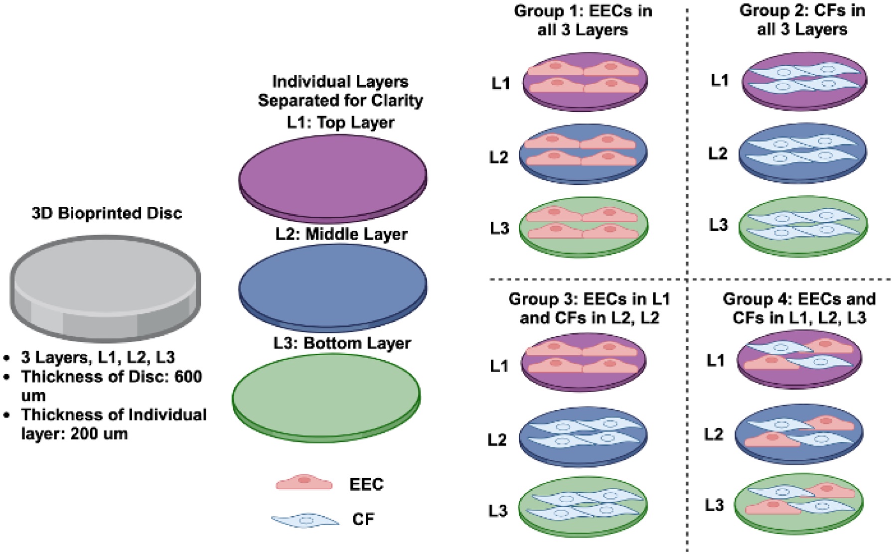
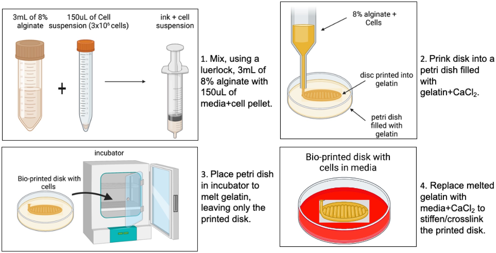
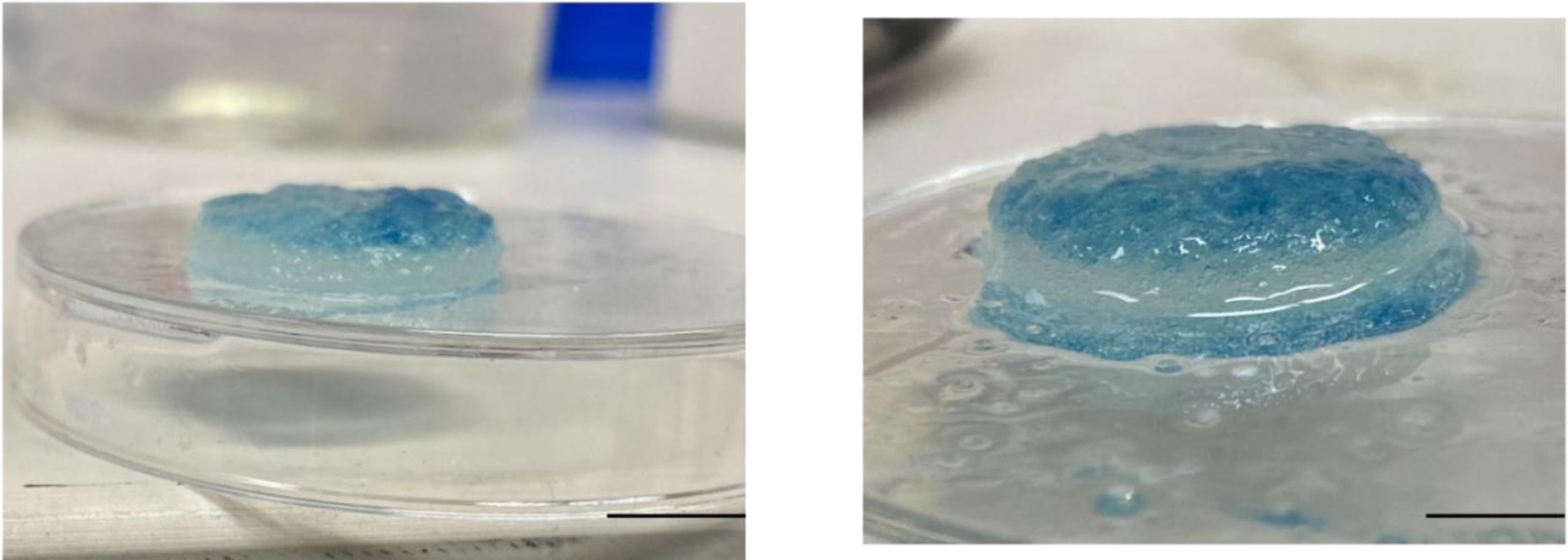
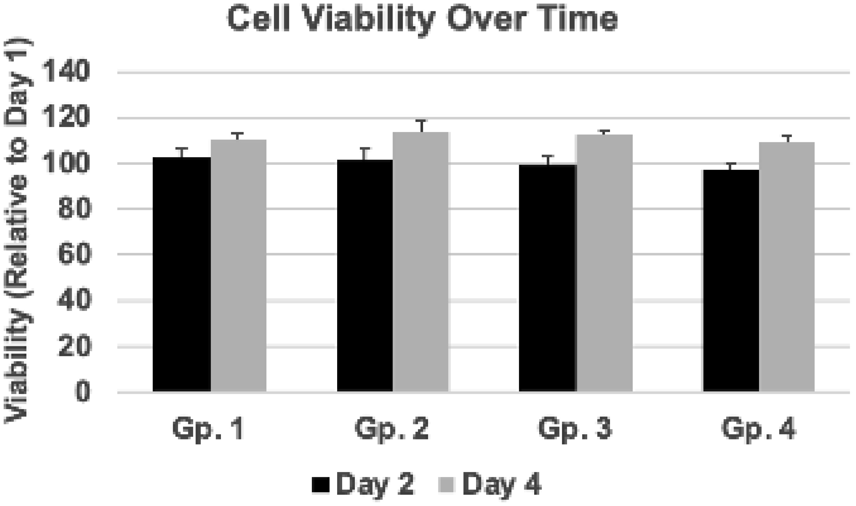
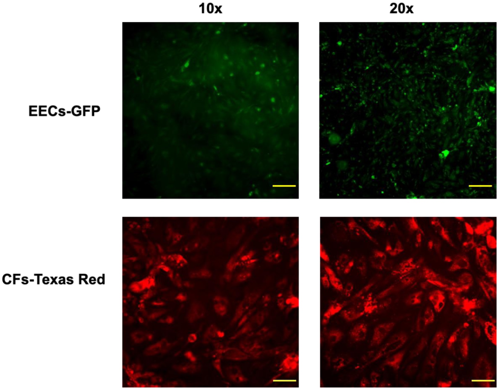
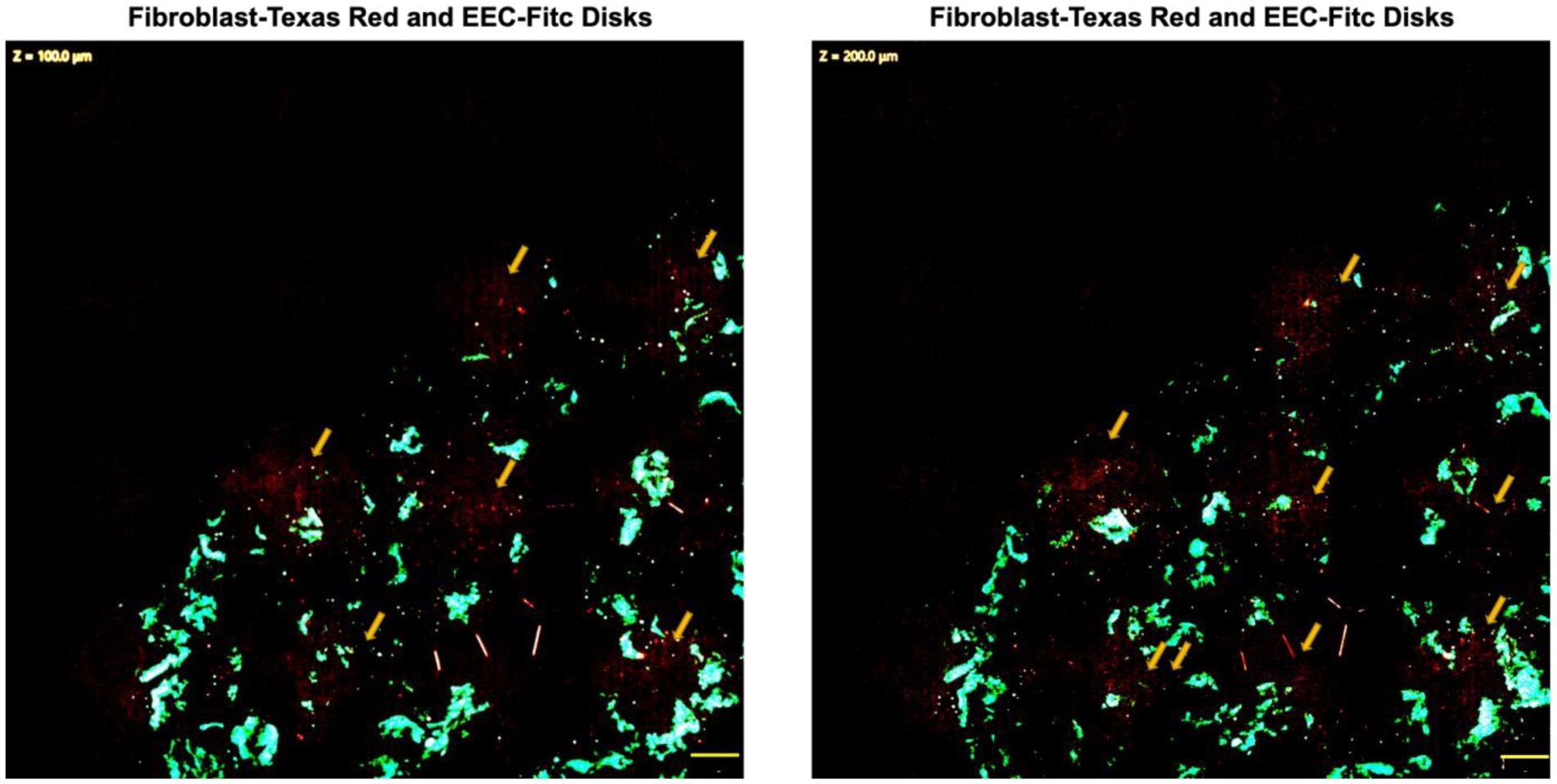
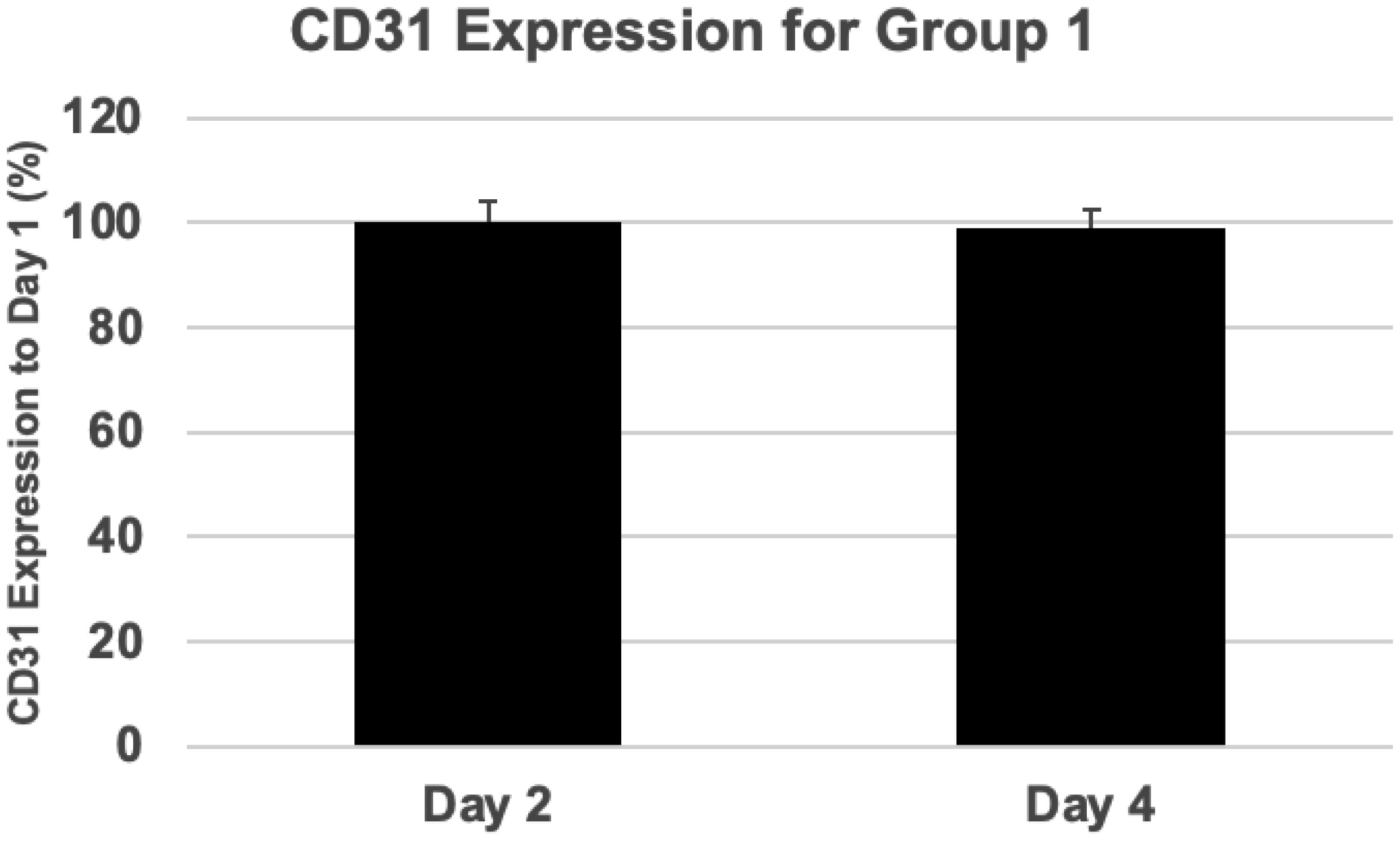
Discrete subaortic stenosis (DSS) is a severe congenital heart condition that results in the formation of a fibrous membrane in the left ventricular outflow track (LVOT). While DSS is surgically treated, it is frequently associated with a high rate of recurrence, necessitating multiple surgeries. The surgical burden of DSS can be reduced by implementing targeted drug therapies based on the underlying molecular mechanisms. There are multiple cell types within the LVOT, consisting of fibroblasts and endocardial endothelial cells (EECs), organized in a complex 3D space. Our objective of this study was to develop a 3D system for the concurrent coculture of fibroblasts and EECs, as a first step in the development of a tool to better understand the cellular communication between these two cell types. To accomplish this objective, we used extrusion-based bioprinting to fabricate 3D discs. Extrusion-based bioprinting was used to generate a 3D disc with fibroblasts and EECs distributed in different configurations within the 3D disc. We demonstrated that the fibroblasts and the EECs maintained viability as a function of time for up to 4 days under static (this is extra spacing here) conditions. Furthermore, to simulate the wall shear stress conditions in the LOVT, a cone and plate bioreactor was used in conjunction with the 3D bioprinted disc for a culture period of up to 24 hours. We demonstrated that EECs maintained CD31 expression for up to 24 hours when cultured within the 3D discs under conditions of elevated shear stress. Collectively, our results demonstrate the initial success of the 3D bioprinted disc model as a potential tool for studying DSS. While additional optimization and validation studies are required, the model described in this study has the potential to provide insight into the underlying molecular mechanism of DSS disease phenotype and lead to the development of targeted therapies for the treatment of this challenging congenital heart condition.
Citation: Pengfei Ji, Sunita Brimmer, Jeffrey S. Heinle, Jane Grande-Allen, Ravi K. Birla, Sundeep G. Keswani. Development of a novel In Vitro Co-culture system for discrete subaortic stenosis[J]. AIMS Bioengineering, 2025, 12(3): 357-369. doi: 10.3934/bioeng.2025016
Discrete subaortic stenosis (DSS) is a severe congenital heart condition that results in the formation of a fibrous membrane in the left ventricular outflow track (LVOT). While DSS is surgically treated, it is frequently associated with a high rate of recurrence, necessitating multiple surgeries. The surgical burden of DSS can be reduced by implementing targeted drug therapies based on the underlying molecular mechanisms. There are multiple cell types within the LVOT, consisting of fibroblasts and endocardial endothelial cells (EECs), organized in a complex 3D space. Our objective of this study was to develop a 3D system for the concurrent coculture of fibroblasts and EECs, as a first step in the development of a tool to better understand the cellular communication between these two cell types. To accomplish this objective, we used extrusion-based bioprinting to fabricate 3D discs. Extrusion-based bioprinting was used to generate a 3D disc with fibroblasts and EECs distributed in different configurations within the 3D disc. We demonstrated that the fibroblasts and the EECs maintained viability as a function of time for up to 4 days under static (this is extra spacing here) conditions. Furthermore, to simulate the wall shear stress conditions in the LOVT, a cone and plate bioreactor was used in conjunction with the 3D bioprinted disc for a culture period of up to 24 hours. We demonstrated that EECs maintained CD31 expression for up to 24 hours when cultured within the 3D discs under conditions of elevated shear stress. Collectively, our results demonstrate the initial success of the 3D bioprinted disc model as a potential tool for studying DSS. While additional optimization and validation studies are required, the model described in this study has the potential to provide insight into the underlying molecular mechanism of DSS disease phenotype and lead to the development of targeted therapies for the treatment of this challenging congenital heart condition.

| [1] |
Masse DD, Shar JA, Brown KN, et al. (2018) Discrete subaortic stenosis: perspective roadmap to a complex disease. Front Cardiovasc Med 5: 122. https://doi.org/10.3389/fcvm.2018.00122 
|
| [2] |
Shar JA, Brown KN, Keswani SG, et al. (2020) Impact of aortoseptal angle abnormalities and discrete subaortic stenosis on left-ventricular outflow tract hemodynamics: preliminary computational assessment. Front Bioeng Biotechnol 8: 114. https://doi.org/10.3389/fbioe.2020.00114
|
| [3] |
Mohan JC, Shukla M, Mohan V, et al. (2016) Acquired discrete subaortic stenosis late after mitral valve replacement. Indian Heart J 68: S105-S109. https://doi.org/10.1016/j.ihj.2016.01.001
|
| [4] |
Birla RK, Williams SK (2020) 3D bioprinting and its potential impact on cardiac failure treatment: An industry perspective. APL Bioeng 4: 010903. https://doi.org/10.1063/1.5128371
|
| [5] |
Birla RK (2020) A methodological nine-step process to bioengineer heart muscle tissue. Tissue Cell 67: 101425. https://doi.org/10.1016/j.tice.2020.101425
|
| [6] |
Abbasgholizadeh R, Islas JF, Navran S, et al. (2020) A highly conductive 3D cardiac patch fabricated using cardiac myocytes reprogrammed from human adipogenic mesenchymal stem cells. Cardiovasc Eng Technol 11: 205-218. https://doi.org/10.1007/s13239-019-00451-0
|
| [7] |
Patel NM, Birla RK (2018) The bioengineered cardiac left ventricle. ASAIO J 64: 56-62. https://doi.org/10.1097/MAT.0000000000000642
|
| [8] |
Hogan M, Souza G, Birla R (2016) Assembly of a functional 3D primary cardiac construct using magnetic levitation. AIMS Bioeng 3: 277-288. https://doi.org/10.3934/bioeng.2016.3.277
|
| [9] |
Williams SK, Birla RK (2020) Tissue engineering solutions to replace contractile function during pediatric heart surgery. Tissue Cell 67: 101452. https://doi.org/10.1016/j.tice.2020.101452
|
| [10] |
Khait L, Hecker L, Blan NR, et al. (2008) Getting to the heart of tissue engineering. J Cardiovasc Transl Res 1: 71-84. https://doi.org/10.1007/s12265-007-9005-x
|
| [11] |
Mohamed MA, Islas JF, Schwartz RJ, et al. (2017) Electrical stimulation of artificial heart muscle: a look into the electrophysiologic and genetic implications. ASAIO J 63: 333-341. https://doi.org/10.1097/MAT.0000000000000486
|
| [12] |
Birla RK, Huang YC, Dennis RG (2007) Development of a novel bioreactor for the mechanical loading of tissue-engineered heart muscle. Tissue Eng 13: 2239-2248. https://doi.org/10.1089/ten.2006.0359
|
| [13] |
Khait L, Hecker L, Radnoti D, et al. (2008) Micro-perfusion for cardiac tissue engineering: development of a bench-top system for the culture of primary cardiac cells. Ann Biomed Eng 36: 713-725. https://doi.org/10.1007/s10439-008-9459-2
|
| [14] |
Brown KN, Phan HKT, Jui EL, et al. (2023) Isolation and characterization of porcine endocardial endothelial cells. Tissue Eng Part C Methods 29: 371-380. https://doi.org/10.1089/ten.TEC.2023.0009
|
| [15] |
Brimmer S, Ji P, Birla RK, et al. (2024) Development of novel 3D spheroids for discrete subaortic stenosis. Cardiovasc Eng Technol 15: 704-715. https://doi.org/10.1007/s13239-024-00746-x
|
| [16] |
Chavarria D, Georges KA, O'Grady BJ, et al. (2025) Modular cone-and-plate device for mechanofluidic assays in Transwell inserts. Front Bioeng Biotechnol 13: 1494553. https://doi.org/10.3389/fbioe.2025.1494553
|
| [17] |
Ye C, Ali S, Sun Q, et al. (2019) Novel cone-and-plate flow chamber with controlled distribution of wall fluid shear stress. Comput Biol Med 106: 140-148. https://doi.org/10.1016/j.compbiomed.2019.01.014
|
| [18] |
Sucosky P, Padala M, Elhammali A, et al. (2008) Design of an ex vivo culture system to investigate the effects of shear stress on cardiovascular tissue. J Biomech Eng 130: 035001. https://doi.org/10.1115/1.2907753
|
| [19] |
Shar JA, Keswani SG, Grande-Allen KJ, et al. (2022) Significance of aortoseptal angle anomalies to left ventricular hemodynamics and subaortic stenosis: a numerical study. Comput Biol Med 146: 105613. https://doi.org/10.1016/j.compbiomed.2022.105613
|
| [20] |
Shar JA, Keswani SG, Grande-Allen KJ, et al. (2021) Computational assessment of valvular dysfunction in discrete subaortic stenosis: a parametric study. Cardiovasc Eng Technol 12: 559-575. https://doi.org/10.1007/s13239-020-00513-8
|
Figures(7)

Pengfei Ji, Sunita Brimmer, Jeffrey S. Heinle, Jane Grande-Allen, Ravi K. Birla, Sundeep G. Keswani. Development of a novel In Vitro Co-culture system for discrete subaortic stenosis[J]. AIMS Bioengineering, 2025, 12(3): 357-369. doi: 10.3934/bioeng.2025016


 DownLoad: