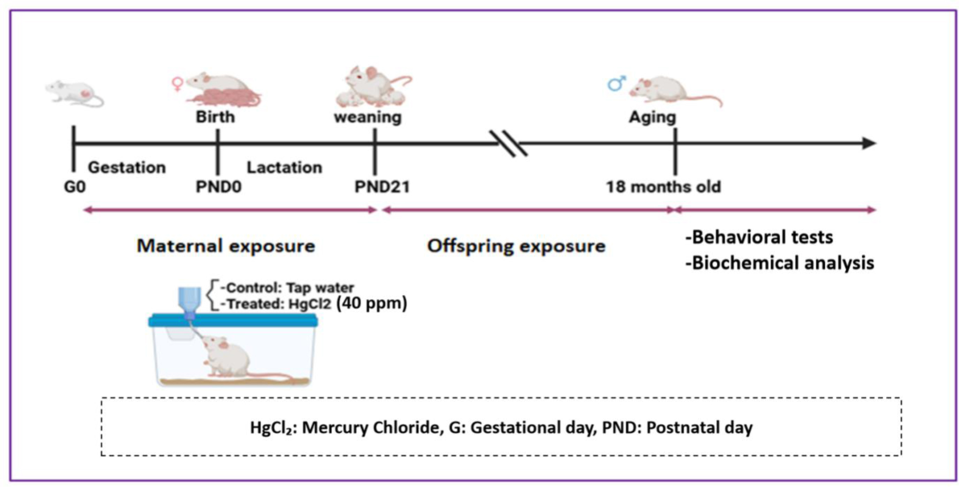
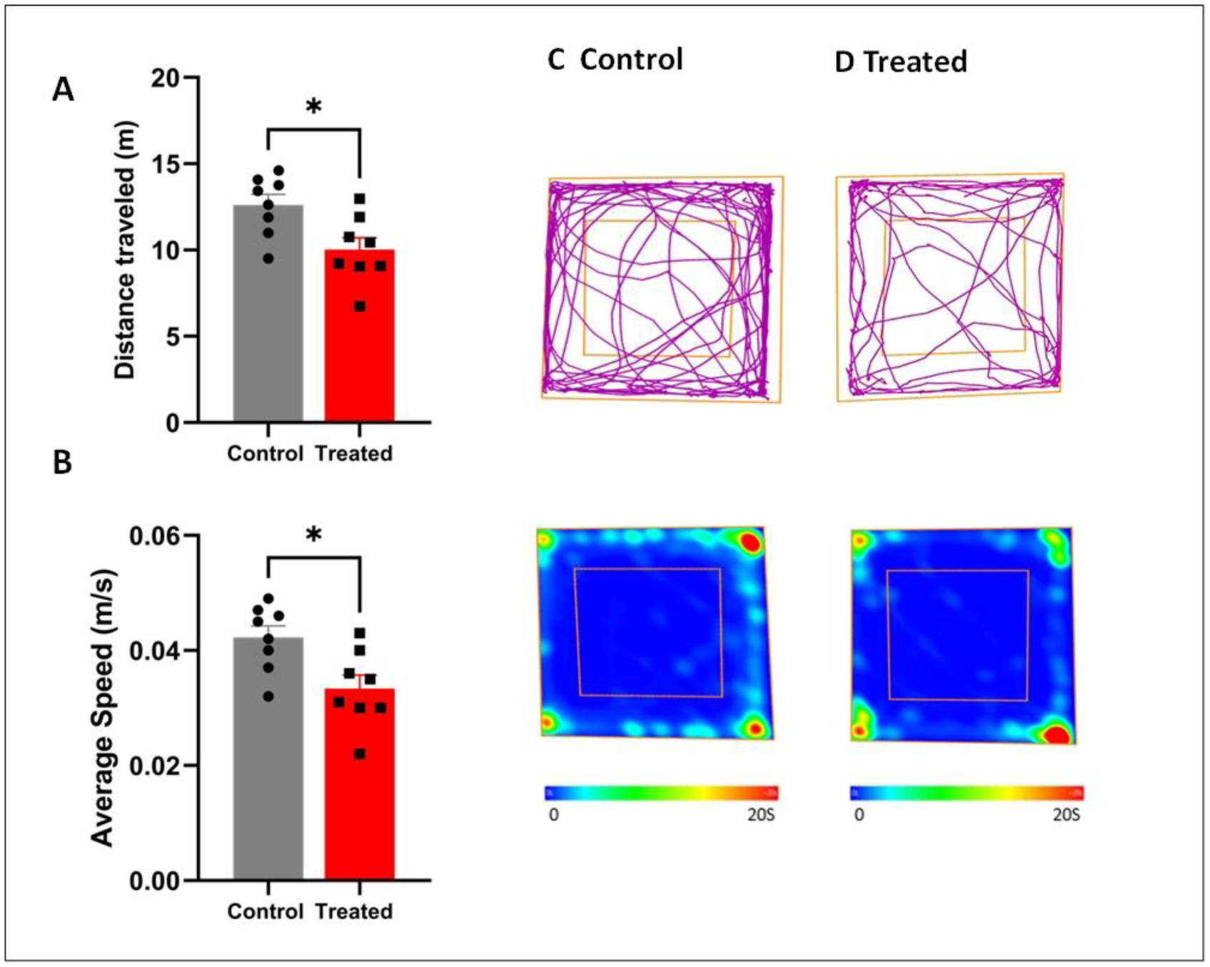
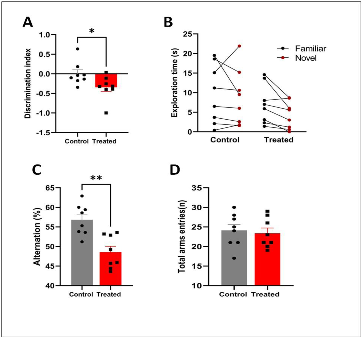
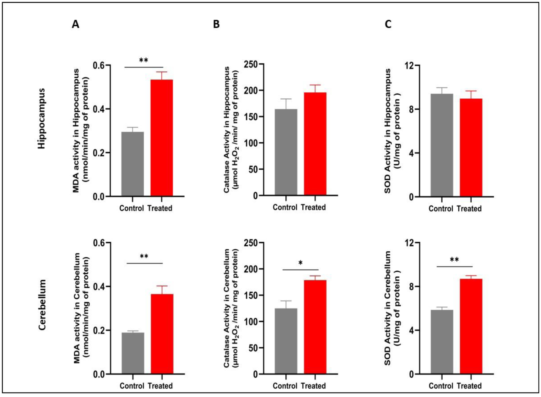
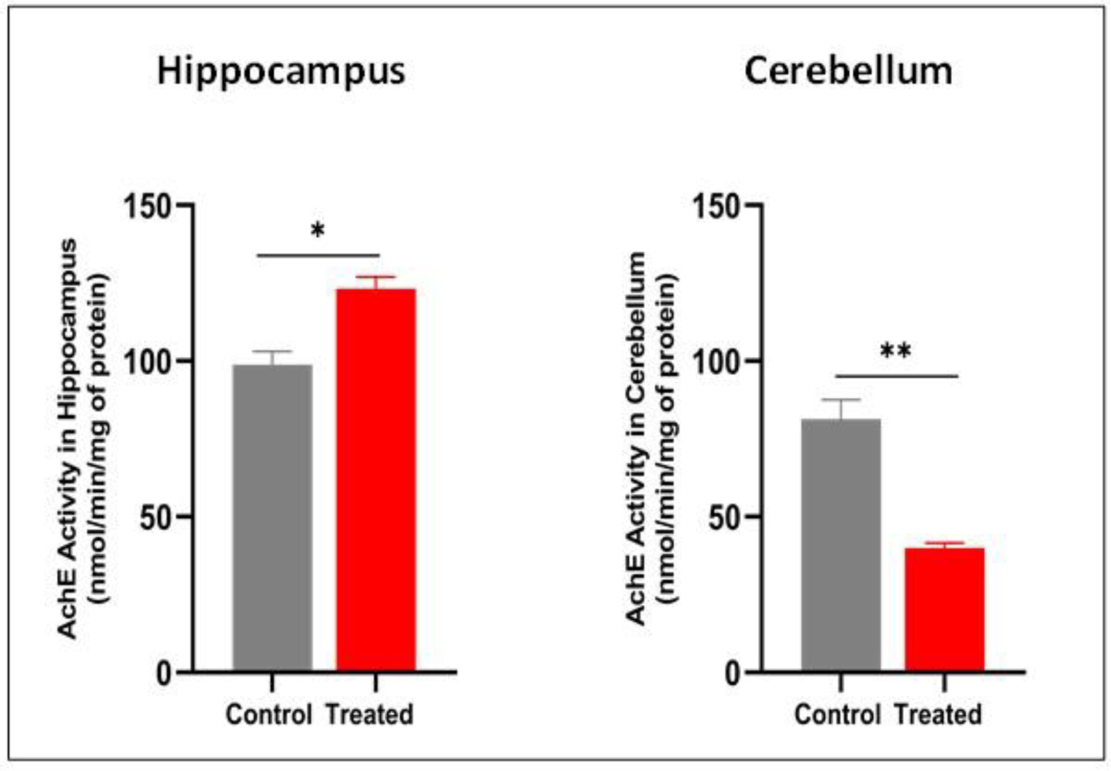
Aging is a multifactorial biological process characterized by the progressive decline of cellular, tissue, and organ functions, leading to increased vulnerability to neurodegenerative disorders. Among the proposed mechanisms, the oxidative stress theory of aging remains one of the most widely accepted, emphasizing the role of reactive oxygen species (ROS) accumulation, particularly in the central nervous system (CNS). Mercury is a persistent environmental pollutant known to induce oxidative damage, but its contribution to age-related neuronal decline remains poorly understood. Our aim of this study was to investigate whether chronic HgCl2 exposure exacerbates age-related decline in memory and locomotor functions in aged male mice. To this end, mice were divided into two groups: A control group, which received only tap water, and an HgCl2-exposed group, which received 40 ppm HgCl2 in their drinking water daily from gestation until 18 months of age. At the end of the exposure, mice were subjected to behavioral tests to assess memory and locomotor activity. Following these behavioral assessments, oxidative stress parameters and acetylcholinesterase (AChE) activity were measured in the hippocampus and cerebellum. Our results showed that HgCl2 impaired locomotor activity in the Open Field test (OF) and memory performance in the Y-maze and Novel Object Recognition (NOR) test. These deficits were accompanied by a significant increase in malondialdehyde (MDA) and acetylcholinesterase (AChE) activity in the hippocampus. Furthermore, mercury exposure led to an increase in MDA and a significant upregulation of catalase (CAT) and superoxide dismutase (SOD) activities in the cerebellum. These changes were also associated with a marked decrease in AChE activity within the same region. Overall, these findings suggest that lifelong mercury exposure accelerates neurobehavioral decline by exacerbating oxidative and cholinergic disturbances, providing insight into how early and sustained exposure to environmental toxicants may promote premature brain aging.
Citation: Hafsa Malqui, Meriem Laaroussi, Hammou Anarghou, Oumaima Essaidi, Laila Berroug, Mounir Cherkaoui, El Hachmi Er-rachdaoui, Latifa Talhaoui, Fatiha Chigr. Developmental and lifelong exposure to mercury chloride exacerbates cognitive and motor decline in aged mice[J]. AIMS Neuroscience, 2026, 13(1): 153-170. doi: 10.3934/Neuroscience.2026007
Aging is a multifactorial biological process characterized by the progressive decline of cellular, tissue, and organ functions, leading to increased vulnerability to neurodegenerative disorders. Among the proposed mechanisms, the oxidative stress theory of aging remains one of the most widely accepted, emphasizing the role of reactive oxygen species (ROS) accumulation, particularly in the central nervous system (CNS). Mercury is a persistent environmental pollutant known to induce oxidative damage, but its contribution to age-related neuronal decline remains poorly understood. Our aim of this study was to investigate whether chronic HgCl2 exposure exacerbates age-related decline in memory and locomotor functions in aged male mice. To this end, mice were divided into two groups: A control group, which received only tap water, and an HgCl2-exposed group, which received 40 ppm HgCl2 in their drinking water daily from gestation until 18 months of age. At the end of the exposure, mice were subjected to behavioral tests to assess memory and locomotor activity. Following these behavioral assessments, oxidative stress parameters and acetylcholinesterase (AChE) activity were measured in the hippocampus and cerebellum. Our results showed that HgCl2 impaired locomotor activity in the Open Field test (OF) and memory performance in the Y-maze and Novel Object Recognition (NOR) test. These deficits were accompanied by a significant increase in malondialdehyde (MDA) and acetylcholinesterase (AChE) activity in the hippocampus. Furthermore, mercury exposure led to an increase in MDA and a significant upregulation of catalase (CAT) and superoxide dismutase (SOD) activities in the cerebellum. These changes were also associated with a marked decrease in AChE activity within the same region. Overall, these findings suggest that lifelong mercury exposure accelerates neurobehavioral decline by exacerbating oxidative and cholinergic disturbances, providing insight into how early and sustained exposure to environmental toxicants may promote premature brain aging.

| [1] |
López-Otín C, Blasco MA, Partridge L, et al. (2023) Hallmarks of aging: An expanding universe. Cell 186: 243-278. https://doi.org/10.1016/j.cell.2022.11.001 
|
| [2] |
Holliday R (2006) Aging is no longer an unsolved problem in biology. Ann N Y Acad Sci 1067: 1-9. https://doi.org/10.1196/annals.1354.002
|
| [3] |
Radak Z, Zhao Z, Goto S, et al. (2011) Age-associated neurodegeneration and oxidative damage to lipids, proteins and DNA. Mol Aspects Med 32: 305-315. https://doi.org/10.1016/j.mam.2011.10.010
|
| [4] |
Rattan SIS (2008) Increased molecular damage and heterogeneity as the basis of aging. Biol Chem 389: 267-272. https://doi.org/10.1515/BC.2008.030
|
| [5] |
de Groot M, Cremers LGM, Ikram MA, et al. (2016) White matter degeneration with aging: Longitudinal diffusion MR imaging analysis. Radiology 279: 532-541. https://doi.org/10.1148/radiol.2015150103
|
| [6] |
Navakkode S, Kennedy BK (2024) Neural ageing and synaptic plasticity: prioritizing brain health in healthy longevity. Front Aging Neurosci 16: 1428244. https://doi.org/10.3389/fnagi.2024.1428244
|
| [7] |
Nyberg L, Salami A, Andersson M, et al. (2010) Longitudinal evidence for diminished frontal cortex function in aging. Proc Natl Acad Sci U S A 107: 22682-22686. https://doi.org/10.1073/pnas.1012651108
|
| [8] |
Raz N, Lindenberger U, Rodrigue KM, et al. (2005) Regional brain changes in aging healthy adults: general trends, individual differences and modifiers. Cereb Cortex 15: 1676-1689. https://doi.org/10.1093/cercor/bhi044
|
| [9] |
Deary IJ, Corley J, Gow AJ, et al. (2009) Age-associated cognitive decline. Br Med Bull 92: 135-152. https://doi.org/10.1093/bmb/ldp033
|
| [10] |
Rönnlund M, Nyberg L, Bäckman L, et al. (2005) Stability, growth, and decline in adult life span development of declarative memory: cross-sectional and longitudinal data from a population-based study. Psychol Aging 20: 3-18. https://doi.org/10.1037/0882-7974.20.1.3
|
| [11] |
Harman D (1956) Aging: A theory based on free radical and radiation chemistry. J Gerontol 11: 298-300. https://doi.org/10.1093/geronj/11.3.298
|
| [12] |
Kregel KC, Zhang HJ (2007) An integrated view of oxidative stress in aging: basic mechanisms, functional effects, and pathological considerations. Am J Physiol Regul Integr Comp Physiol 292: R18-R36. https://doi.org/10.1152/ajpregu.00327.2006
|
| [13] |
Bonda DJ, Wang X, Lee HG, et al. (2014) Neuronal failure in Alzheimer's disease: a view through the oxidative stress looking-glass. Neurosci Bull 30: 243-252. https://doi.org/10.1007/s12264-013-1424-x
|
| [14] | Wang X, Michaelis EK (2010) Selective neuronal vulnerability to oxidative stress in the brain. Front Aging Neurosci 2: 12. https://doi.org/10.3389/fnagi.2010.00012 |
| [15] |
Grimm A, Eckert A (2017) Brain aging and neurodegeneration: from a mitochondrial point of view. J Neurochem 143: 418-431. https://doi.org/10.1111/jnc.14037
|
| [16] |
Mecocci P, Boccardi V, Cecchetti R, et al. (2018) A long journey into aging, brain aging, and Alzheimer's disease following the oxidative stress tracks. J Alzheimers Dis 62: 1319-1335. https://doi.org/10.3233/JAD-170732
|
| [17] |
Nuran Ercal BSP, Hande Gurer-Orhan BSP, Nukhet Aykin-Burns BSP (2001) Toxic metals and oxidative stress part I: Mechanisms involved in me-tal induced oxidative damage. Curr Top Med Chem 1: 529-539. https://doi.org/10.2174/1568026013394831
|
| [18] |
Gogna T, Housden BE, Houldsworth A (2024) Exploring the role of reactive oxygen species in the pathogenesis and pathophysiology of Alzheimer's and Parkinson's disease and the efficacy of antioxidant treatment. Antioxidants (Basel) 13: 1138. https://doi.org/10.3390/antiox13091138
|
| [19] |
Bjørklund G, Chirumbolo S, Dadar M, et al. (2019) Mercury exposure and its effects on fertility and pregnancy outcome. Basic Clin Pharmacol Toxicol 125: 317-327. https://doi.org/10.1111/bcpt.13264
|
| [20] |
Laks DR (2010) Luteinizing hormone provides a causal mechanism for mercury associated disease. Med Hypotheses 74: 698-701. https://doi.org/10.1016/j.mehy.2009.10.036
|
| [21] |
Pavithra KG, SundarRajan P, Kumar PS, et al. (2023) Mercury sources, contaminations, mercury cycle, detection and treatment techniques: A review. Chemosphere 312: 137314. https://doi.org/10.1016/j.chemosphere.2022.137314
|
| [22] |
Mello-Carpes PB, Barros W, Borges S, et al. (2013) Chronic exposure to low mercury chloride concentration induces object recognition and aversive memories deficits in rats. Int J Dev Neurosci 31: 468-472. https://doi.org/10.1016/j.ijdevneu.2013.05.009
|
| [23] |
Peixoto NC, Serafim MA, Flores EMM, et al. (2007) Metallothionein, zinc, and mercury levels in tissues of young rats exposed to zinc and subsequently to mercury. Life Sci 81: 1264-1271. https://doi.org/10.1016/j.lfs.2007.08.038
|
| [24] |
Malqui H, Anarghou H, Ouardi FZ, et al. (2018) Continuous exposure to inorganic mercury affects neurobehavioral and physiological parameters in mice. J Mol Neurosci 66: 291-305. https://doi.org/10.1007/s12031-018-1176-1
|
| [25] |
Picciotto MR, Higley MJ, Mineur YS (2012) Acetylcholine as a neuromodulator: cholinergic signaling shapes nervous system function and behavior. Neuron 76: 116-129. https://doi.org/10.1016/j.neuron.2012.08.036
|
| [26] |
Teles-Grilo Ruivo L, Mellor J (2013) Cholinergic modulation of hippocampal network function. Front Synaptic Neurosci 5: 2. https://doi.org/10.3389/fnsyn.2013.00002
|
| [27] |
Pickford J, Iosif CI, Bashir ZI, et al. (2024) Inhibiting cholinergic signalling in the cerebellar interpositus nucleus impairs motor behaviour. Eur J Neurosci 59: 2208-2224. https://doi.org/10.1111/ejn.16066
|
| [28] |
Zhang C, Zhou P, Yuan T (2016) The cholinergic system in the cerebellum: from structure to function. Rev Neurosci 27: 769-776. https://doi.org/10.1515/revneuro-2016-0008
|
| [29] |
Dumas JA, Newhouse PA (2011) The cholinergic hypothesis of cognitive aging revisited again: cholinergic functional compensation. Pharmacol Biochem Behav 99: 254-261. https://doi.org/10.1016/j.pbb.2011.02.022
|
| [30] |
Frasco MF, Colletier JP, Weik M, et al. (2007) Mechanisms of cholinesterase inhibition by inorganic mercury. FEBS J 274: 1849-1861. https://doi.org/10.1111/j.1742-4658.2007.05732.x
|
| [31] |
Luo H, Cheng Q, Pan X (2020) Photochemical behaviors of mercury (Hg) species in aquatic systems: A systematic review on reaction process, mechanism, and influencing factor. Sci Total Environ 720: 137540. https://doi.org/10.1016/j.scitotenv.2020.137540
|
| [32] |
Ennaceur A, Delacour J (1988) A new one-trial test for neurobiological studies of memory in rats. 1: Behavioral data. Behav Brain Res 31: 47-59. https://doi.org/10.1016/0166-4328(88)90157-X
|
| [33] |
Nagahara AH, McGaugh JL (1992) Muscimol infused into the medial septal area impairs long-term memory but not short-term memory in inhibitory avoidance, water maze place learning and rewarded alternation tasks. Brain Res 591: 54-61. https://doi.org/10.1016/0006-8993(92)90977-H
|
| [34] |
Kraeuter AK, Guest PC, Sarnyai Z (2019) The Y-Maze for assessment of spatial working and reference memory in mice. Methods Mol Biol 1916: 105-111. https://doi.org/10.1007/978-1-4939-8994-2_10
|
| [35] |
Lowry OH, Rosebrough NJ, Farr AL, et al. (1951) Protein measurement with the Folin phenol reagent. J Biol Chem 193: 265-275. https://doi.org/10.1016/S0021-9258(19)52451-6
|
| [36] |
Ellman GL, Courtney KD, Andres V, et al. (1961) A new and rapid colorimetric determination of acetylcholinesterase activity. Biochem Pharmacol 7: 88-95. https://doi.org/10.1016/0006-2952(61)90145-9
|
| [37] |
Buege JA, Aust SD (1978) Microsomal lipid peroxidation. Methods Enzymol 52: 302-310. https://doi.org/10.1016/S0076-6879(78)52032-6
|
| [38] |
Aebi H (1974) Catalase. Methods of enzymatic analysis : 673-684. https://doi.org/10.1016/B978-0-12-091302-2.50032-3
|
| [39] |
Asada K, Takahashi M, Nagate M (1974) Assay and inhibitors of spinach superoxide dismutase. Agric Biol Chem 38: 471-473. https://doi.org/10.1080/00021369.1974.10861178
|
| [40] |
Pollard KM, Cauvi DM, Toomey CB, et al. (2019) Mercury-induced inflammation and autoimmunity. Biochim Biophys Acta Gen Subj 1863: 129299. https://doi.org/10.1016/j.bbagen.2019.02.001
|
| [41] |
Stohs S (1995) Oxidative mechanisms in the toxicity of metal ions. Free Radic Biol Med 18: 321-336. https://doi.org/10.1016/0891-5849(94)00159-H
|
| [42] |
Franceschi C, Garagnani P, Morsiani C, et al. (2018) The continuum of aging and age-related diseases: Common mechanisms but different rates. Front Med (Lausanne) 5: 61. https://doi.org/10.3389/fmed.2018.00061
|
| [43] |
Kennedy BK, Berger SL, Brunet A, et al. (2014) Geroscience: Linking aging to chronic disease. Cell 159: 709-713. https://doi.org/10.1016/j.cell.2014.10.039
|
| [44] |
Candelario-Jalil E, Mhadu NH, Al-Dalain SM, et al. (2001) Time course of oxidative damage in different brain regions following transient cerebral ischemia in gerbils. Neurosci Res 41: 233-241. https://doi.org/10.1016/S0168-0102(01)00282-6
|
| [45] | Sarnowska A (2002) Application of organotypic hippocampal culture for study of selective neuronal death. Folia Neuropathol 40: 101-106. |
| [46] |
Wang X, Pal R, Chen X-w, et al. (2005) High intrinsic oxidative stress may underlie selective vulnerability of the hippocampal CA1 region. Brain Res Mol Brain Res 140: 120-126. https://doi.org/10.1016/j.molbrainres.2005.07.018
|
| [47] |
Aragão WAB, Teixeira FB, Fagundes NCF, et al. (2018) Hippocampal dysfunction provoked by mercury chloride exposure: evaluation of cognitive impairment, oxidative stress, tissue injury and nature of cell death. Oxid Med Cell Longev 2018: 7878050. https://doi.org/10.1155/2018/7878050
|
| [48] |
Behzadfar L, Hassani S, Feizpour H, et al. (2020) Effects of mercuric chloride on spatial memory deficit-induced by beta-amyloid and evaluation of mitochondrial function markers in the hippocampus of rats. Metallomics 12: 144-153. https://doi.org/10.1039/c9mt00161a
|
| [49] |
Leaderbrand K, Chen HJ, Corcoran KA, et al. (2016) Muscarinic acetylcholine receptors act in synergy to facilitate learning and memory. Learn Mem 23: 631-638. https://doi.org/10.1101/lm.043133.116
|
| [50] |
Mineur YS, Mose TN, Vanopdenbosch L, et al. (2022) Hippocampal acetylcholine modulates stress-related behaviors independent of specific cholinergic inputs. Mol Psychiatry 27: 1829-1838. https://doi.org/10.1038/s41380-021-01404-7
|
| [51] |
Melo JB, Agostinho P, Oliveira CR (2003) Involvement of oxidative stress in the enhancement of acetylcholinesterase activity induced by amyloid beta-peptide. Neurosci Res 45: 117-127. https://doi.org/10.1016/s0168-0102(02)00201-8
|
| [52] |
Franciscato C, Goulart FR, Lovatto NM, et al. (2009) ZnCl2 exposure protects against behavioral and acetylcholinesterase changes induced by HgCl2. Int J Dev Neurosci 27: 459-468. https://doi.org/10.1016/j.ijdevneu.2009.05.002
|
| [53] |
Moretto MB, Lermen CL, Morsch VM, et al. (2004) Effect of subchronic treatment with mercury chloride on NTPDase, 5′-nucleotidase and acetylcholinesterase from cerebral cortex of rats. J Trace Elem Med Biol 17: 255-260. https://doi.org/10.1016/S0946-672X(04)80027-0
|
| [54] |
Flora G, Gupta D, Tiwari A (2012) Toxicity of lead: A review with recent updates. Interdiscip Toxicol 5: 47-58. https://doi.org/10.2478/v10102-012-0009-2
|
| [55] |
Agrawal S, Bhatnagar P, Flora SJS (2015) Changes in tissue oxidative stress, brain biogenic amines and acetylcholinesterase following co-exposure to lead, arsenic and mercury in rats. Food Chem Toxicol 86: 208-216. https://doi.org/10.1016/j.fct.2015.10.013
|
| [56] |
Ab E, Ca I (2022) Cadmium and mercury exposure: Oxidative, neurobehavioural and histological alterations to the cerebellum of Wistar rats. Ibom Med J 15: 141-147. https://doi.org/10.61386/imj.v15i2.252
|
| [57] |
Bellum S, Bawa B, Thuett KA, et al. (2007) Changes in biochemical processes in cerebellar granule cells of mice exposed to methylmercury. Int J Toxicol 26: 261-269. https://doi.org/10.1080/10915810701369758
|
| [58] |
Bellum S, Thuett KA, Bawa B, et al. (2013) The effect of methylmercury exposure on behavior and cerebellar granule cell physiology in aged mice. J Appl Toxicol 33: 959-969. https://doi.org/10.1002/jat.2786
|
| [59] |
Belyaeva EA, Sokolova TV, Emelyanova LV, et al. (2012) Mitochondrial electron transport chain in heavy metal-induced neurotoxicity: Effects of cadmium, mercury, and copper. ScientificWorldJournal 2012: 136063. https://doi.org/10.1100/2012/136063
|
| [60] |
Siqueira IR, Fochesatto C, de Andrade A, et al. (2005) Total antioxidant capacity is impaired in different structures from aged rat brain. Int J Dev Neurosci 23: 663-671. https://doi.org/10.1016/j.ijdevneu.2005.03.001
|
| [61] | Mesquita M, Pedroso TF, Oliveira CS, et al. (2016) Effects of zinc against mercury toxicity in female rats 12 and 48 hours after HgCl2 exposure. EXCLI J 15: 256-267. https://doi.org/10.17179/excli2015-709 |
| [62] | Egba SI, Famurewa AC, Omoruyi LE (2022) Buchholzia coriacea seed extract attenuates mercury-induced cerebral and cerebellar oxidative neurotoxicity via NO signaling and suppression of oxidative stress, adenosine deaminase and acetylcholinesterase activities in rats. Avicenna J Phytomed 12: 42-53. https://doi.org/10.22038/AJP.2021.18262 |
| [63] |
Young A (1997) Ageing and physiological functions. Philos Trans R Soc Lond B Biol Sci 352: 1837-1843. https://doi.org/10.1098/rstb.1997.0169
|
| [64] |
Falluel-Morel A, Sokolowski K, Sisti HM, et al. (2007) Developmental mercury exposure elicits acute hippocampal cell death, reductions in neurogenesis, and severe learning deficits during puberty. J Neurochem 103: 1968-1981. https://doi.org/10.1111/j.1471-4159.2007.04882.x
|
| [65] |
Rahimi-Balaei M, Bergen H, Kong J, et al. (2018) Neuronal migration during development of the cerebellum. Front Cell Neurosci 12: 484. https://doi.org/10.3389/fncel.2018.00484
|
| [66] |
Feng W, Wang M, Li B, et al. (2004) Mercury and trace element distribution in organic tissues and regional brain of fetal rat after in utero and weaning exposure to low dose of inorganic mercury. Toxicol Lett 152: 223-234. https://doi.org/10.1016/j.toxlet.2004.05.001
|
| [67] |
Cace IB, Milardovic A, Prpic I, et al. (2011) Relationship between the prenatal exposure to low-level of mercury and the size of a newborn's cerebellum. Med Hypotheses 76: 514-516. https://doi.org/10.1016/j.mehy.2010.12.005
|
| [68] |
Stern S, Cox C, Cernichiari E, et al. (2001) Perinatal and lifetime exposure to methylmercury in the mouse: Blood and brain concentrations of mercury to 26 months of age. Neurotoxicology 22: 467-477. https://doi.org/10.1016/s0161-813x(01)00047-x
|
Figures(5)

Hafsa Malqui, Meriem Laaroussi, Hammou Anarghou, Oumaima Essaidi, Laila Berroug, Mounir Cherkaoui, El Hachmi Er-rachdaoui, Latifa Talhaoui, Fatiha Chigr. Developmental and lifelong exposure to mercury chloride exacerbates cognitive and motor decline in aged mice[J]. AIMS Neuroscience, 2026, 13(1): 153-170. doi: 10.3934/Neuroscience.2026007


 DownLoad: