The calcium-dependent enzyme Transglutaminase 2 (TG2) (E.C. 2.3.2.13), which can promote post-translational modifications of proteins, is involved in several physiological processes, including development, neuronal cell death, and differentiation, as well as synaptic plasticity and transmission in the central nervous system (CNS). Several studies highlight the potential role of the TG2/NF-κB activation pathway in neurodegenerative diseases, including Multiple Sclerosis (MS), and the neuroinflammation that is associated with these conditions. The cross-linking activity of TG2, facilitating the formation of isopeptide bonds between glutamine and lysine residues, appears to be involved in forming protein aggregate deposits in these pathological conditions. Specifically, in the chronic neuroinflammation of MS, TG2 seems to play a central role in the fibrotic process of the lesion. Several potential biomarkers have been investigated for the prognosis and monitoring of MS, but no researchers have explored the presence of potential inflammatory signals in peripheral blood mononuclear cells (PBMCs) during the presymptomatic stage of MS, known as Radiologically Isolated Syndrome (RIS), on account of the lack of information regarding its pathological aspects. Since researchers have demonstrated a correlation between TG2 mRNA levels in PBMCs and the clinical and radiological progression of MS, we aimed to evaluate the expression levels of TG2 in RIS patients, comparing them with those in relapsing-remitting MS (RRMS) patients and healthy controls (HCs) using real-time PCR analysis. Preliminary data showed that RIS patients exhibit lower TG2 mRNA expression levels compared to RRMS patients, while no difference in TG2 mRNA expression being observed between RIS patients and HCs. This suggests that RIS patients exhibit a lower neuroinflammation grade than RRMS patients and that TG2 may represent a potential biochemical marker for assessing neuroinflammation associated with this disease. Future investigations may include longitudinal assessments of the potential role of TG2 mRNA blood levels in predicting or monitoring the progression from RIS to MS.
Citation: Rosa Giacca, Miriana Conte, Alessandro d'Ambrosio, Alvino Bisecco, Renato Docimo, Mario Risi, Manuela Altieri, Riccardo Borgo, Rosario Domenico Melisi, Vittorio Gentile, Antonio Gallo. Use of Transglutaminase 2 mRNA expression in peripheral blood mononuclear cells in patients with Radiologically Isolated Syndrome as a neuroinflammation biomarker: A preliminary study[J]. AIMS Neuroscience, 2025, 12(2): 284-290. doi: 10.3934/Neuroscience.2025015
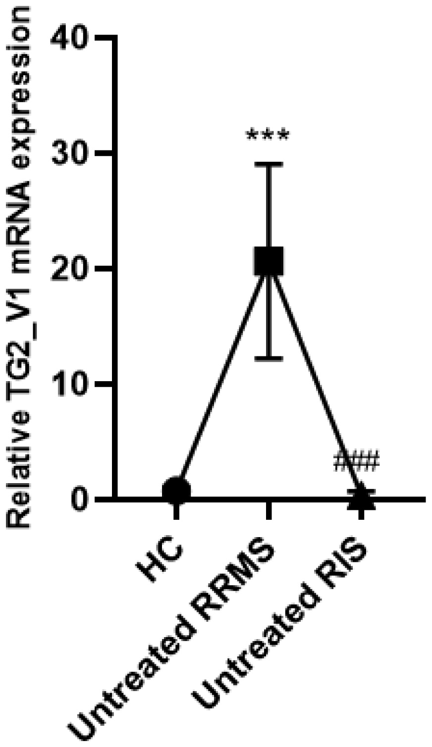
The calcium-dependent enzyme Transglutaminase 2 (TG2) (E.C. 2.3.2.13), which can promote post-translational modifications of proteins, is involved in several physiological processes, including development, neuronal cell death, and differentiation, as well as synaptic plasticity and transmission in the central nervous system (CNS). Several studies highlight the potential role of the TG2/NF-κB activation pathway in neurodegenerative diseases, including Multiple Sclerosis (MS), and the neuroinflammation that is associated with these conditions. The cross-linking activity of TG2, facilitating the formation of isopeptide bonds between glutamine and lysine residues, appears to be involved in forming protein aggregate deposits in these pathological conditions. Specifically, in the chronic neuroinflammation of MS, TG2 seems to play a central role in the fibrotic process of the lesion. Several potential biomarkers have been investigated for the prognosis and monitoring of MS, but no researchers have explored the presence of potential inflammatory signals in peripheral blood mononuclear cells (PBMCs) during the presymptomatic stage of MS, known as Radiologically Isolated Syndrome (RIS), on account of the lack of information regarding its pathological aspects. Since researchers have demonstrated a correlation between TG2 mRNA levels in PBMCs and the clinical and radiological progression of MS, we aimed to evaluate the expression levels of TG2 in RIS patients, comparing them with those in relapsing-remitting MS (RRMS) patients and healthy controls (HCs) using real-time PCR analysis. Preliminary data showed that RIS patients exhibit lower TG2 mRNA expression levels compared to RRMS patients, while no difference in TG2 mRNA expression being observed between RIS patients and HCs. This suggests that RIS patients exhibit a lower neuroinflammation grade than RRMS patients and that TG2 may represent a potential biochemical marker for assessing neuroinflammation associated with this disease. Future investigations may include longitudinal assessments of the potential role of TG2 mRNA blood levels in predicting or monitoring the progression from RIS to MS.

| [1] |
Lorand L, Graham RM (2003) Transglutaminases: crosslinking enzymes with pleiotropic functions. Nat Rev Mol Cell Biol 4: 140-156. https://doi.org/10.1038/nrm1014 
|
| [2] |
Chrobok NL, Bol JGJM, Wilhelmus MMM, et al. (2019) Tissue Transglutaminase Appears in Monocytes and Macrophages but Not in Lymphocytes in White Matter Multiple Sclerosis Lesions. J Neuropathol Exp Neurol 78: 492-500. https://doi.org/10.1093/jnen/nlz030
|
| [3] |
Sestito C, Leurs CE, Steenwijk MD, et al. (2021) Tissue Transglutaminase Expression Associates With Progression of Multiple Sclerosis. Neurol Neuroimmunol Neuroinflamm 8: 1-10. https://doi.org/10.1212/NXI.0000000000000998
|
| [4] | Xavier X (18–20 September 2024) The 40th Congress of the European Committee for Treatment and Research in Multiple Sclerosis; 2024 Sep 18–20; Bella Center Copenhagen, Copenhagen, Denmark. |
| [5] |
De Stefano N, Giorgio A, Tintoré M, et al. (2018) Radiologically isolated syndrome or subclinical multiple sclerosis: MAGNIMS consensus recommendations. Mult Scler 24: 214-221. https://doi.org/10.1177/1352458517717808
|
| [6] |
Lebrun-Frenay C (2025) The confavreux lecture: The radiologically isolated syndrome diagnosis, prognosis and perspectives. Mult Scler 31: 249-256. https://doi.org/10.1177/13524585241311217
|
| [7] |
Mandrioli J, Sola P, Bedin R, et al. (2008) A multifactorial prognostic index in multiple sclerosis. Cerebrospinal fluid IgM oligoclonal bands and clinical features to predict the evolution of the disease. J Neurol 255: 1023-1031. https://doi.org/10.1007/s00415-008-0827-5
|
| [8] |
Chomczynski P, Sacchi N (1987) Single-step method of RNA isolation by acid guanidinium thiocyanate-phenolchloroform extraction. Anal Biochem 162: 156-159. https://doi.org/10.1016/0003-2697(87)90021-2
|
| [9] |
Livak KJ, Schmittgen TD (2001) Analysis of relative gene expression data using real-time quantitative PCR and the 2(-Delta Delta C(T)) Method. Methods 25: 402-408. https://doi.org/10.1006/meth.2001.1262
|
| [10] |
Koini M, Pirpamer L, Hofer E, et al. (2021) Factors influencing serum neurofilament light chain levels in normal aging. Aging (Albany NY) 13: 25729-25738. https://doi.org/10.18632/aging.203790
|
Figures(1) / Tables(1)

Rosa Giacca, Miriana Conte, Alessandro d'Ambrosio, Alvino Bisecco, Renato Docimo, Mario Risi, Manuela Altieri, Riccardo Borgo, Rosario Domenico Melisi, Vittorio Gentile, Antonio Gallo. Use of Transglutaminase 2 mRNA expression in peripheral blood mononuclear cells in patients with Radiologically Isolated Syndrome as a neuroinflammation biomarker: A preliminary study[J]. AIMS Neuroscience, 2025, 12(2): 284-290. doi: 10.3934/Neuroscience.2025015


 DownLoad: