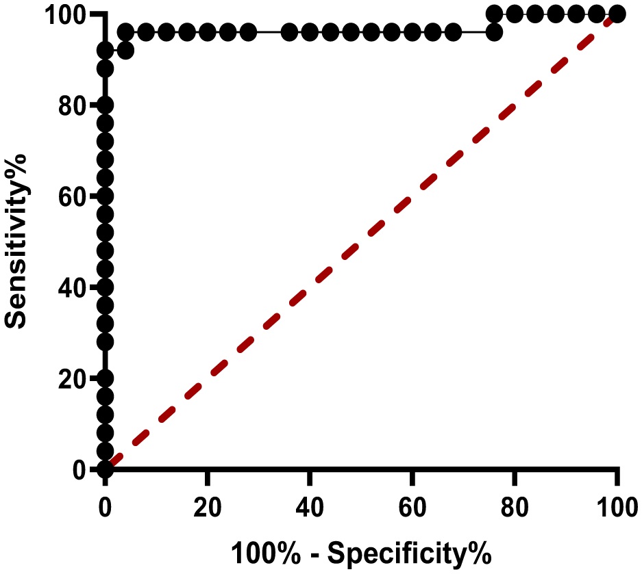
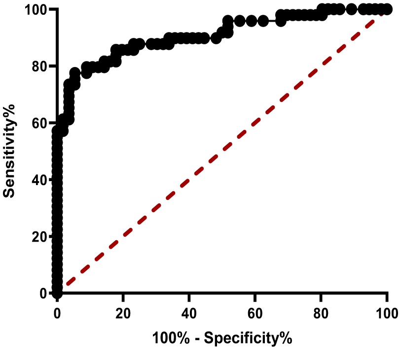
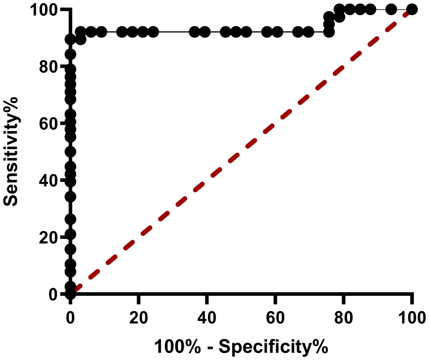
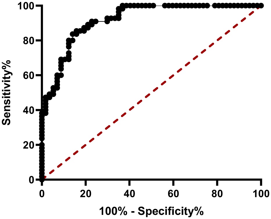
This study sought to identify potential biomarkers of thyroid cancer, such as visfatin, resistin, IL-17, and IL-23 levels, alongside investigating their involvement in the progression of the disease. Methods: This study included 56 patients that have thyroid cancer and 47 healthy people whose sexes and ages were matched to the healthy group. The sandwich enzyme-linked immunosorbent assay (ELISA) kit, which is widely available and dependable, was utilized to measure the level of interleukin-17, interleukin-23, visfatin, and resistin within the serum of the patient and control groups. These biochemical values were plotted against a receiver operator characteristic (ROC) curve to determine their potential diagnostic use. Results: The serum levels of Interleukin-17 (255 ± 25.82 pg/mL), IL-23 (461.03 ± 29.97 pg/mL), visfatin (19.42 ± 2.91 ng/mL), and resistin (25.3 ± 1.9 µg/L) were all noticeably higher in the thyroid cancer patients; the ROC analysis indicated that these serum concentrations may be used as potential biomarkers for thyroid cancer diagnoses. Conclusion: In contrast to IL-17 and IL-23, which demonstrated stronger specificities (94.1% and 83.7%, respectively) and closer associations with thyroid cancer pathogenesis, the resistin and visfatin levels were significantly elevated in patients with thyroid cancer and showed diagnostic potential in the ROC analysis (AUC > 0.8). However, their non-specific pro-inflammatory roles and wider variability in metabolic conditions (e.g., obesity, insulin resistance) may limit their reliability as early diagnostic biomarkers. Therefore, resistin and visfatin need to be further validated in bigger cohorts to evaluate the confounding impacts, while IL-17 and IL-23 may be more suited for early identification.
Citation: Hazhar M. Balaky, Parween Abdulsamad Ismail. Serum resistin, visfatin, IL-17 and IL-23 as novel diagnostic biomarkers for thyroid carcinoma[J]. AIMS Allergy and Immunology, 2025, 9(3): 166-179. doi: 10.3934/Allergy.2025013
This study sought to identify potential biomarkers of thyroid cancer, such as visfatin, resistin, IL-17, and IL-23 levels, alongside investigating their involvement in the progression of the disease. Methods: This study included 56 patients that have thyroid cancer and 47 healthy people whose sexes and ages were matched to the healthy group. The sandwich enzyme-linked immunosorbent assay (ELISA) kit, which is widely available and dependable, was utilized to measure the level of interleukin-17, interleukin-23, visfatin, and resistin within the serum of the patient and control groups. These biochemical values were plotted against a receiver operator characteristic (ROC) curve to determine their potential diagnostic use. Results: The serum levels of Interleukin-17 (255 ± 25.82 pg/mL), IL-23 (461.03 ± 29.97 pg/mL), visfatin (19.42 ± 2.91 ng/mL), and resistin (25.3 ± 1.9 µg/L) were all noticeably higher in the thyroid cancer patients; the ROC analysis indicated that these serum concentrations may be used as potential biomarkers for thyroid cancer diagnoses. Conclusion: In contrast to IL-17 and IL-23, which demonstrated stronger specificities (94.1% and 83.7%, respectively) and closer associations with thyroid cancer pathogenesis, the resistin and visfatin levels were significantly elevated in patients with thyroid cancer and showed diagnostic potential in the ROC analysis (AUC > 0.8). However, their non-specific pro-inflammatory roles and wider variability in metabolic conditions (e.g., obesity, insulin resistance) may limit their reliability as early diagnostic biomarkers. Therefore, resistin and visfatin need to be further validated in bigger cohorts to evaluate the confounding impacts, while IL-17 and IL-23 may be more suited for early identification.

| [1] | Xi C, Zhang GQ, Sun ZK., et al. (2020) Interleukins in thyroid cancer: From basic researches to applications in clinical practice. Front Immunol 11: 1124. https://doi.org/10.3389/fimmu.2020.01124 |
| [2] | Banerjee S, Nahar U, Dahiya D, et al. (2023) IL-17 A correlates with disease progression in papillary thyroid carcinoma. Diagn Pathol1 8: 93. https://doi.org/10.1186/s13000-023-01362-4 |
| [3] | Zhao J, Zhang Q, Yang Y, et al. (2021) High prevalence of thyroid carcinoma in patients with insulin resistance: A meta-analysis of case-control studies. Aging 13: 22232-22241. https://doi.org/10.18632/aging.203529 |
| [4] | Steppan CM, Lazar MA (2002) Resistin and obesity-associated insulin resistance. Trends Endocrinol Metab 13: 18-23. https://doi.org/10.1016/S1043-2760(01)00522-7 |
| [5] | Zhou L, Song K, Luo W (2023) Association between circulating resistin levels and thyroid dysfunction: A systematic review and meta-analysis. Front Endocrinol 13: 1071922. https://doi.org/10.3389/fendo.2022.1071922 |
| [6] | Xu F, Ning X, Zhao T, et al. (2022) Visfatin is negatively associated with coronary artery lesions in subjects with impaired fasting glucose. Open Med 17: 1405-1411. https://doi.org/10.1515/med-2022-0540 |
| [7] | Uçan B, Kebapçı N, Uslu S, et al. (2018) Plasma visfatin concentrations in hypothyroid patients and its relationship with thyroid autoimmunity and atherosclerosis. Ortadoğu Tıp Derg 10: 498-505. https://doi.org/10.21601/ortadogutipdergisi.364345 |
| [8] | Mohammadi M, Mianabadi F, Mehrad-Majd H (2019) Circulating visfatin levels and cancers risk: A systematic review and meta-analysis. J cell Physiol 234: 5011-5022. https://doi.org/10.1002/jcp.27302 |
| [9] | Martins MB, Marcello MA, de Assis Batista F, et al. (2018) Serum interleukin measurement may help identify thyroid cancer patients with active disease. Clin Biochem 52: 1-7. https://doi.org/10.1016/j.clinbiochem.2017.10.003 |
| [10] | Yao Z, Fanslow WC, Seldin MF, et al. (1995) Herpesvirus Saimiri encodes a new cytokine, IL-17, which binds to a novel cytokine receptor. Immunity 3: 811-821. https://doi.org/10.1016/1074-7613(95)90070-5 |
| [11] | Okada S, Puel A, Casanova JL, et al. (2016) Chronic mucocutaneous candidiasis disease associated with inborn errors of IL-17 immunity. Clin Transl Immunol 5: e114. https://doi.org/10.1038/cti.2016.71 |
| [12] | de Morales JM, Puig L, Daudén E, et al. (2020) Critical role of interleukin (IL)-17 in inflammatory and immune disorders: An updated review of the evidence focusing in controversies. Autoimmun Revi 19: 102429. https://doi.org/10.1016/j.autrev.2019.102429 |
| [13] | Li S, Li S, Lin M, et al. (2022) Interleukin-17 and vascular endothelial growth factor: New biomarkers for the diagnosis of papillary thyroid carcinoma in patients with Hashimoto's thyroiditis. J Int Med Res 50. https://doi.org/10.1177/03000605211067121 |
| [14] | Bankir M, Yanardag Acik D (2021) IL-17 and IL-23 levels in patients with early-stage chronic lymphocytic leukemia. North Clin Istanbul 8. https://doi.org/10.14744/nci.2020.02997 |
| [15] | Bayraktar N, Eren MA, Bayraktar M, et al. (2023) Analysis of Interleukin-17, Interleukin-23, neopterin and Nesfatin-1 levels in the sera of Hashimoto patients. J Med Biochem 42: 460-468. https://doi.org/10.5937/jomb0-40683 |
| [16] | Zheng T, Xu C, Mao C, et al. (2018) Increased interleukin-23 in Hashimoto's thyroiditis disease induces autophagy suppression and reactive oxygen species accumulation. Front Immunol 9: 96. https://doi.org/10.3389/fimmu.2018.00096 |
| [17] | McGeachy MJ, Chen Y, Tato CM, et al. (2009) The interleukin 23 receptor is essential for the terminal differentiation of interleukin 17–producing effector T helper cells in vivo. Nat Immunol 10: 314-324. https://doi.org/10.1038/ni.1698 |
| [18] | Galano Carvalho DF, Zanetti BR, Miranda L, et al. (2017) High IL-17 expression is associated with an unfavorable prognosis in thyroid cancer. Oncol Lett 13: 1925-1931. https://doi.org/10.3892/ol.2017.5638 |
| [19] | Pan Y, Xu Y, Fan C, et al. (2024) The role of neck adipose tissue in lymph node metastasis of head and neck cancer. Front Oncol 14: 1390824. https://doi.org/10.3389/fonc.2024.1390824 |
| [20] | Nieman KM, Romero IL, Van Houten B, et al. (2013) Adipose tissue and adipocytes support tumorigenesis and metastasis. Biochim Biophys Acta Mol Cell Biol Lipids 1831: 1533-1541. https://doi.org/10.1016/j.bbalip.2013.02.010 |
| [21] | Yang CC, Chang SF, Chao JK, et al. (2014) Activation of AMP-activated protein kinase attenuates hepatocellular carcinoma cell adhesion stimulated by adipokine resistin. BMC cancer 14: 1-9. https://doi.org/10.1186/1471-2407-14-112 |
| [22] | Tian W, Zhu Y, Wang Y, et al. (2013) Visfatin, a potential biomarker and prognostic factor for endometrial cancer. Gynecol Oncol 129: 505-512. https://doi.org/10.1016/j.ygyno.2013.02.022 |
| [23] | Mohammadi M, Hedayati M (2022) Visfatin: An adipokine that plays a crucial role in increasing the risk of cancer. Iran J Blood Cancer 14: 68-74. https://doi.org/10.58209/ijbc.14.2.68 |
| [24] | Pazgan-Simon M, Kukla M, Zuwała-Jagiełło J, et al. (2020) Serum visfatin and vaspin levels in hepatocellular carcinoma (HCC). PLoS One 15: e0227459. https://doi.org/10.1371/journal.pone.0227459 |
| [25] | Bułdak RJ, Bułdak Ł, Polaniak R, et al. (2013) Visfatin affects redox adaptative responses and proliferation in Me45 human malignant melanoma cells: An in vitro study. Oncol Rep 29: 771-778. https://doi.org/10.3892/or.2012.2175 |
| [26] | Adya R, Tan BK, Punn A, et al. (2008) Visfatin induces human endothelial VEGF and MMP-2/9 production via MAPK and PI3K/Akt signalling pathways: Novel insights into visfatin-induced angiogenesis. Cardiovasc Res 78: 356-365. https://doi.org/10.1093/cvr/cvm111 |
| [27] | Schuettfort VM, Pradere B, Trinh QD, et al. (2022) Impact of preoperative plasma levels of interleukin 6 and interleukin 6 soluble receptor on disease outcomes after radical cystectomy for bladder cancer. Cancer Immunol Immunother 71: 1-11. https://doi.org/10.1007/s00262-021-02953-0 |
| [28] | Barilla RM, Diskin B, Caso RC, et al. (2019) Specialized dendritic cells induce tumor-promoting IL-10+ IL-17+ FoxP3neg regulatory CD4+ T cells in pancreatic carcinoma. Nat commun 10: 1424. https://doi.org/10.1038/s41467-019-09416-2 |
| [29] | Hong YR, Lee SH, Lim DJ, et al. (2017) The stratification of patient risk depending on the size and ratio of metastatic lymph nodes in papillary thyroid carcinoma. World J Surg Oncol 15: 1-9. https://doi.org/10.1186/s12957-017-1141-4 |
| [30] | Hughes NM, Nae A, Barry J, et al. (2017) Sonographic differences between conventional and follicular variant papillary thyroid carcinoma. Eur Arch Oto-Rhino-Laryngol 274: 2907-2913. https://doi.org/10.1007/s00405-017-4557-0 |
| [31] | Li H, Tsokos MG, Bhargava R, et al. (2021) IL-23 reshapes kidney resident cell metabolism and promotes local kidney inflammation. J Clin Invest 131. https://doi.org/10.1172/JCI142428 |
| [32] | Yan J, Smyth MJ, Teng MW (2018) Interleukin (IL)-12 and IL-23 and their conflicting roles in cancer. Cold Spring Harb Perspect Biol 10: a028530. https://doi.org/10.1101/cshperspect.a028530 |
| [33] | de Mel S, Hue SS, Jeyasekharan AD, et al. (2019) Molecular pathogenic pathways in extranodal NK/T cell lymphoma. J Hematol Oncol 12: 1-18. https://doi.org/10.1186/s13045-019-0716-7 |
| [34] | Hou Y, Zhu L, Tian H, et al. (2018) IL-23-induced macrophage polarization and its pathological roles in mice with imiquimod-induced psoriasis. Protein Cell 9: 1027-1038. https://doi.org/10.1007/s13238-018-0505-z |
| [35] | Wu F, Fan J, He Y, et al. (2021) Single-cell profiling of tumor heterogeneity and the microenvironment in advanced non-small cell lung cancer. Nat commun 12: 2540. https://doi.org/10.1038/s41467-021-22801-0 |
| [36] | Figueroa-Vega N, Alfonso-Perez M, Benedicto I, et al. (2010) Increased circulating pro-inflammatory cytokines and Th17 lymphocytes in Hashimoto's thyroiditis. J Clin Endocrinol Metab 95: 953-962. https://doi.org/10.1210/jc.2009-1719 |
| [37] | Kwon H, Chang Y, Cho A, et al. (2019) Metabolic obesity phenotypes and thyroid cancer risk: A cohort study. Thyroid 29: 349-358. https://doi.org/10.1089/thy.2018.0327 |
| [38] | Almquist M, Johansen D, Björge T, et al. (2011) Metabolic factors and risk of thyroid cancer in the Metabolic syndrome and Cancer project (Me-Can). Cancer causes control 22: 743-751. https://doi.org/10.1007/s10552-011-9747-2 |
| [39] | Karabulut S, Usul Afsar C, Karabulut M, et al. (2016) Clinical significance of serum interleukin-17 levels in colorectal cancer patients. J BUON 21: 1137-1145. Available from: https://pubmed.ncbi.nlm.nih.gov/27837615/ |
| [40] | Sawicka-Gutaj N, Ziółkowska P, Derwich A, et al. (2022) Is eNAMPT/visfatin a potential serum marker of papillary thyroid cancer?. Ther Adv Endocrinol Metab 13. https://doi.org/10.1177/20420188221090005 |
Figures(4) / Tables(5)

Hazhar M. Balaky, Parween Abdulsamad Ismail. Serum resistin, visfatin, IL-17 and IL-23 as novel diagnostic biomarkers for thyroid carcinoma[J]. AIMS Allergy and Immunology, 2025, 9(3): 166-179. doi: 10.3934/Allergy.2025013


 DownLoad: