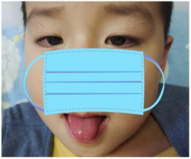
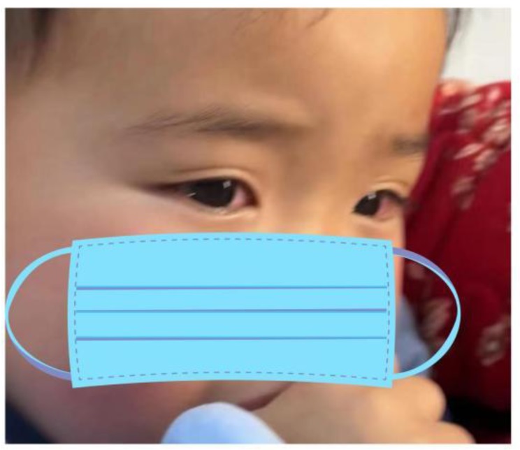
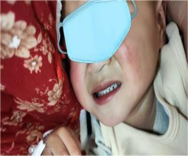
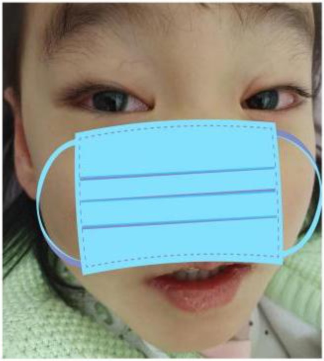
Recurrent Kawasaki Disease (RKD) is defined as the return of clinical symptoms, signs, and related laboratory indicators to normal after the last Kawasaki disease (KD) treatment, with an interval of at least 2 months from the last episode. Compared with initial KD, patients with RKD have a higher risk of developing refractory KD and coronary artery lesions (CALs). Our purpose of this study was to summarize the pathogenesis of RKD so clinicians can achieve early recognition and treatment. In this report, we described 4 children who developed KD for the second time. Case 1: A boy with 4 months between the two episodes of KD was diagnosed with refractory KD and received a second treatment of intravenous immunoglobulin (IVIG). Case 2: A girl with a family history of KD, with 20 months between the two episodes. She was diagnosed with Complete KD (CKD) and received first-line treatment. Case 3: A girl with 2 years between the two episodes was diagnosed with Incomplete KD (IKD), and underwent targeted sequencing of multiple pathogens in the upper respiratory tract due to 3 pathogenic infections and received first-line treatment. Case 4: A boy with 5 years between onset of the disease and diagnosis of IKD. C-reactive protein was significantly elevated at the time of admission, and he received IVIG combined with aspirin and methylprednisolone. All 4 children recovered after treatment without CALs. Children with a history of KD or infections with pathogens strongly associated with KD recurrence need to be assessed for RKD, regardless of the time interval.
Citation: Hui-Min Zhang, Qi-Ling Yin, You-Qiong Liu, Ya-Le Zhang, Wei-Hua Zhang. Recurrent Kawasaki disease in children: Four case reports[J]. AIMS Allergy and Immunology, 2025, 9(2): 123-135. doi: 10.3934/Allergy.2025009
Recurrent Kawasaki Disease (RKD) is defined as the return of clinical symptoms, signs, and related laboratory indicators to normal after the last Kawasaki disease (KD) treatment, with an interval of at least 2 months from the last episode. Compared with initial KD, patients with RKD have a higher risk of developing refractory KD and coronary artery lesions (CALs). Our purpose of this study was to summarize the pathogenesis of RKD so clinicians can achieve early recognition and treatment. In this report, we described 4 children who developed KD for the second time. Case 1: A boy with 4 months between the two episodes of KD was diagnosed with refractory KD and received a second treatment of intravenous immunoglobulin (IVIG). Case 2: A girl with a family history of KD, with 20 months between the two episodes. She was diagnosed with Complete KD (CKD) and received first-line treatment. Case 3: A girl with 2 years between the two episodes was diagnosed with Incomplete KD (IKD), and underwent targeted sequencing of multiple pathogens in the upper respiratory tract due to 3 pathogenic infections and received first-line treatment. Case 4: A boy with 5 years between onset of the disease and diagnosis of IKD. C-reactive protein was significantly elevated at the time of admission, and he received IVIG combined with aspirin and methylprednisolone. All 4 children recovered after treatment without CALs. Children with a history of KD or infections with pathogens strongly associated with KD recurrence need to be assessed for RKD, regardless of the time interval.

| [1] | Kawasaki T (1967) Acute febrile mucocutaneous syndrome with lymphoid involvement with specific desquamation of the fingers and toes in children. Arerugi 16: 178-222. |
| [2] | Jone PN, Tremoulet A, Choueiter N, et al. (2024) Update on diagnosis and management of Kawasaki disease: A scientific statement from the American heart association. Circulation 150: e481-e500. https://doi.org/10.1161/CIR.0000000000001295 |
| [3] |
Hirata S, Nakamura Y, Yanagawa H (2001) Incidence rate of recurrent Kawasaki disease and related risk factors: From the results of nationwide surveys of Kawasaki disease in Japan. Acta Paediatr 90: 40-44. https://doi.org/10.1080/080352501750064851 
|
| [4] |
Saha A, Sarkar S (2018) Recurrent Kawasaki disease. Indian J Pediatr 85: 693-694. https://doi.org/10.1007/s12098-017-2567-y
|
| [5] |
Nakamura Y, Yanagawa H, Ojima T, et al. (1998) Cardiac sequelae of Kawasaki disease among recurrent cases. Arch Dis Child 78: 163-165. https://doi.org/10.1136/adc.78.2.163
|
| [6] |
Kamal S, Khan MA, Altorok N (2016) Recurrent Kawasaki disease: Mind the age, but it does not matter. J Clin Rheumatol 22: 223-224. https://doi.org/10.1097/RHU.0000000000000411
|
| [7] |
Hayashida K Ae R, Masuda H, Kosami K, et al. (2021) Clinical characteristics of patients with Kawasaki disease whose siblings had the same disease. Pediatr Infect Dis J 40: 531-536. https://doi.org/10.1097/INF.0000000000003074
|
| [8] |
Hamada H, Sekizuka T, Oba K, et al. (2016) Comprehensive pathogen detection associated with four recurrent episodes of Kawasaki disease in a patient during a single year using next-generation sequencing. JMM Case Rep 3: e005019. https://doi.org/10.1099/jmmcr.0.005019
|
| [9] |
Yang HM, Du ZD, Fu PP (2013) Clinical features of recurrent Kawasaki disease and its risk factors. Eur J Pediatr 172: 1641-1647. https://doi.org/10.1007/s00431-013-2101-9
|
| [10] |
Yang TJ, Lin MT, Lu CY, et al. (2018) The prevention of coronary arterial abnormalities in Kawasaki disease: A meta-analysis of the corticosteroid effectiveness. J Microbiol Immunol Infect 51: 321-331. https://doi.org/10.1016/j.jmii.2017.08.012
|
| [11] |
Sudo D, Nakamura Y (2017) Nationwide surveys show that the incidence of recurrent Kawasaki disease in Japan has hardly changed over the last 30 years. Acta Paediatr 106: 796-800. https://doi.org/10.1111/apa.13773
|
| [12] |
Newburger JW, Takahashi M, Gerber MA, et al. (2004) Diagnosis, treatment, and long-term management of Kawasaki disease: A statement for health professionals from the committee on rheumatic fever, endocarditis and Kawasaki disease, council on cardiovascular disease in the young, American heart association. Circulation 110: 2747-2771. https://doi.org/10.1161/01.CIR.0000145143.19711.78
|
| [13] |
Kaya Akca U, Arslanoglu Aydin E, Aykan HH, et al. (2022) Comparison of IVIG resistance predictive models in Kawasaki disease. Pediatr Res 91: 621-626. https://doi.org/10.1038/s41390-021-01459-w
|
| [14] |
Furukawa T, Kishiro M, Akimoto K, et al. (2008) Effects of steroid pulse therapy on immunoglobulin-resistant Kawasaki disease. Arch Dis Child 93: 142-146. https://doi.org/10.1136/adc.2007.126144
|
| [15] |
Chen S, Dong Y, Yin Y, et al. (2013) Intravenous immunoglobulin plus corticosteroid to prevent coronary artery abnormalities in Kawasaki disease: A meta-analysis. Heart 99: 76-82. https://doi.org/10.1136/heartjnl-2012-302126
|
| [16] |
Davies S, Sutton N, Blackstock S, et al. (2015) Predicting IVIG resistance in UK Kawasaki disease. Arch Dis Child 100: 366-368. https://doi.org/10.1136/archdischild-2014-307397
|
| [17] |
Balasubramanian S, Ganesh R (2009) Recurrent Kawasaki disease. Indian J Pediatr 76: 848-849. https://doi.org/10.1007/s12098-009-0157-3
|
| [18] |
Friedman KG, Jone PN (2020) Update on the management of Kawasaki disease. Pediatr Clin North Am 67: 811-819. https://doi.org/10.1016/j.pcl.2020.06.002
|
| [19] |
Che X, Gao L, Zhen Z, et al. (2022) Risk factors and predictive models for intravenous immunoglobulin resistance in children with recurrent Kawasaki disease. J inflammation res 15: 2877-2889. https://doi.org/10.2147/JIR.S360802
|
| [20] |
Guleria S, Pilania RK, Jindal AK, et al. (2019) Recurrent Kawasaki disease at a tertiary care center in Chandigarh, North West India: 24 years of clinical experience. Int J Rheum Dis 22: 1183-1187. https://doi.org/10.1111/1756-185X.13519
|
| [21] |
Agarwal S, Agrawal DK (2017) Kawasaki disease: Etiopathogenesis and novel treatment strategies. Expert Rev Clin Immunol 13: 247-258. https://doi.org/10.1080/1744666X.2017.1232165
|
| [22] |
McCrindle BW, Rowley AH, Newburger JW, et al. (2017) Diagnosis, treatment, and long-term management of Kawasaki disease: A scientific statement for health professionals from the American heart association. Circulation 135: e927-e999. https://doi.org/10.1161/CIR.0000000000000484
|
| [23] |
Printz BF, Sleeper LA, Newburger JW, et al. (2011) Noncoronary cardiac abnormalities are associated with coronary artery dilation and with laboratory inflammatory markers in acute Kawasaki disease. J Am Col Cardio 57: 86-92. https://doi.org/10.1016/j.jacc.2010.08.619
|
Figures(5) / Tables(8)

Hui-Min Zhang, Qi-Ling Yin, You-Qiong Liu, Ya-Le Zhang, Wei-Hua Zhang. Recurrent Kawasaki disease in children: Four case reports[J]. AIMS Allergy and Immunology, 2025, 9(2): 123-135. doi: 10.3934/Allergy.2025009


 DownLoad: