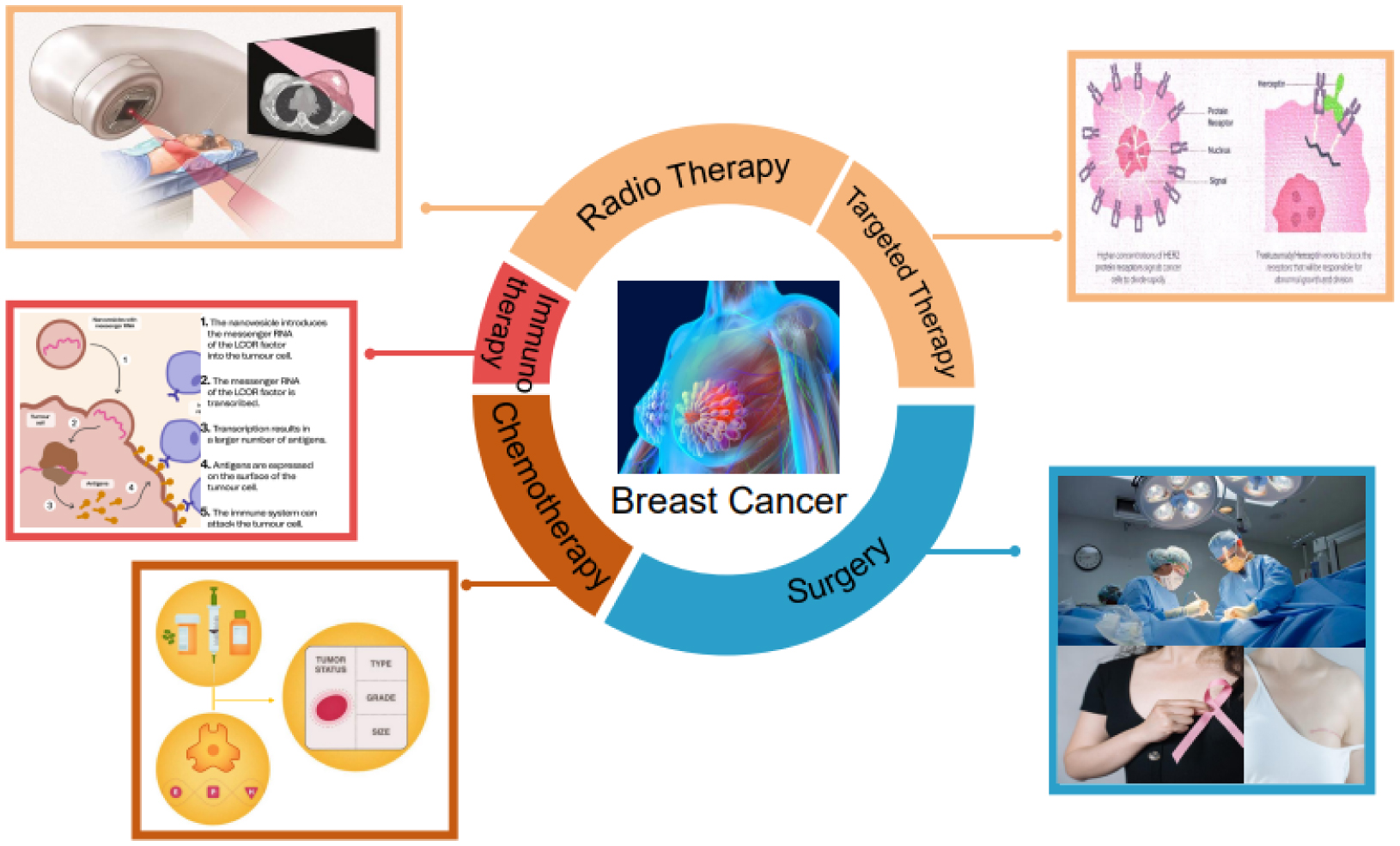
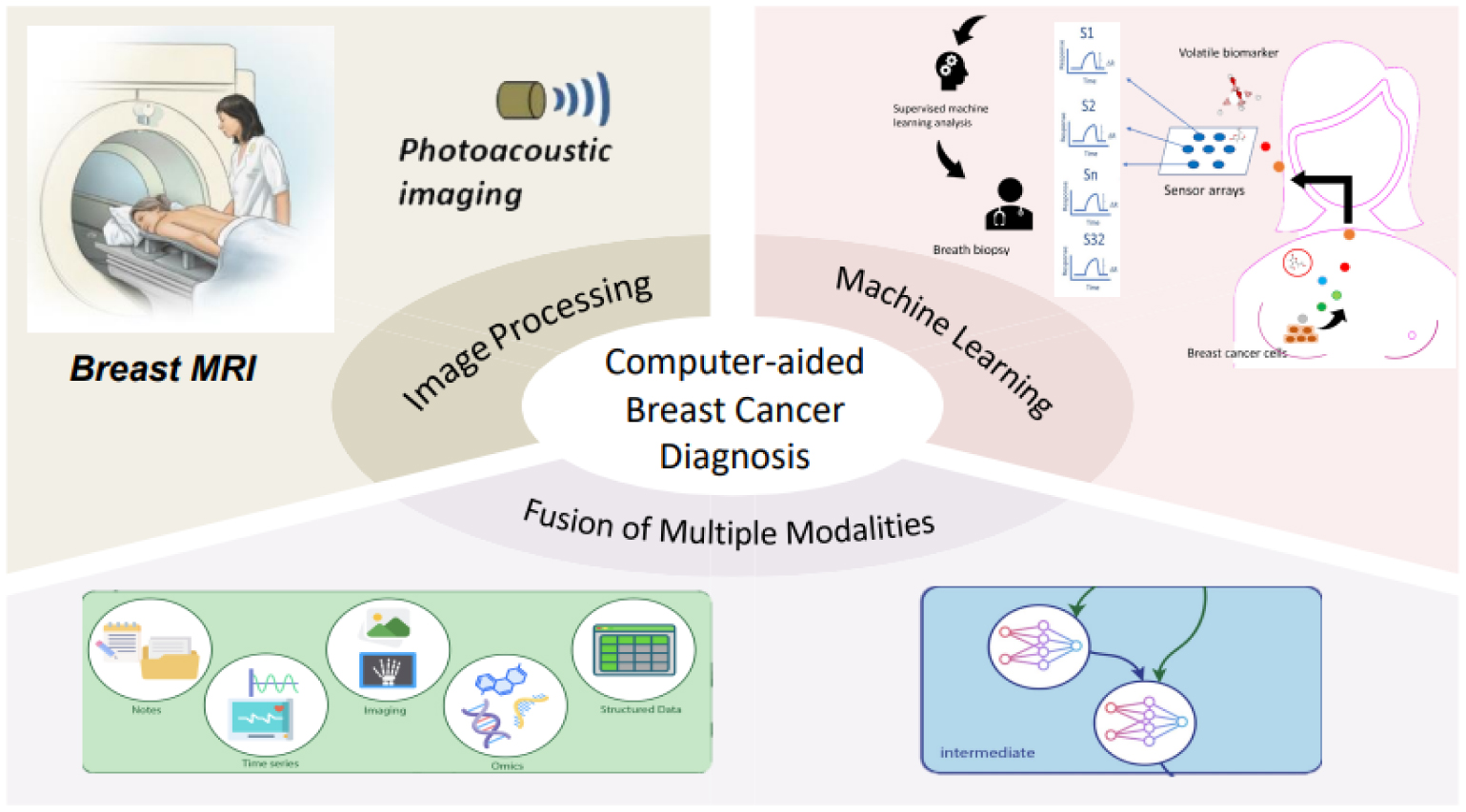
Breast cancer remains a significant public health issue, being a leading cause of cancer-related mortality among women globally. Timely diagnosis and efficient treatment are crucial for enhancing patient outcomes, reducing healthcare burdens and advancing community health. This systematic review, following the PRISMA guidelines, aims to comprehensively synthesize the recent advancements in computer-aided diagnosis and treatment for breast cancer. The study covers the latest developments in image analysis and processing, machine learning and deep learning algorithms, multimodal fusion techniques and radiation therapy planning and simulation. The results of the review suggest that machine learning, augmented and virtual reality and data mining are the three major research hotspots in breast cancer management. Moreover, this paper discusses the challenges and opportunities for future research in this field. The conclusion highlights the importance of computer-aided techniques in the management of breast cancer and summarizes the key findings of the review.
Citation: Kai Cheng, Jiangtao Wang, Jian Liu, Xiangsheng Zhang, Yuanyuan Shen, Hang Su. Public health implications of computer-aided diagnosis and treatment technologies in breast cancer care[J]. AIMS Public Health, 2023, 10(4): 867-895. doi: 10.3934/publichealth.2023057
Breast cancer remains a significant public health issue, being a leading cause of cancer-related mortality among women globally. Timely diagnosis and efficient treatment are crucial for enhancing patient outcomes, reducing healthcare burdens and advancing community health. This systematic review, following the PRISMA guidelines, aims to comprehensively synthesize the recent advancements in computer-aided diagnosis and treatment for breast cancer. The study covers the latest developments in image analysis and processing, machine learning and deep learning algorithms, multimodal fusion techniques and radiation therapy planning and simulation. The results of the review suggest that machine learning, augmented and virtual reality and data mining are the three major research hotspots in breast cancer management. Moreover, this paper discusses the challenges and opportunities for future research in this field. The conclusion highlights the importance of computer-aided techniques in the management of breast cancer and summarizes the key findings of the review.

| [1] |
Rangayyan R M, Ayres F J, Desautels J L (2007) A review of computer-aided diagnosis of breast cancer: Toward the detection of subtle signs. J Franklin Inst 344: 312-348. https://doi.org/10.1016/j.jfranklin.2006.09.003 
|
| [2] |
Chen K, Lu P, Beeraka NM, et al. (2022) Mitochondrial mutations and mitoepigenetics: Focus on regulation of oxidative stress-induced responses in breast cancers. Semin Cancer Biol 83: 556-569. https://doi.org/10.1016/j.semcancer.2020.09.012
|
| [3] |
Lord S, Lei W, Craft P, et al. (2007) A systematic review of the effectiveness of magnetic resonance imaging (MRI) as an addition to mammography and ultrasound in screening young women at high risk of breast cancer. Eur J Cancer 43: 1905-1917. https://doi.org/10.1016/j.ejca.2007.06.007
|
| [4] |
Kuhl C K, Schrading S, Leutner C C, et al. (2005) Mammography, breast ultrasound, and magnetic resonance imaging for surveillance of women at high familial risk for breast cancer. J Clin Oncol 23: 8469-8476. https://doi.org/10.1200/JCO.2004.00.4960
|
| [5] |
Esteva F J, Hubbard-Lucey V M, Tang J, et al. (2019) Immunotherapy and targeted therapy combinations in metastatic breast cancer. Lancet Oncol 20: e175-e186. https://doi.org/10.1016/S1470-2045(19)30026-9
|
| [6] |
Korde L A, Somerfield M R, Carey L A, et al. (2021) Neoadjuvant chemotherapy, endocrine therapy, and targeted therapy for breast cancer: Asco guideline. J Clin Oncol 39: 1485-1505. https://doi.org/10.1200/JCO.20.03399
|
| [7] |
Hughes P E, Caenepeel S, Wu L C (2016) Targeted therapy and checkpoint immunotherapy combinations for the treatment of cancer. Trends Immunol 37: 462-476. https://doi.org/10.1016/j.it.2016.04.010
|
| [8] |
Al-Antari M A, Al-Masni M A, Choi M T, et al. (2018) A fully integrated computer-aided diagnosis system for digital x-ray mammograms via deep learning detection, segmentation, and classification. Int J Med Inform 117: 44-54. https://doi.org/10.1016/j.ijmedinf.2018.06.003
|
| [9] |
Yassin N I, Omran S, El Houby E M, et al. (2018) Machine learning techniques for breast cancer computer aided diagnosis using different image modalities: A systematic review. Comput Methods Programs Biomed 156: 25-45. https://doi.org/10.1016/j.cmpb.2017.12.012
|
| [10] |
Tian C, Xu Z, Wang L, et al. (2023) Arc fault detection using artificial intelligence: Challenges and benefits. Math Biosci Eng 20: 12404-12432. https://doi.org/10.3934/mbe.2023552
|
| [11] |
Shi Y, Li H, Fu X, et al. (2023) Self-powered difunctional sensors based on sliding contact-electrification and tribovoltaic effects for pneumatic monitoring and controlling. Nano Energy 110: 108339. https://doi.org/10.1016/j.nanoen.2023.108339
|
| [12] |
Shi Y, Li L, Yang J, et al. (2023) Center-based transfer feature learning with classifier adaptation for surface defect recognition. Mech Syst Signal Process 188: 110001. https://doi.org/10.1016/j.ymssp.2022.110001
|
| [13] | Wang D, Khosla A, Gargeya R, et al. (2016) Deep learning for identifying metastatic breast cancer. arXiv preprint arXiv:1606.05718 . https://doi.org/10.48550/arXiv.1606.05718 |
| [14] |
Houssein E H, Emam M M, Ali A A, et al. (2021) Deep and machine learning techniques for medical imaging-based breast cancer: A comprehensive review. Expert Syst Appl 167: 114161. https://doi.org/10.1016/j.eswa.2020.114161
|
| [15] |
Wang Y, Liu Z, Xu J, et al. (2022) Heterogeneous network representation learning approach for ethereum identity identification. IEEE Trans Comput Soc Syst 10: 890-899. https://doi.org/10.1109/TCSS.2022.3164719
|
| [16] |
Zhao J, Lv Y (2023) Output-feedback robust tracking control of uncertain systems via adaptive learning. Int J Control Autom Syst 21: 1108-1118. https://doi.org/10.1007/s12555-021-0882-6
|
| [17] | Ramesh K, Kumar G K, Swapna K, et al. (2021) A review of medical image segmentation algorithms. EAI Endorsed Trans. Pervasive Health Technol 7: e6-e6. https://doi.org/10.4108/eai.12-4-2021.169184 |
| [18] |
Kamnitsas K, Ledig C, Newcombe V F, et al. (2017) Efficient multi-scale 3D CNN with fully connected CRF for accurate brain lesion segmentation. Med Image Anal 36: 61-78. https://doi.org/10.1016/j.media.2016.10.004
|
| [19] |
Qi W, Aliverti A (2019) A multimodal wearable system for continuous and real-time breathing pattern monitoring during daily activity. IEEE J Biomed Health Inform 24: 2199-2207. https://doi.org/10.1109/JBHI.2019.2963048
|
| [20] |
Mokni R, Gargouri N, Damak A, et al. (2021) An automatic computer-aided diagnosis system based on the multimodal fusion of breast cancer (mf-cad). Biomed Signal Process Control 69: 102914. https://doi.org/10.1016/j.bspc.2021.102914
|
| [21] |
Qi W, Ovur S E, Li Z, et al. (2021) Multi-sensor guided hand gesture recognition for a teleoperated robot using a recurrent neural network. IEEE Robot Autom Lett 6: 6039-6045. https://doi.org/10.1109/LRA.2021.3089999
|
| [22] |
Schlachter M, Raidou R G, Muren L P, et al. (2019) State-of-the-art report: Visual computing in radiation therapy planning. Comput Graph Forum 38: 753-779. https://doi.org/10.1111/cgf.13726
|
| [23] |
Rodby K A, Turin S, Jacobs R J, et al. (2014) Advances in oncologic head and neck reconstruction: systematic review and future considerations of virtual surgical planning and computer aided design/computer aided modeling. J Plast Reconstr Aesthet Surg 67: 1171-1185. https://doi.org/10.1016/j.bjps.2014.04.038
|
| [24] |
Su H, Mariani A, Ovur S E, et al. (2021) Toward teaching by demonstration for robot-assisted minimally invasive surgery. IEEE Trans Autom Sci Eng 18: 484-494. https://doi.org/10.1109/TASE.2020.3045655
|
| [25] |
Su H, Qi W, Chen J, Zhang D (2022) Fuzzy approximation-based task-space control of robot manipulators with remote center of motion constraint. IEEE Trans Fuzzy Syst 30: 1564-1573. https://doi.org/10.1109/TFUZZ.2022.3157075
|
| [26] |
Rivenbark A G, O'Connor S M, Coleman W B (2013) Molecular and cellular heterogeneity in breast cancer: challenges for personalized medicine. Am J Pathol 183: 1113-1124. https://doi.org/10.1016/j.ajpath.2013.08.002
|
| [27] | Kajala A, Jain V (2020) Diagnosis of breast cancer using machine learning algorithms-a review. 2020 International Conference on Emerging Trends in Communication, Control and Computing (ICONC3) : 1-5. https://doi.org/10.1109/ICONC345789.2020.9117320 |
| [28] | Sheth D, Giger M L (2020) Artificial intelligence in the interpretation of breast cancer on MRI. J Magn Reson 51: 1310-1324. https://doi.org/10.1002/jmri.26878 |
| [29] | Charan S, Khan M J, Khurshid K (2018) Breast cancer detection in mammograms using convolutional neural network. in 2018 International Conference on Computing, Mathematics and Engineering Technologies (iCoMET) : 1-5. https://doi.org/10.1109/ICOMET.2018.8346384 |
| [30] |
Tsochatzidis L, Costaridou L, Pratikakis I (2019) Deep learning for breast cancer diagnosis from mammograms—a comparative study. Journal of Imaging 5: 37. https://doi.org/10.3390/jimaging5030037
|
| [31] |
Shen L, Margolies L R, Rothstein J H, et al. (2019) Deep learning to improve breast cancer detection on screening mammography. Sci Rep 9: 12495. https://doi.org/10.1038/s41598-019-48995-4
|
| [32] |
Salama W M, Aly M H (2021) Deep learning in mammography images segmentation and classification: Automated cnn approach. Alex Eng J 60: 4701-4709. https://doi.org/10.1016/j.aej.2021.03.048
|
| [33] |
Budak Ü, Cömert Z, Rashid Z N, et al. (2019) Computer-aided diagnosis system combining fcn and bi-lstm model for efficient breast cancer detection from histopathological images. Appl Soft Comput 85: 105765. https://doi.org/10.1016/j.asoc.2019.105765
|
| [34] |
Aljuaid H, Alturki N, Alsubaie N, et al. (2022) Computer-aided diagnosis for breast cancer classification using deep neural networks and transfer learning. Comput Methods Programs Biomed 223: 106951. https://doi.org/10.1016/j.cmpb.2022.106951
|
| [35] |
Eltrass A S, Salama M S (2020) Fully automated scheme for computer-aided detection and breast cancer diagnosis using digitised mammograms. IET Image Process 14: 495-505. https://doi.org/10.1049/iet-ipr.2018.5953
|
| [36] | Mehmood M, Ayub E, Ahmad F, et al. (2021) Machine learning enabled early detection of breast cancer by structural analysis of mammograms. Comput Mater Contin 67: 641-657. https://doi.org/10.32604/cmc.2021.013774 |
| [37] |
Jasti V D P, Zamani A S, Arumugam K, et al. (2022) Computational technique based on machine learning and image processing for medical image analysis of breast cancer diagnosis. Secur Commun Netw 2022: 1-7. https://doi.org/10.1155/2022/1918379
|
| [38] |
Qi W, Su H (2022) A cybertwin based multimodal network for ecg patterns monitoring using deep learning. IEEE Trans Industr Inform 18: 6663-6670. https://doi.org/10.1109/TII.2022.3159583
|
| [39] | Liu Q, Liu Z, Yong S, et al. (2020) Computer-aided breast cancer diagnosis based on image segmentation and interval analysis. Automatica (Oxf) 61: 496-506. https://doi.org/10.1080/00051144.2020.1785784 |
| [40] |
Maqsood S, Damaševičius R, Maskeliūnas R (2022) Ttcnn: A breast cancer detection and classification towards computer-aided diagnosis using digital mammography in early stages. Appl Sci 12: 3273. https://doi.org/10.3390/app12073273
|
| [41] |
Younis Y S, Ali A H, Alhafidhb O K, et al. (2022) Early diagnosis of breast cancer using image processing techniques. J Nanomater 2022: 1-6. https://doi.org/10.1155/2022/2641239
|
| [42] |
Pavithra S, Vanithamani R, Justin J (2020) Computer aided breast cancer detection using ultrasound images. Mater Today Proc 33: 4802-4807. https://doi.org/10.1016/j.matpr.2020.08.381
|
| [43] |
Lozano A, Hayes J C, Compton L M, et al. (2020) Determining the thermal characteristics of breast cancer based on high-resolution infrared imaging, 3d breast scans, and magnetic resonance imaging. Sci Rep 10: 1-14. https://doi.org/10.1038/s41598-020-66926-6
|
| [44] |
Liu Z, Yang D, Wang Y, et al. (2023) Egnn: Graph structure learning based on evolutionary computation helps more in graph neural networks. Appl Soft Comput 135: 110040. https://doi.org/10.1016/j.asoc.2023.110040
|
| [45] |
Chaudhury S, Krishna A N, Gupta S, et al. (2022) Effective image processing and segmentation-based machine learning techniques for diagnosis of breast cancer. Comput Math Methods Med 2022. https://doi.org/10.1155/2022/6841334
|
| [46] |
Zhang H, Han L, Chen K, et al. (2020) Diagnostic efficiency of the breast ultrasound computer-aided prediction model based on convolutional neural network in breast cancer. J Digit Imaging 33: 1218-1223. https://doi.org/10.1007/s10278-020-00357-7
|
| [47] |
Pourasad Y, Zarouri E, Salemizadeh Parizi M, et al. (2021) Presentation of novel architecture for diagnosis and identifying breast cancer location based on ultrasound images using machine learning. Diagnostics 11: 1870. https://doi.org/10.3390/diagnostics11101870
|
| [48] |
Bourouis S, Band S S, Mosavi A, et al. (2022) Meta-heuristic algorithm-tuned neural network for breast cancer diagnosis using ultrasound images. Front Oncoly 12: 834028. https://doi.org/10.3389/fonc.2022.834028
|
| [49] |
Qi X, Yi F, Zhang L, et al. (2022) Computer-aided diagnosis of breast cancer in ultrasonography images by deep learning. Neurocomputing 472: 152-165. https://doi.org/10.1016/j.neucom.2021.11.047
|
| [50] |
Ji Y, Li H, Edwards A V, et al. (2019) Independent validation of machine learning in diagnosing breast cancer on magnetic resonance imaging within a single institution. Cancer Imaging 19: 1-11. https://doi.org/10.1186/s40644-019-0252-2
|
| [51] |
Tahmassebi A, Wengert G J, Helbich T H, et al. (2019) Impact of machine learning with multiparametric magnetic resonance imaging of the breast for early prediction of response to neoadjuvant chemotherapy and survival outcomes in breast cancer patients. Invest Radiol 54: 110. https://doi.org/10.1097/RLI.0000000000000518
|
| [52] |
Adachi M, Fujioka T, Mori M, et al. (2020) Detection and diagnosis of breast cancer using artificial intelligence based assessment of maximum intensity projection dynamic contrast-enhanced magnetic resonance images. Diagnostics 10: 330. https://doi.org/10.3390/diagnostics10050330
|
| [53] |
Zheng J, Lin D, Gao Z, et al. (2020) Deep learning assisted efficient adaboost algorithm for breast cancer detection and early diagnosis. IEEE Access 8: 96946-96954. https://doi.org/10.1109/ACCESS.2020.2993536
|
| [54] | Sultan L R, Schultz S M, Cary T W, et al. (2018) Machine learning to improve breast cancer diagnosis by multimodal ultrasound. IEEE International Ultrason Symp (IUS) : 1-4. https://doi.org/10.1109/ultsym.2018.8579953 |
| [55] |
Mohammed M A, Al-Khateeb B, Rashid A N, et al. (2018) Neural network and multi-fractal dimension features for breast cancer classification from ultrasound images. Comput Electr Eng 70: 871-882. https://doi.org/10.1016/j.compeleceng.2018.01.033
|
| [56] |
Huang R, Lin Z, Dou H, et al. (2021) Aw3m: An auto-weighting and recovery framework for breast cancer diagnosis using multi-modal ultrasound. Med Image Anal 72: 102137. https://doi.org/10.1016/j.media.2021.102137
|
| [57] |
Yan R, Zhang F, Rao X, et al. (2021) Richer fusion network for breast cancer classification based on multimodal data. BMC Medical Inform Decis Mak 21: 1-15. https://doi.org/10.1186/s12911-020-01340-6
|
| [58] | Duanmu H, Huang P B, Brahmavar S, et al. (2020) Prediction of pathological complete response to neoadjuvant chemotherapy in breast cancer using deep learning with integrative imaging, molecular and demographic data. in Medical Image Computing and Computer Assisted Intervention–MICCAI 2020: 23rd International Conference, Lima, Peru, October 4–8, 2020, Proceedings, Part II 23 : 242-252. https://doi.org/10.1007/978-3-030-59713-9_24 |
| [59] |
Byra M, Dobruch-Sobczak K, Klimonda Z, et al. (2020) Early prediction of response to neoadjuvant chemotherapy in breast cancer sonography using siamese convolutional neural networks. IEEE J Biomed Health Inform 25: 797-805. https://doi.org/10.1109/JBHI.2020.3008040
|
| [60] |
De Boer L L, Bydlon T M, Van Duijnhoven F, et al. (2018) Towards the use of diffuse reflectance spectroscopy for real-time in vivo detection of breast cancer during surgery. J Transl Med 16: 1-14. https://doi.org/10.1186/s12967-018-1747-5
|
| [61] |
Kulkarni S A, Kulkarni K, Schacht D, et al. (2021) High-resolution full-3d specimen imaging for lumpectomy margin assessment in breast cancer. Ann Surg Oncoly 28: 5513-5524. https://doi.org/10.1245/s10434-021-10499-9
|
| [62] |
Chaurasia V, Pal S, Tiwari B (2018) Prediction of benign and malignant breast cancer using data mining techniques. J Algorithm Comput Technol 12: 119-126. https://doi.org/10.1177/1748301818756225
|
| [63] |
Ösz Á, Lánczky A, Györffy B (2021) Survival analysis in breast cancer using proteomic data from four independent datasets. Sci Rep 11: 16787. https://doi.org/10.1038/s41598-021-96340-5
|
| [64] |
Hong J C, Rahimy E, Gross C P, et al. (2018) Radiation dose and cardiac risk in breast cancer treatment: An analysis of modern radiation therapy including community settings. Pract Radiat Oncol 8: e79-e86. https://doi.org/10.1016/j.prro.2017.07.005
|
| [65] |
Sager O, Dincoglan F, Uysal B, et al. (2018) Evaluation of adaptive radiotherapy (art) by use of replanning the tumor bed boost with repeated computed tomography (ct) simulation after whole breast irradiation (wbi) for breast cancer patients having clinically evident seroma. Jpn J Radiol 36: 401-406. https://doi.org/10.1007/s11604-018-0735-2
|
| [66] |
Jung J W, Lee C, Mosher E G, et al. (2019) Automatic segmentation of cardiac structures for breast cancer radiotherapy. Physics Imaging Radiat Oncol 12: 44-48. https://doi.org/10.1016/j.phro.2019.11.007
|
| [67] |
Enderling H, Alfonso J C L, Moros E, et al. (2019) Integrating mathematical modeling into the roadmap for personalized adaptive radiation therapy. Trends Cancer 5: 467-474. https://doi.org/10.1016/j.trecan.2019.06.006
|
| [68] |
Yang L, Fu B, Li Y, et al. (2020) Prediction model of the response to neoadjuvant chemotherapy in breast cancers by a naive bayes algorithm. Comput Methods Programs Biomed 192: 105458. https://doi.org/10.1016/j.cmpb.2020.105458
|
| [69] |
Lan L, Xia Y, Li R, et al. (2018) A fiber optoacoustic guide with augmented reality for precision breast-conserving surgery. Light Sci Appl 7: 2. https://doi.org/10.1038/s41377-018-0006-0
|
| [70] |
Gouveia P F, Costa J, Morgado P, et al. (2021) Breast cancer surgery with augmented reality. Breast 56: 14-17. https://doi.org/10.1016/j.breast.2021.01.004
|
| [71] | Allison B, Ye X, Janan F (2020) Breast3D: An augmented reality system for breast CT and MRI. IEEE Int. Conf. Artif. Intell. Virtual Real. (AIVR) : 247-251. https://doi.org/10.1109/AIVR50618.2020.00052 |
| [72] |
Chan H H, Haerle S K, Daly M J, et al. (2021) An integrated augmented reality surgical navigation platform using multi-modality imaging for guidance. PLoS One 16: e0250558. https://doi.org/10.1371/journal.pone.0250558
|
| [73] |
Duraes M, Crochet P, Pagès E, et al. (2019) Surgery of nonpalpable breast cancer: First step to a virtual per-operative localization? first step to virtual breast cancer localization. Breast J 25: 874-879. https://doi.org/10.1111/tbj.13379. Epub 2019 Jun 9
|
| [74] |
Chirico A, Maiorano P, Indovina P, et al. (2020) Virtual reality and music therapy as distraction interventions to alleviate anxiety and improve mood states in breast cancer patients during chemotherapy. J Cell Physiol 235: 5353-5362. https://doi.org/10.1002/jcp.29422
|
| [75] |
Feyzioğlu Ö, Dinçer S, Akan A, et al. (2020) Is xbox 360 kinect-based virtual reality training as effective as standard physiotherapy in patients undergoing breast cancer surgery?. Support Care Cancer 28: 4295-4303. https://doi.org/10.1007/s00520-019-05287-x
|
| [76] |
Stamatelos S K, Bhargava A, Kim E, et al. (2019) Tumor ensemble-based modeling and visualization of emergent angiogenic heterogeneity in breast cancer. Sci Rep 9: 1-14. https://doi.org/10.1038/s41598-019-40888-w
|
| [77] |
DiCorpo D, Tiwari A, Tang R, et al. (2020) The role of micro-ct in imaging breast cancer specimens. Breast Cancer Res Treat 180: 343-357. https://doi.org/10.1007/s10549-020-05547-z
|
| [78] |
Li L, Patil D, Petruncio G, et al. (2021) Integration of multitargeted polymer-based contrast agents with photoacoustic computed tomography: an imaging technique to visualize breast cancer intratumor heterogeneity. ACS Nano 15: 2413-2427. https://doi.org/10.1021/acsnano.0c05893
|
| [79] |
Mosayebi A, Mojaradi B, Bonyadi Naeini A, et al. (2020) Modeling and comparing data mining algorithms for prediction of recurrence of breast cancer. PloS one 15: e0237658. https://doi.org/10.1371/journal.pone.0237658. eCollection 2020
|
| [80] |
Simsek S, Kursuncu U, Kibis E, et al. (2020) A hybrid data mining approach for identifying the temporal effects of variables associated with breast cancer survival. Expert Syst Appl 139: 112863. https://doi.org/10.1016/j.eswa.2019.112863
|
| [81] |
Denkert C, von Minckwitz G, Darb-Esfahani S, et al. (2018) Tumour-infiltrating lymphocytes and prognosis in different subtypes of breast cancer: a pooled analysis of 3771 patients treated with neoadjuvant therapy. Lancet Oncol 19: 40-50. https://doi.org/10.1016/S1470-2045(17)30904-X
|
| [82] |
Lindsay W D, Ahern C A, Tobias J S, et al. (2019) Automated data extraction and ensemble methods for predictive modeling of breast cancer outcomes after radiation therapy. Med Phys 46: 1054-1063. https://doi.org/10.1002/mp.13314
|
| [83] |
McKenna M T, Weis J A, Brock A, et al. (2018) Precision medicine with imprecise therapy: computational modeling for chemotherapy in breast cancer. Transl Oncol 11: 732-742. https://doi.org/10.1016/j.tranon.2018.03.009
|
| [84] |
Ming C, Viassolo V, Probst-Hensch N, et al. (2019) Machine learning techniques for personalized breast cancer risk prediction: comparison with the bcrat and boadicea models. Breast Cancer Res 21: 1-1. https://doi.org/10.1186/s13058-019-1158-4
|
| [85] |
Acconcia F (2022) Evaluation of the sensitivity of breast cancer cell lines to cardiac glycosides unveils atp1b3 as a possible biomarker for the personalized treatment of erα expressing breast cancers. Int J Mol Sci 23: 11102. https://doi.org/10.3390/ijms231911102
|
| [86] |
Coombes R C, Page K, Salari R, et al. (2019) Personalized detection of circulating tumor dna antedates breast cancer metastatic recurrencepersonalized ctdna detection of breast cancer recurrence. Clin Cancer Res 25: 4255-4263. https://doi.org/10.1158/1078-0432.CCR-18-3663
|
| [87] |
McDonald B R, Contente-Cuomo T, Sammut S-J, et al. (2019) Personalized circulating tumor dna analysis to detect residual disease after neoadjuvant therapy in breast cancer. Sci Transl Med 11: eaax7392. https://doi.org/10.1126/scitranslmed.aax7392
|
| [88] |
Zhong X, Luo T, Deng L, et al. (2020) Multidimensional machine learning personalized prognostic model in an early invasive breast cancer population-based cohort in china: algorithm validation study. JMIR Med Inform 8: e19069. https://doi.org/10.2196/19069
|
| [89] |
Veta M, Van Diest P J, Jiwa M, et al. (2016) Mitosis counting in breast cancer: Object-level interobserver agreement and comparison to an automatic method. PloS one 11: e0161286. https://doi.org/10.1371/journal.pone.0161286
|
| [90] |
Holmes C E, Muss H B (2003) Diagnosis and treatment of breast cancer in the elderly. CA Cancer J Clin 53: 227-244. https://doi.org/10.3322/canjclin.53.4.227
|
| [91] | Sharma G N, Dave R, Sanadya J, et al. (2010) Various types and management of breast cancer: an overview. J Adv Pharm Technol Res 1: 109. |
| [92] |
Tang J, Rangayyan R M, Xu J, et al. (2009) Computer-aided detection and diagnosis of breast cancer with mammography: recent advances. IEEE Trans Inf Technol Biomed 13: 236-251. https://doi.org/10.1109/TITB.2008.2009441
|
| [93] |
Su H, Qi W, Schmirander Y, et al. (2022) A human activity-aware shared control solution for medical human–robot interaction. Assem Autom 42: 388-394. https://doi.org/10.1108/AA-12-2021-0174
|
| [94] |
Mehta D S, Thapa P, Singh V, et al. (2023) Multimodal and multispectral diagnostic devices for oral and breast cancer screening in low resource settings. Curr Opin Biomed Eng 28: 100485. https://doi.org/10.1016/j.cobme.2023.100485
|


Figures(13) / Tables(2)

Kai Cheng, Jiangtao Wang, Jian Liu, Xiangsheng Zhang, Yuanyuan Shen, Hang Su. Public health implications of computer-aided diagnosis and treatment technologies in breast cancer care[J]. AIMS Public Health, 2023, 10(4): 867-895. doi: 10.3934/publichealth.2023057


 DownLoad: