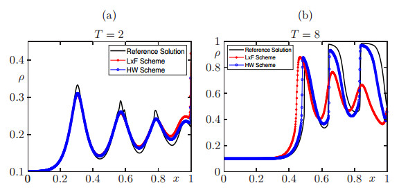
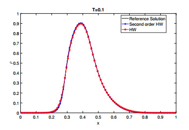
The simulation model proposed in [M. Hilliges and W. Weidlich. A phenomenological model for dynamic traffic flow in networks. Transportation Research Part B: Methodological, 29 (6): 407–431, 1995] can be understood as a simple method for approximating solutions of scalar conservation laws whose flux is of density times velocity type, where the density and velocity factors are evaluated on neighboring cells. The resulting scheme is monotone and converges to the unique entropy solution of the underlying problem. The same idea is applied to devise a numerical scheme for a class of one-dimensional scalar conservation laws with nonlocal flux and initial and boundary conditions. Uniqueness of entropy solutions to the nonlocal model follows from the Lipschitz continuous dependence of a solution on initial and boundary data. By various uniform estimates, namely a maximum principle and bounded variation estimates, along with a discrete entropy inequality, the sequence of approximate solutions is shown to converge to an entropy weak solution of the nonlocal problem. The improved accuracy of the proposed scheme in comparison to schemes based on the Lax-Friedrichs flux is illustrated by numerical examples. A second-order scheme based on MUSCL methods is presented.
Citation: Raimund Bürger, Harold Deivi Contreras, Luis Miguel Villada. A Hilliges-Weidlich-type scheme for a one-dimensional scalar conservation law with nonlocal flux[J]. Networks and Heterogeneous Media, 2023, 18(2): 664-693. doi: 10.3934/nhm.2023029
The simulation model proposed in [M. Hilliges and W. Weidlich. A phenomenological model for dynamic traffic flow in networks. Transportation Research Part B: Methodological, 29 (6): 407–431, 1995] can be understood as a simple method for approximating solutions of scalar conservation laws whose flux is of density times velocity type, where the density and velocity factors are evaluated on neighboring cells. The resulting scheme is monotone and converges to the unique entropy solution of the underlying problem. The same idea is applied to devise a numerical scheme for a class of one-dimensional scalar conservation laws with nonlocal flux and initial and boundary conditions. Uniqueness of entropy solutions to the nonlocal model follows from the Lipschitz continuous dependence of a solution on initial and boundary data. By various uniform estimates, namely a maximum principle and bounded variation estimates, along with a discrete entropy inequality, the sequence of approximate solutions is shown to converge to an entropy weak solution of the nonlocal problem. The improved accuracy of the proposed scheme in comparison to schemes based on the Lax-Friedrichs flux is illustrated by numerical examples. A second-order scheme based on MUSCL methods is presented.

| [1] |
D. Amadori, W. Shen, An integro-differential conservation law arising in a model of granular flow, J. Hyperbolic Differ. Equ., 9 (2012), 105–131. https://doi.org/10.1142/S0219891612500038 doi: 10.1142/S0219891612500038

|
| [2] |
P. Amorim, R. Colombo, A. Teixeira, On the numerical integration of scalar nonlocal conservation laws, ESAIM M2AN, 49 (2015), 19–37. http://dx.doi.org/10.1051/m2an/2014023 doi: 10.1051/m2an/2014023
|
| [3] |
D. Armbruster, P. Degond, C. Ringhofer, A model for the dynamics of large queuing networks and supply chains, SIAM J. Appl. Math., 66 (2006), 896–920. https://doi.org/10.1137/040604625 doi: 10.1137/040604625
|
| [4] |
C. Bardos, A. Y. le Roux, J. C. Nédélec, First order quasilinear equations with boundary conditions, Commun. Partial. Differ. Equ., 4 (1979), 1017–1034. https://doi.org/10.1080/03605307908820117 doi: 10.1080/03605307908820117
|
| [5] |
F. Betancourt, R. Bürger, K. H. Karlsen, E. M. Tory, On nonlocal conservation laws modelling sedimentation, Nonlinearity, 24 (2011), 855–885. https://doi.org/10.1088/0951-7715/24/3/008 doi: 10.1088/0951-7715/24/3/008
|
| [6] |
R. Bürger, A. García, K. Karlsen, J. Towers, A family of numerical schemes for kinematic flows with discontinuous flux, J Eng Math, 60 (2008), 387–425. https://doi.org/10.1007/s10665-007-9148-4 doi: 10.1007/s10665-007-9148-4
|
| [7] |
C. Chalons, P. Goatin, L. M. Villada, High-order numerical schemes for one-dimensional nonlocal conservation laws, SIAM J. Sci. Comput., 40 (2018), A288–A305. https://doi.org/10.1137/16M110825X doi: 10.1137/16M110825X
|
| [8] |
R. M. Colombo, M. Garavello, M. Lécureux-Mercier, A class of nonlocal models for pedestrian traffic, Math Models Methods Appl Sci, 22 (2012), 1150023. https://doi.org/10.1142/S0218202511500230 doi: 10.1142/S0218202511500230
|
| [9] |
R. M. Colombo, M. Herty, M. Mercier, Control of the continuity equation with a non local flow, ESAIM Control Optim. Calc. Var., 17 (2011), 353–379. https://doi.org/10.1051/cocv/2010007 doi: 10.1051/cocv/2010007
|
| [10] |
R. M. Colombo, M. Lécureux-Mercier, Nonlocal crowd dynamics models for several populations, Acta Math. Sci. Ser. B Engl. Ed., 32 (2012), 177–196. https://doi.org/10.1016/S0252-9602(12)60011-3 doi: 10.1016/S0252-9602(12)60011-3
|
| [11] |
R. M. Colombo, E. Rossi, Rigorous estimates on balance laws in bounded domains, Acta Math. Sci. Ser. B Engl. Ed., 35 (2015), 906–944. https://doi.org/10.1016/S0252-9602(15)30028-X doi: 10.1016/S0252-9602(15)30028-X
|
| [12] |
R. M. Colombo, E. Rossi, Nonlocal conservation laws in bounded domains, Math Models Methods Appl Sci, 50 (2018), 4041–4065. https://doi.org/10.1137/18M1171783 doi: 10.1137/18M1171783
|
| [13] |
M. G. Crandall, A. Majda, Monotone difference approximations for scalar conservation laws, Math. Comp., 34 (1980), 1–21. https://doi.org/10.1090/S0025-5718-1980-0551288-3 doi: 10.1090/S0025-5718-1980-0551288-3
|
| [14] |
C. De Filippis, P. Goatin, The initial–boundary value problem for general non-local scalar conservation laws in one space dimension, Nonlinear Analysis, 161 (2017), 131–156. https://doi.org/10.1016/j.na.2017.05.017 doi: 10.1016/j.na.2017.05.017
|
| [15] |
F. A. Chiarello, P. Goatin, Global entropy weak solutions for general non-local traffic flow models with anisotropic kernel, ESAIM: M2AN, 52 (2018), 163–180. https://doi.org/10.1051/m2an/2017066 doi: 10.1051/m2an/2017066
|
| [16] |
J. Friedrich, O. Kolb, S. Göttlich, A Godunov type scheme for a class of LWR traffic flow models with non-local flux, Netw. Heterog. Media, 13 (2018), 531–547. https://doi.org/10.3934/nhm.2018024 doi: 10.3934/nhm.2018024
|
| [17] |
P. Goatin, E. Rossi, Well-posedness of IBVP for 1D scalar non-local conservation laws, Z. Angew. Math. Mech., 99 (2019), e201800318. https://doi.org/10.1002/zamm.201800318 doi: 10.1002/zamm.201800318
|
| [18] |
P. Goatin, S. Scialanga, Well-posedness and finite volume approximations of the LWR traffic flow model with non-local velocity, Netw. Heterog. Media, 11 (2016), 107–121. https://doi.org/10.3934/nhm.2016.11.107 doi: 10.3934/nhm.2016.11.107
|
| [19] |
M. Hilliges, W. Weidlich, A phenomenological model for dynamic traffic flow in networks, TRANSPORT RES B-METH, 29 (1995), 407–431. https://doi.org/10.1016/0191-2615(95)00018-9 doi: 10.1016/0191-2615(95)00018-9
|
| [20] |
E. Rossi, Definitions of solutions to the IBVP for multi-dimensional scalar balance laws, J. Hyperbolic Differ. Equ., 15 (2018), 349–374. https://doi.org/10.1142/S0219891618500133 doi: 10.1142/S0219891618500133
|
| [21] |
E. Rossi, Well-posedness of general 1d initial boundary value problems for scalar balance laws, Discrete Contin Dyn Syst Ser A, 39 (2019), 3577–3608. https://doi.org/10.3934/dcds.2019147 doi: 10.3934/dcds.2019147
|
Figures(5) / Tables(2)
Raimund Bürger, Harold Deivi Contreras, Luis Miguel Villada. A Hilliges-Weidlich-type scheme for a one-dimensional scalar conservation law with nonlocal flux[J]. Networks and Heterogeneous Media, 2023, 18(2): 664-693. doi: 10.3934/nhm.2023029


 DownLoad: