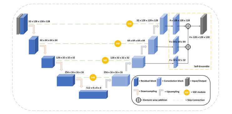
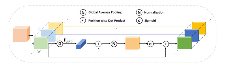
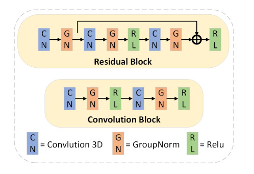
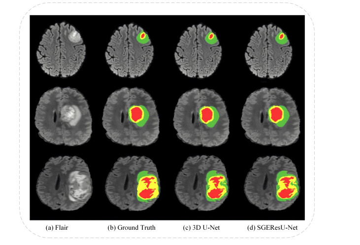
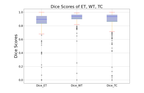
The precise segmentation of tumor regions plays a pivotal role in the diagnosis and treatment of brain tumors. However, due to the variable location, size, and shape of brain tumors, the automatic segmentation of brain tumors is a relatively challenging application. Recently, U-Net related methods, which largely improve the segmentation accuracy of brain tumors, have become the mainstream of this task. Following merits of the 3D U-Net architecture, this work constructs a novel 3D U-Net model called SGEResU-Net to segment brain tumors. SGEResU-Net simultaneously embeds residual blocks and spatial group-wise enhance (SGE) attention blocks into a single 3D U-Net architecture, in which SGE attention blocks are employed to enhance the feature learning of semantic regions and reduce possible noise and interference with almost no extra parameters. Besides, the self-ensemble module is also utilized to improve the segmentation accuracy of brain tumors. Evaluation experiments on the Brain Tumor Segmentation (BraTS) Challenge 2020 and 2021 benchmarks demonstrate the effectiveness of the proposed SGEResU-Net for this medical application. Moreover, it achieves DSC values of 83.31, 91.64 and 86.85%, as well as Hausdorff distances (95%) of 19.278, 5.945 and 7.567 for the enhancing tumor, whole tumor, and tumor core on BraTS 2021 dataset, respectively.
Citation: Dongwei Liu, Ning Sheng, Tao He, Wei Wang, Jianxia Zhang, Jianxin Zhang. SGEResU-Net for brain tumor segmentation[J]. Mathematical Biosciences and Engineering, 2022, 19(6): 5576-5590. doi: 10.3934/mbe.2022261
The precise segmentation of tumor regions plays a pivotal role in the diagnosis and treatment of brain tumors. However, due to the variable location, size, and shape of brain tumors, the automatic segmentation of brain tumors is a relatively challenging application. Recently, U-Net related methods, which largely improve the segmentation accuracy of brain tumors, have become the mainstream of this task. Following merits of the 3D U-Net architecture, this work constructs a novel 3D U-Net model called SGEResU-Net to segment brain tumors. SGEResU-Net simultaneously embeds residual blocks and spatial group-wise enhance (SGE) attention blocks into a single 3D U-Net architecture, in which SGE attention blocks are employed to enhance the feature learning of semantic regions and reduce possible noise and interference with almost no extra parameters. Besides, the self-ensemble module is also utilized to improve the segmentation accuracy of brain tumors. Evaluation experiments on the Brain Tumor Segmentation (BraTS) Challenge 2020 and 2021 benchmarks demonstrate the effectiveness of the proposed SGEResU-Net for this medical application. Moreover, it achieves DSC values of 83.31, 91.64 and 86.85%, as well as Hausdorff distances (95%) of 19.278, 5.945 and 7.567 for the enhancing tumor, whole tumor, and tumor core on BraTS 2021 dataset, respectively.

| [1] | J. Liu, M. Li, J. Wang, F. Wu, Y. Pan, A survey of MRI-based brain tumor segmentation methods, Tsinghua Sci. Technol., 19 (2014), 578–595. https://doi.org/1007-0214-19-6-578 |
| [2] |
S. Bauer, R. Wiest, L. P. Nolte, M. Reyes, A survey of MRI-based medical image analysis for brain tumor studies, Phys. Med. Biol., 58 (2013), R97. https://doi.org/10.1088/0031-9155/58/13/R97 doi: 10.1088/0031-9155/58/13/R97

|
| [3] | O. Ronneberger, P. Fischer, T. Brox, U-Net: convolutional networks for biomedical image segmentation, in Medical Image Computing and Computer-Assisted Intervention - MICCAI 2015-18th International Conference Munich, Lecture Notes in Computer Science, Springer, (2015), 234–241. https://doi.org/10.1007/978-3-319-24574-4_28 |
| [4] | A. Kermi, I. Mahmoudi, M. T. Khadir, Deep convolutional neural networks using U-Net for automatic brain tumor segmentation in multimodal MRI volumes, in Brainlesion: Glioma, Multiple Sclerosis, Stroke and Traumatic Brain Injuries - 4th International Workshop, Springer, (2019), 37–48. https://doi.org/10.1007/978-3-030-11726-9_4 |
| [5] |
J. X. Zhang, Z. K. Jiang, J. Dong, Y. Q. Hou, B, Liu, Attention gate ResU-Net for automatic MRI brain tumor segmentation, IEEE Access, 8 (2020), 58533–58545. https://doi.org/10.1109/ACCESS.2020.2983075 doi: 10.1109/ACCESS.2020.2983075
|
| [6] | A. Albiol, A. Albiol, F. Albiol, Extending 2D deep learning architectures to 3D image segmentation problems, in Brainlesion: Glioma, Multiple Sclerosis, Stroke and Traumatic Brain Injuries - 4th International Workshop, Springer, (2019), 73–82. https://doi.org/10.1007/978-3-030-11726-9_7 |
| [7] | H. Jia, W. Cai, H. Huang, Y. Xia, H2NF-Net for brain tumor segmentation using multimodal MR imaging: 2nd place solution to BraTS Challenge 2020 Segmentation Task, in Brainlesion: Glioma, Multiple Sclerosis, Stroke and Traumatic Brain Injuries - 6th International Workshop, Springer, (2021), 58–68. https://doi.org/10.1007/978-3-030-72087-2_6 |
| [8] | X. Zhang, W. Jian, K. Cheng, 3D dense U-nets for brain tumor segmentation, in Pre-Conference Proceedings of the 7th MICCAI BraTS Challenge, (2018), 562–570. |
| [9] |
P. Liu, Q. Dou, Q. Wang, P. A. Heng, An encoder-decoder neural network with 3D squeeze-and-excitation and deep supervision for brain tumor segmentation, IEEE Access, 8 (2020), 34029–34037. https://doi.org/10.1109/ACCESS.2020.2973707 doi: 10.1109/ACCESS.2020.2973707
|
| [10] | A. Myronenko, 3D MRI brain tumor segmentation using autoencoder regularization, in Brainlesion: Glioma, Multiple Sclerosis, Stroke and Traumatic Brain Injuries - 4th International Workshop, Springer, (2019), 311–320. https://doi.org/10.1007/978-3-030-11726-9_28 |
| [11] | Y. Zhao, Y. Zhang, C. Liu, Bag of tricks for 3D MRI brain tumor segmentation, in Brainlesion: Glioma, Multiple Sclerosis, Stroke and Traumatic Brain Injuries - 5th International Workshop, Springer, (2020), 210–220. https://doi.org/10.1007/978-3-030-46640-4_20 |
| [12] | Z. Jiang, C. Ding, M. Liu, Two-Stage cascaded U-Net: 1st place solution to BraTS challenge 2019 segmentation task, in Brainlesion: Glioma, Multiple Sclerosis, Stroke and Traumatic Brain Injuries - 5th International Workshop, Springer, (2019), 231–241. https://doi.org/10.1007/978-3-030-46640-4_22 |
| [13] | F. Isensee, P. F. Jager, P. M. Full, P. Vollmuth, K. H. Maier-Hein, nnU-Net for brain tumor segmentation. in Brainlesion: Glioma, Multiple Sclerosis, Stroke and Traumatic Brain Injuries - 6th International Workshop, Springer, (2021), 118–132. https://doi.org/10.1007/978-3-030-72087-2_11 |
| [14] | F. Isensee, P. Kickingereder, W. Wick, M. Bendszus, K. H. Maier-Hein, No new-net, in Brainlesion: Glioma, Multiple Sclerosis, Stroke and Traumatic Brain Injuries - 4th International Workshop, Springer, (2019), 234–244. https://doi.org/10.1007/978-3-030-11726-9_21 |
| [15] | X. Li, X. L. Hu, J. Yang Li, Spatial group-wise enhance: Improving semantic feature learning in convolutional networks, preprint, arXiv: 1905.09646. |
| [16] | K. He, X. Zhang, S. Ren, J. Sun, Deep residual learning for image recognition, in 2016 IEEE Conference on Computer Vision and Pattern Recognition, CVPR 2016, IEEE Computer Society, (2016), 770–778. https://doi.org/10.1109/CVPR.2016.90 |
| [17] | U. Baid, S. Ghodasara, S. Mohan, M. Bilello, E. Calabrese, E. Colak, et al., The RSNA-ASNR-MICCAI BraTS 2021 Benchmark on brain tumor segmentation and radiogenomic classification, preprint, arXiv: 2107.02314. |
| [18] |
B. H. Menze, A. Jakab, S. Bauer, J. Kalpathy-Cramer, K. Farahani, J. Kirby, et al., The multimodal brain tumor image segmentation benchmark (BraTS), IEEE Trans. Med. Imaging, 34 (2015), 1993–2024. https://doi.org/10.1109/TMI.2014.2377694 doi: 10.1109/TMI.2014.2377694
|
| [19] |
S. Bakas, H. Akbari, A. Sotiras, M. Bilello, M. Rozycki, J. S. Kirby, et al, Advancing the cancer genome atlas glioma MRI collections with expert segmentation labels and radiomic features, Sci. Data, 4 (2017), 170117. https://doi.org/10.1038/sdata.2017.117 doi: 10.1038/sdata.2017.117
|
| [20] | S. Bakas, H. Akbari, A. Sotiras, M. Bilello, M. Rozycki, J. S. Kirby, et al, Segmentation labels and radiomic features for the preoperative scans of the TCGAGBM collection, Cancer Imaging Arch., 2017. https://doi.org/10.7937/K9/TCIA.2017.KLXWJJ1Q |
| [21] | S. Bakas, H. Akbari, A. Sotiras, M. Bilello, M. Rozycki, J. S. Kirby, et al, Segmentation labels and radiomic features for the preoperative scans of the TCGALGG collection, Cancer Imaging Arch., 2017. https://doi.org/10.7937/K9/TCIA.2017.GJQ7R0EF |
| [22] | S. Bakas, M. Reyes, A. Jakab, S. Bauer, M. Rempfler, A. Crimi, et al., Identifying the best machine learning algorithms for brain tumor segmentation, progression assessment, and overall survival prediction in the BRATS challenge, preprint, arXiv: 1811.02629. |
| [23] | J. Tang, T. Li, H. Shu, H. Zhu, Variational-Autoencoder regularized 3D MultiResUNet for the BraTS 2020 brain tumor segmentation, in Brainlesion: Glioma, Multiple Sclerosis, Stroke and Traumatic Brain Injuries - 6th International Workshop, Springer, (2021), 431–440. https://doi.org/10.1007/978-3-030-72087-2_38 |
| [24] | K. Cheng, C. Hu, P. Yin, et al. Glioma sub-region segmentation on Multi-parameter MRI with label dropout, in Brainlesion: Glioma, Multiple Sclerosis, Stroke and Traumatic Brain Injuries - 6th International Workshop, Springer, (2021), 420–430. https://doi.org/10.1007/978-3-030-72087-2_37 |
| [25] | W. B. Zhang, G. Yang, H. Huang, W. J. Yang, X. M. Xu, Y. K. Liu, et al., ME-Net: Multi-encoder net framework for brain tumor segmentation. Int. J. Imag. Syst. Tech., 31 (2021), 1834–1848. https://doi.org/10.1002/ima.22571 |
| [26] | W. Wang, C. Chen, M. Ding, H. Yu, S. Zha, J. Li, TransBTS: Multimodal brain tumor segmentation using transformer, in Medical Image Computing and Computer Assisted Intervention – MICCAI 2021, 24th International Conference, Springer, (2021), 109–119. https://doi.org/10.1007/978-3-030-87193-2_11 |
| [27] | V. Sundaresan, L. Griffanti, M. Jenkinson, Brain tumour segmentation using a triplanar ensemble of U-Nets on MR images, in Brainlesion: Glioma, Multiple Sclerosis, Stroke and Traumatic Brain Injuries - 6th International Workshop, Springer, (2021), 340–353. https://doi.org/10.1007/978-3-030-72084-1_31 |
| [28] |
Y. Fang, H. Huang, W. J. Yang, X. M. Xu, W. W. Jiang, X. B. Lai, Nonlocal convolutional block attention module VNet for gliomas automatic segmentation, Int. J. Imag. Syst. Tech., 32 (2022), 528–543. https://doi.org/10.1002/ima.22639 doi: 10.1002/ima.22639
|
| [29] |
H. Huang, G. Yang, W. B. Zhang, X. M. Xu, W. J. Yang, W. W. Jiang, et al., A deep multi-task learning framework for brain tumor segmentation, Front Oncol., 11 (2021), 690244. https://doi.org/10.3389/fonc.2021.690244 doi: 10.3389/fonc.2021.690244
|
| [30] | J. X. Zhang, Z. K. Jiang, D. W. Liu, Q. L. Sun, Y. Q. Hou, B. Liu, 3D asymmetric expectation-maximization attention network for brain tumor segmentation. NMR Biomd., (2021), e4657. https://doi.org/10.1002/nbm.4657 |
Figures(6) / Tables(5)

Dongwei Liu, Ning Sheng, Tao He, Wei Wang, Jianxia Zhang, Jianxin Zhang. SGEResU-Net for brain tumor segmentation[J]. Mathematical Biosciences and Engineering, 2022, 19(6): 5576-5590. doi: 10.3934/mbe.2022261


 DownLoad: