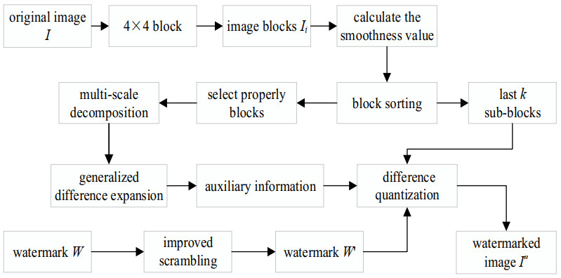
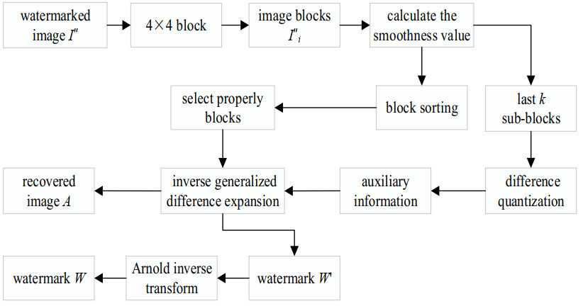
Aiming at solving the problems of bad imperceptibility and low embedding rate of existing algorithms, a novel large-capacity reversible image watermarking based on improved difference expansion (DE) is proposed. Firstly, the smoothness calculation algorithm is used to calculate and sort the smoothness values of the divided image sub-blocks; then, the scrambled watermark is embedded into the sub-blocks with less smoothness after removing the abrupt point by using the generalized difference expansion (GDE); finally, the absolute difference operation is applied to the generated overflow pixels to make their pixel values within a reasonable range for embedding watermark information. Under the premise of ensuring a certain visual quality, multiple watermark embedding can effectively improve the embedding rate. The simulation results show that this algorithm not only realizes blind extraction, but also recovers the original images without loss. At the same time, this algorithm achieves a high embedding rate (the average embedding rate is as high as 77.91 dB) without decreasing the visual quality.
Citation: Shaozhang Xiao, Xingyuan Zuo, Zhengwei Zhang, Fenfen Li. Large-capacity reversible image watermarking based on improved DE[J]. Mathematical Biosciences and Engineering, 2022, 19(2): 1108-1127. doi: 10.3934/mbe.2022051
Aiming at solving the problems of bad imperceptibility and low embedding rate of existing algorithms, a novel large-capacity reversible image watermarking based on improved difference expansion (DE) is proposed. Firstly, the smoothness calculation algorithm is used to calculate and sort the smoothness values of the divided image sub-blocks; then, the scrambled watermark is embedded into the sub-blocks with less smoothness after removing the abrupt point by using the generalized difference expansion (GDE); finally, the absolute difference operation is applied to the generated overflow pixels to make their pixel values within a reasonable range for embedding watermark information. Under the premise of ensuring a certain visual quality, multiple watermark embedding can effectively improve the embedding rate. The simulation results show that this algorithm not only realizes blind extraction, but also recovers the original images without loss. At the same time, this algorithm achieves a high embedding rate (the average embedding rate is as high as 77.91 dB) without decreasing the visual quality.

| [1] |
Z. Zhang, L. Wu, S. Xiao, S. Gao, Adaptive reversible image watermarking algorithm based on IWT and level set, EURASIP J. Adv. Signal Process., 15 (2017), 1–16. doi: 10.1186/s13634-017-0450-7. doi: 10.1186/s13634-017-0450-7

|
| [2] |
J. Tian, Reversible data embedding using a difference expansion, IEEE Trans. Circuits Syst. Video Tech., 8 (2003), 890–896. doi: 10.1109/TCSVT.2003.815962. doi: 10.1109/TCSVT.2003.815962
|
| [3] |
X. L. Li, J. Li, B. Li, High-fidelity reversible data hiding scheme based on pixel-value ordering and prediction-error expansion, Signal Process., 1 (2012), 198–205. doi: 10.1016/j.sigpro.2012.07.025. doi: 10.1016/j.sigpro.2012.07.025
|
| [4] |
J. X. Wang, J. Q. Ni, J. W. Pan, A high capacity reversible data hiding scheme based on generalized prediction-error expansion and adaptive embedding, Signal Process., 5 (2014), 370–380. doi: 10.1016/j.sigpro.2013.12.005. doi: 10.1016/j.sigpro.2013.12.005
|
| [5] |
Z. W. Zhang, L. F. Wu, Y. Y. Yan, S. Z. Xiao, An improved reversible image watermarking algorithm based on difference expansion, Int. J. Distr. Sens. Net., 1 (2017), 1–15. doi: 10.1177/1550147716686577. doi: 10.1177/1550147716686577
|
| [6] |
Z. W. Zhang, M. J. Zhang, L. Y. Wang, Reversible image watermarking algorithm based on quadratic difference expansion, Math. Prob. Eng., 1 (2020), 1–8. doi: 10.1155/2020/1806024. doi: 10.1155/2020/1806024
|
| [7] |
H. K. Maity, S. P. Maity, Reversible image watermarking using modified difference expansion, Emerging Applications of Information Technology (EAIT), in 2012 Third International Conference on IEEE, 3 (2012), 320–323. doi: 10.1109/EAIT.2012.6407936. doi: 10.1109/EAIT.2012.6407936
|
| [8] | S. L. Lin, C. F. Huang. M. H. Liou, C. Y. Chen, Improving histogram based reversible information hiding by an optimal weight-based prediction scheme, J. Inf. Hid. Multimed. Signal Process., 1 (2013), 19–33. |
| [9] |
A. Muhammad, S. Q. Aqsa, K. Asifullah, Protection of medical images and patient related information in healthcare: Using an intelligent and reversible watermarking technique, Appl. Soft Comput., 51 (2017), 168–179. doi: 10.1016/j.asoc.2016.11.044. doi: 10.1016/j.asoc.2016.11.044
|
| [10] |
K. C. Vinoth, V. Natarajan, Hybrid local prediction error based difference expansion reversible watermarking for medical images, Comput. Electr. Eng., 53 (2016), 333–345. doi: 10.1016/j.compeleceng.2015.11.033. doi: 10.1016/j.compeleceng.2015.11.033
|
| [11] |
Y. J. Jia, Z. X. Yin, X. P. Zhang, Reversible data hiding based on reducing invalid shifting of pixels in histogram shifting, Signal Process., 163 (2019), 238–246. doi: 10.1016/j.sigpro.2019.05.020. doi: 10.1016/j.sigpro.2019.05.020
|
| [12] |
M. Ntahobari, T. Ahmad, Protecting data by improving quality of stego image based on enhanced reduced diference expansion, Int. J. Electr. Comput. Eng., 4 (2018), 2468–2476. doi: 10.11591/ijece.v8i4. doi: 10.11591/ijece.v8i4
|
| [13] |
X. Yu, X. Wang, Q. Q. Pei, Reversible watermarking based on multi-dimensional Prediction-error expansion, Multimed. Tools Appl., 14 (2018), 18085–18104. doi: 10.1007/s11042-018-5794-y. doi: 10.1007/s11042-018-5794-y
|
| [14] | W. G. Su, Y. L. Shen, X. Wang, Two-layer reversible watermarking algorithm using difference expansion, J. Comput. Res. Develop., 7 (2019), l498–1505. |
| [15] | Y. Q. Li, J. X. Li, Y. H. Liang, Image watermarking algorithm based on image characteristics and huffman coding, Comput. Appl. Soft., 30 (2013), 128–130. |
| [16] | N. K. Anil, M. Haribabu, B. C. Hima, Novel image watermarking algorithm with DWT-SVD, Int. J. Comput. Appl., 1 (2014), 12–17. |
| [17] |
S. E. Hala, S. F. Elzoghdy, S. F. Osama, Adaptive difference expansion-based reversible data hiding scheme for digital images, Arabian J. Sci. Eng., 41 (2016), 1091–1107. doi: 10.1007/s13369-015-1956-7. doi: 10.1007/s13369-015-1956-7
|
| [18] |
Z. W. Zhang, L. F. Wu, Y. Y. Yan, An improved reversible image watermarking algorithm based on difference expansion, Int. J. Distr. Sens. Net., 1 (2017), 1–15. doi: 10.1177/1550147716686577. doi: 10.1177/1550147716686577
|
| [19] |
M. Arsalan, S. A. Malik, A. Khan, Intelligent reversible watermarking in integer wavelet domain for medical images, J. Syst. Soft., 4 (2014), 883–894. doi: 10.1016/j.jss.2011.11.005. doi: 10.1016/j.jss.2011.11.005
|
| [20] |
O. M. Al-Osamah, E. K. Bee, Two-dimensional difference expansion (2D-DE) scheme with a characteristics-based threshold, Signal Process., 1 (2015), 154–162. doi: 10.1016/j.sigpro.2013.12.005. doi: 10.1016/j.sigpro.2013.12.005
|
| [21] |
B. Lei, E. L. Tan, S. Chen, N. Dong, T. Wang, H. Lei, Reversible watermarking scheme for medical image based on differential evolution, Expert Syst. Appl., 7 (2016), 3178–3188. doi: 10.1016/j.eswa.2013.11.019 doi: 10.1016/j.eswa.2013.11.019
|
| [22] |
K. Swaraja, Medical image region based watermarking for secured telemedicine, Multimed. Tools Appl., 21 (2018), 28249–28280. doi: 10.1007/s11042-018-6020-7. doi: 10.1007/s11042-018-6020-7
|
| [23] |
H. A. Zheng, C. A. Wang, J. B. Wang, S. C. Xiang, A new reversible watermarking scheme using the content-adaptive block size for prediction, Signal Process., 164 (2019), 74–83. doi: 10.1016/j.sigpro.2019.05.035. doi: 10.1016/j.sigpro.2019.05.035
|


Figures(8) / Tables(7)

Shaozhang Xiao, Xingyuan Zuo, Zhengwei Zhang, Fenfen Li. Large-capacity reversible image watermarking based on improved DE[J]. Mathematical Biosciences and Engineering, 2022, 19(2): 1108-1127. doi: 10.3934/mbe.2022051


 DownLoad: