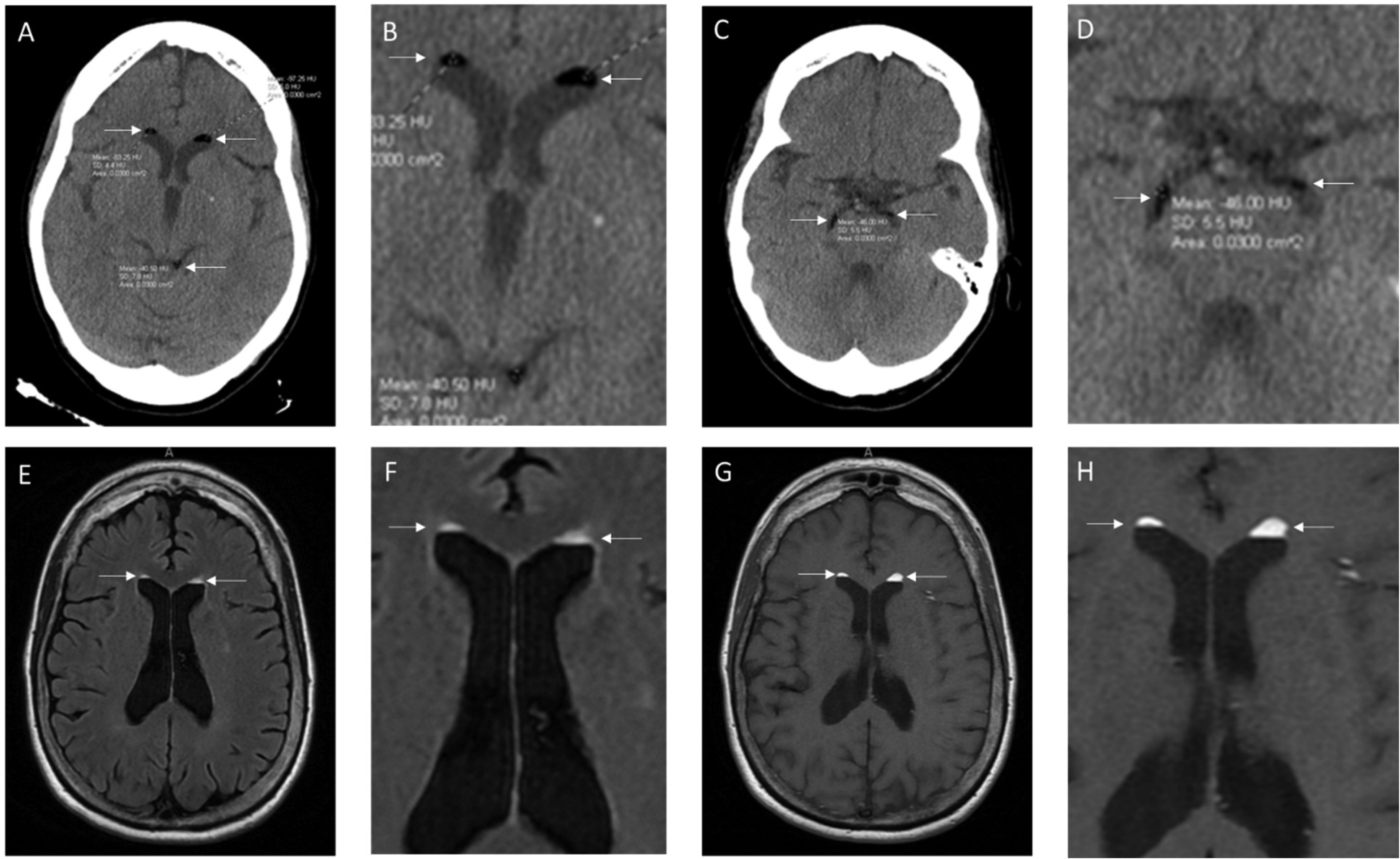
It is rare to find free floating fat droplets in the cerebral spinal fluid (CSF) spaces of the brain. When fat droplets are seen in the CSF spaces, the most common cause is the rupture of a dermoid cyst. Dermoid cysts are congenital inclusion cysts that form during the neural tube closure between the third and fifth weeks of embryogenesis. In this case report, we describe a case of a 74-year-old, right-handed female who presented with an acute onset of visual disturbances and left-hand numbness. Computed tomography (CT) and magnetic resonance imaging (MRI) of the head revealed hypodense “lesions” in the lateral ventricles and basal cisterns. The CT Hounsfield unit was between −41 to −83 Hounsfield Units, which is compatible with fat rather than air. The T1 weighted and FLAIR MRI showed hyperintense lesions “floating” on top of the CSF in the lateral ventricles, which is typical for fat droplets, presumably caused by a ruptured dermoid cyst. This case emphasizes the importance of analyzing Hounsfield Units to distinguish lesions by density, where fat ranges from −50 to −150 Hounsfield Units and air is −1000 Hounsfield Units. Pneumocephalus is the presence of air in the epidural, subdural, or subarachnoid space and can cause confusion, nausea, seizures and focal neurological symptoms. A careful analysis of the neuroimaging findings in the CT with or without MRI is important in making a correct diagnosis of a ruptured dermoid cyst versus pneumocephalus.
Citation: Mark Reed, Christopher Miller, Cortney Connor, Jason S. Chang, Forshing Lui. Fat droplets in the cerebrospinal fluid (CSF) spaces of the brain[J]. AIMS Neuroscience, 2024, 11(4): 484-489. doi: 10.3934/Neuroscience.2024029
It is rare to find free floating fat droplets in the cerebral spinal fluid (CSF) spaces of the brain. When fat droplets are seen in the CSF spaces, the most common cause is the rupture of a dermoid cyst. Dermoid cysts are congenital inclusion cysts that form during the neural tube closure between the third and fifth weeks of embryogenesis. In this case report, we describe a case of a 74-year-old, right-handed female who presented with an acute onset of visual disturbances and left-hand numbness. Computed tomography (CT) and magnetic resonance imaging (MRI) of the head revealed hypodense “lesions” in the lateral ventricles and basal cisterns. The CT Hounsfield unit was between −41 to −83 Hounsfield Units, which is compatible with fat rather than air. The T1 weighted and FLAIR MRI showed hyperintense lesions “floating” on top of the CSF in the lateral ventricles, which is typical for fat droplets, presumably caused by a ruptured dermoid cyst. This case emphasizes the importance of analyzing Hounsfield Units to distinguish lesions by density, where fat ranges from −50 to −150 Hounsfield Units and air is −1000 Hounsfield Units. Pneumocephalus is the presence of air in the epidural, subdural, or subarachnoid space and can cause confusion, nausea, seizures and focal neurological symptoms. A careful analysis of the neuroimaging findings in the CT with or without MRI is important in making a correct diagnosis of a ruptured dermoid cyst versus pneumocephalus.
Hounsfield Units
Computed Tomography
Magnetic Resonance Image
Fluid Attenuated Inversion Recovery
Cerebrospinal Fluid

| [1] |
Jacków J, Tse G, Martin A, et al. (2018) Ruptured intracranial dermoid cysts: a pictorial review. Pol J Radiol 83: e465-e470. https://doi.org/10.5114/pjr.2018.80206 
|
| [2] |
Ray M, Barnett D, Snipes G, et al. (2012) Ruptured intracranial dermoid cyst. Baylor University Medical Center Proceedings 25: 23-25. https://doi.org/10.1080/08998280.2012.11928775
|
| [3] |
Delgado-Muñoz P, Oliver-Ricart M, Labat-Alvarez E (2022) A Rare Case of an Intracranial Dermoid Cyst with Atypical Appearance on Computed Tomography and Magnetic Resonance Imaging. Am J Case Rep 23: e935115. https://doi.org/10.12659/AJCR.935115
|
| [4] |
Zhang MH, Feng Q, Zhu HL, et al. (2021) Asymptomatic traumatic rupture of an intracranial dermoid cyst: A case report. World J Clin Cases 9: 4046-4051. https://doi.org/10.12998/wjcc.v9.i16.4046
|
| [5] |
Zimny A, Zinskia L, Bladowska J, et al. (2013) Intracranial lesions with high signal intensity on T1-weighted MR images - review of pathologies. Pol J Radiol 78: 36-46. https://doi.org/10.12659/PJR.889663
|
| [6] |
Gorissen Z, Hakvoort K, Boogaart M, et al. (2019) Pneumocephalus: a rare and life-threatening, but reversible, complication after penetrating lumbar injury. Acta Neurochir 161: 361-365. https://doi.org/10.1007/s00701-018-03796-y
|
| [7] | DenOtter T, Schubert J (2023) Hounsfield Unit. StatPearls . Treasure Island: StatPearls Publishing. Available from: https://www.ncbi.nlm.nih.gov/books/NBK547721/ |
| [8] |
Sood S, Gupta R (2014) Susceptibility Artifacts in Ruptured Intracranial Dermoid Cysts: A Poorly Understood but Important Phenomenon. Neuroradiol J 27: 677-684. https://doi.org/10.15274/NRJ-2014-10090
|
| [9] |
Picardo M, Ottaviani M, Camera E, et al. (2009) Sebaceous gland lipids. Dermato-Endocrinology 1: 68-71. https://doi.org/10.4161/derm.1.2.8472
|
| [10] |
Osborn A, Preece M (2006) Intracranial Cysts: Radiologic-Pathologic Correlation and Imaging Approach. Radiology 239: 650-664. https://doi.org/10.1148/radiol.2393050823
|
| [11] |
Mikami T, Maeda C, Aoki F, et al. (2021) A dermoid cyst misdiagnosed as a lipoma due to atypical magnetic resonance images: a case report. J Med Case Rep 15: 99. https://doi.org/10.1186/s13256-020-02584-6
|
| [12] |
El-Bahy K, Kotb A, Galal A, et al. (2006) Ruptured intracranial dermoid cysts. Acta Neurochir 148: 457-462. https://doi.org/10.1007/s00701-005-0722-0
|
| [13] | Das J, Bajaj J (2024) Pneumocephalus. StatPearls . Treasure Island: StatPearls Publishing. Available from: https://www.ncbi.nlm.nih.gov/books/NBK535412/ |
| [14] |
Ray M, Barnett D, Snipes G, et al. (2012) Ruptured intracranial dermoid cyst. Baylor University Medical Center Proceedings 1: 23-25. https://doi.org/10.1080/08998280.2012.11928775
|
| [15] |
Blitz SE, Bernstock JD, Dmytriw AA, et al. (2021) Ruptured suprasellar dermoid cyst treated with lumbar drain to prevent postoperative hydrocephalus: Case report and Focused Review of Literature. Front Surg 8. https://doi.org/10.3389/fsurg.2021.714771
|
Figures(1)

Mark Reed, Christopher Miller, Cortney Connor, Jason S. Chang, Forshing Lui. Fat droplets in the cerebrospinal fluid (CSF) spaces of the brain[J]. AIMS Neuroscience, 2024, 11(4): 484-489. doi: 10.3934/Neuroscience.2024029


 DownLoad: