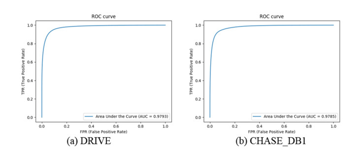
The segmentation of blood vessels from retinal images is an important and challenging task in medical analysis and diagnosis. This paper proposes a new architecture of the U-Net network for retinal blood vessel segmentation. Adding dense block to U-Net network makes each layer's input come from the all previous layer's output which improves the segmentation accuracy of small blood vessels. The effectiveness of the proposed method has been evaluated on two public datasets (DRIVE and CHASE_DB1). The obtained results (DRIVE: Acc = 0.9559, AUC = 0.9793, CHASE_DB1: Acc = 0.9488, AUC = 0.9785) demonstrate the better performance of the proposed method compared to the state-of-the-art methods. Also, the results show that our method achieves better results for the segmentation of small blood vessels and can be helpful to evaluate related ophthalmic diseases.
Citation: Yinlin Cheng, Mengnan Ma, Liangjun Zhang, ChenJin Jin, Li Ma, Yi Zhou. Retinal blood vessel segmentation based on Densely Connected U-Net[J]. Mathematical Biosciences and Engineering, 2020, 17(4): 3088-3108. doi: 10.3934/mbe.2020175
The segmentation of blood vessels from retinal images is an important and challenging task in medical analysis and diagnosis. This paper proposes a new architecture of the U-Net network for retinal blood vessel segmentation. Adding dense block to U-Net network makes each layer's input come from the all previous layer's output which improves the segmentation accuracy of small blood vessels. The effectiveness of the proposed method has been evaluated on two public datasets (DRIVE and CHASE_DB1). The obtained results (DRIVE: Acc = 0.9559, AUC = 0.9793, CHASE_DB1: Acc = 0.9488, AUC = 0.9785) demonstrate the better performance of the proposed method compared to the state-of-the-art methods. Also, the results show that our method achieves better results for the segmentation of small blood vessels and can be helpful to evaluate related ophthalmic diseases.

| [1] |
S. Moccia, E. De Momi, S. El Hadji, L. S. Mattos, Blood vessel segmentation algorithms—review of methods, datasets and evaluation metrics, Comput. Methods Programs Biomed., 158 (2018), 71-91. doi: 10.1016/j.cmpb.2018.02.001

|
| [2] |
H. K. Li, M. Horton, S. E. Bursell, J. Cavallerano, I. Zimmer-Galler, M. Tennant, Telehealth practice recommendations for diabetic retinopathy, Telemed. E. Health, 17 (2011), 814-837. doi: 10.1089/tmj.2011.0075
|
| [3] |
M. M. Fraz, P. Remagnino, A. Hoppe, B. Uyyanonvara, A. R. Rudnicka, G. C. Owen, et al., Blood vessel segmentation methodologies in retinal images-a survey, Comput. Methods Programs Biomed., 108 (2012), 407-433. doi: 10.1016/j.cmpb.2012.03.009
|
| [4] | F. Xu, X. C. Wang, M. Q. Zhou, Z. K. Wu, X. Y. Liu, Segmentation algorithm of brain vessel image based on SEM statistical mixture model, 2010 Seventh International Conference on Fuzzy Systems and Knowledge Discovery IEEE, 1830-1833. Available from: https://ieeexplore.ieee.org/abstract/document/5569429. |
| [5] |
M. S. Hassouna, A. A. Farag, S. Hushek, T. Moriarity, Cerebrovascular segmentation from TOF using stochastic models, Med. Image Anal., 10 (2006), 2-18. doi: 10.1016/j.media.2004.11.009
|
| [6] |
D. A. Oliveira, R. Q. Feitosa, M. M. Correia, Segmentation of liver, its vessels and lesions from CT images for surgical planning, Biomed. Eng. Online, 10 (2011), 30. doi: 10.1186/1475-925X-10-30
|
| [7] |
E. Goceri, Z. K. Shah, M. N. Gurcan, Vessel segmentation from abdominal magnetic resonance images: Adaptive and reconstructive approach, Int. J. Numer. Method Biomed. Eng., 33 (2017), e2811. doi: 10.1002/cnm.2811
|
| [8] |
T. Mapayi, J. R. Tapamo, S. Viriri, Retinal vessel segmentation: A comparative study of fuzzy C-means and sum entropy information on phase congruency, Int. J. Adv. Rob. Syst., 12 (2015), 133. doi: 10.5772/60581
|
| [9] |
R. Nekovei, Y. Sun, Back-propagation network and its configuration for blood vessel detection in angiograms, IEEE Trans. Neural Networks, 6 (1995), 64-72. doi: 10.1109/72.363449
|
| [10] |
J. V. Soares, J. J. Leandro, R. M. Cesar, H. F. Jelinek, M. J. Cree, Retinal vessel segmentation using the 2-D Gabor wavelet and supervised classification, IEEE Trans. Med. Imaging, 25 (2006), 1214-1222. doi: 10.1109/TMI.2006.879967
|
| [11] |
J. Staal, M. D. Abràmoff, M. Niemeijer, M. A. Viergever, B. Ginneken, Ridge-based vessel segmentation in color images of the retina, IEEE Trans. Med. Imaging, 23 (2004), 501-509. doi: 10.1109/TMI.2004.825627
|
| [12] | S. Hanaoka, Y. Nomura, M. Nemoto, S. Miki, T. Yoshikawa, N. Hayashi, er al., Hotpig: A novel geometrical feature for vessel morphometry and its application to cerebral aneurysm detection, International Conference on Medical Image Computing and Computer-Assisted Intervention, 2015,103-110. Available from: https://link.springer.com/chapter/10.1007/978-3-319-24571-3_13. |
| [13] | J. I. Orlando, E. Prokofyeva, M. B. Blaschko, A discriminatively trained fully connected conditional random field model for blood vessel segmentation in fundus images, IEEE Trans. Biomed. Eng., 64 (2016), 16-27. |
| [14] |
S. Chaudhuri, S. Chatterjee, N. Katz, M. Nelson, M. Goldbaum, Detection of blood vessels in retinal images using two-dimensional matched filters, IEEE Trans. Med. Imaging, 8 (1989), 263-269. doi: 10.1109/42.34715
|
| [15] |
F. Zana, J. C. Klein, Segmentation of vessel-like patterns using mathematical morphology and curvature evaluation, IEEE Trans. Image Process., 10 (2001), 1010-1019. doi: 10.1109/83.931095
|
| [16] |
M. M. Fraz, S. A. Barman, P. Remagnino, A. Hoppe, A. Basit, B. Uyyanonvara, et al., An approach to localize the retinal blood vessels using bit planes and centerline detection, Comput. Methods Programs Biomed., 108 (2012), 600-616. doi: 10.1016/j.cmpb.2011.08.009
|
| [17] |
Z. Jiang, H. Zhang, Y. Wang, S. B. Ko, Retinal blood vessel segmentation using fully convolutional network with transfer learning, Comput. Med. Imaging Graphics, 68 (2018), 1-15. doi: 10.1016/j.compmedimag.2018.04.005
|
| [18] | O. Ronneberger, P. Fischer, T. Brox, U-net: Convolutional networks for biomedical image segmentation; International Conference on Medical Image Computing and Computer-Assisted Intervention, 2015,234-241. Available from: https://link.springer.com/chapter/10.1007/978-3-319-24574-4_28. |
| [19] | O. Oktay, J. Schlemper, L. L. Folgoc, M. Lee, M. Heinrich, K. Misawa, et al., Attention u-net: Learning where to look for the pancreas, arXiv preprint arXiv: 1804.03999 (2018). |
| [20] |
L. Rundo, C. Han, Y. Nagano, J. Zhang, R. Hataya, C. Militello, et al., USE-Net: Incorporating Squeeze-and-Excitation blocks into U-Net for prostate zonal segmentation of multi-institutional MRI datasets, Neurocomputing, 365 (2019), 31-43. doi: 10.1016/j.neucom.2019.07.006
|
| [21] | X. C. Wang, W. Li, B. Y. Miao, H. Jing, Z. W. Jiang, W. Xu, et al., Retina blood vessel segmentation using a U-net based Convolutional neural network, International Conference on Data Science, 2018. Available from: https://researchbank.swinburne.edu.au/file/fce08160-bebd-44ff-b445-6f3d84089ab2/1/2018-xianchneng-retina_blood_vessel.pdf. |
| [22] | S. Kumawat, S. Raman, Local Phase U-net for Fundus Image Segmentation, ICASSP 2019 - 2019 IEEE International Conference on Acoustics, Speech and Signal Processing (ICASSP), 2019. 1209-1213. Available from: https://ieeexplore.ieee.org/abstract/document/8683390. |
| [23] |
L. Xu, S. Luo, A novel method for blood vessel detection from retinal images, Biomed. Eng. Online, 9 (2010), 14. doi: 10.1186/1475-925X-9-14
|
| [24] |
S. L. Wang, Y. L. Yin, G. B. Cao, B. Z. Wei, Y. J. Zheng, G. P. Yang, Hierarchical retinal blood vessel segmentation based on feature and ensemble learning, Neurocomputing, 149 (2015), 708-717. doi: 10.1016/j.neucom.2014.07.059
|
| [25] | T. A. Soomro, A. J. Afifi, J. Gao, O. Hellwich, M. A. U. Khan, M. Paul, et al., Boosting sensitivity of a retinal vessel segmentation algorithm with convolutional neural network, 2017 International Conference on Digital Image Computing: Techniques and Applications (DICTA), 2017. Available from: https://ieeexplore.ieee.org/abstract/document/8227413/. |
| [26] | K. He, X. Zhang, S. Ren, J. Sun, Delving deep into rectifiers: Surpassing human-level performance on imagenet classification, Proceedings of the IEEE international conference on computer vision. 2015, 1026-1034. Available from: https://www.cv-foundation.org/openaccess/content_iccv_2015/html/He_Delving_Deep_into_ICCV_2015_paper.html. |
| [27] | T. A. Qureshi, M. Habib, A. Hunter, B. Al-Diri, A manually-labeled, artery/vein classified benchmark for the DRIVE dataset, Proceedings of the 26th IEEE International Symposium on Computer-Based Medical Systems, 2013. Available from: https://ieeexplore.ieee.org/abstract/document/6627847. |
| [28] |
C. G. Owen, A. R. Rudnicka, R. Mullen, S. A. Barman, D. Monekosso, P. H. Whincup, et al., Measuring retinal vessel tortuosity in 10-year-old children: Validation of the computer-assisted image analysis of the retina (CAIAR) program, Invest. Ophthalmol. Visual Sci., 50 (2009), 2004-2010. doi: 10.1167/iovs.08-3018
|
| [29] | F. Calimeri, A. Marzullo, C. Stamile, G. Terracina, Blood Vessel Segmentation in Retinal Fundus Images Using Hypercube NeuroEvolution of Augmenting Topologies (HyperNEAT); WIRN 2017, Quantifying and Processing Biomedical and Behavioral Signals, 173-183. Available from: https://link.springer.com/chapter/10.1007/978-3-319-95095-2_17. |
| [30] |
M. G. Cinsdikici, D. Aydın, Detection of blood vessels in ophthalmoscope images using MF/ant (matched filter/ant colony) algorithm, Comput. Methods Programs Biomed., 96 (2009), 85-95. doi: 10.1016/j.cmpb.2009.04.005
|
| [31] | K. S. Sreejini, V. K. Govindan, Improved multiscale matched filter for retina vessel segmentation using PSO algorithm, Egypt. Inf. J., 16 (2015), 253-260. |
| [32] |
B. Zhang, L. Zhang, L. Zhang, F. Karraya, Retinal vessel extraction by matched filter with first-order derivative of Gaussian, Comput. Biol. Med., 40 (2010), 438-445. doi: 10.1016/j.compbiomed.2010.02.008
|
| [33] |
A. Hoover, V. Kouznetsova, M. Goldbaum, Locating blood vessels in retinal images by piecewise threshold probing of a matched filter response, IEEE Trans. Med. Imaging, 19 (2000), 203-210. doi: 10.1109/42.845178
|
| [34] |
M. Vlachos, E. Dermatas, Multi-scale retinal vessel segmentation using line tracking, Comput. Med. Imaging Graphics, 34 (2010), 213-227. doi: 10.1016/j.compmedimag.2009.09.006
|
| [35] |
F. K. Quek, C. Kirbas, Vessel extraction in medical images by wave-propagation and traceback, IEEE Trans. Med. Imaging, 20 (2001), 117-131. doi: 10.1109/42.913178
|
| [36] |
B. S. Lam, Y. Gao and A. W. C. Liew, General retinal vessel segmentation using regularization-based multiconcavity modeling, IEEE Trans. Med. Imaging, 29 (2010), 1369-1381. doi: 10.1109/TMI.2010.2043259
|
| [37] |
B. S. Y. Lam, H. Yan, A novel vessel segmentation algorithm for pathological retina images based on the divergence of vector fields, IEEE Trans. Med. Imaging, 27 (2008), 237-246. doi: 10.1109/TMI.2007.909827
|
| [38] | L. Espona, M. J. Carreira, M. Penedo, M. Ortega, Retinal vessel tree segmentation using a deformable contour model, 2008 19th International Conference on Pattern Recognition, 2018. Available from: https://ieeexplore.ieee.org/abstract/document/4761762. |
| [39] |
A. M. Reza, Realization of the contrast limited adaptive histogram equalization (CLAHE) for real-time image enhancement, J. VLSI Signal Process. Syst. Signal Image Video Technol., 38 (2004), 35-44. doi: 10.1023/B:VLSI.0000028532.53893.82
|
| [40] |
L. Rundo, A. Tangherloni, M. S. Nobile, C. Militello, D. Besozzi, G. Mauri, et al., MedGA: A novel evolutionary method for image enhancement in medical imaging systems, Expert Syst. Appl., 119 (2019), 387-399. doi: 10.1016/j.eswa.2018.11.013
|
| [41] |
M. Zhou, K. Jin, S. Wang, J. Ye, D. Qian, Color retinal image enhancement based on luminosity and contrast adjustment, IEEE Trans. Biomed. Eng., 65 (2018), 521-527. doi: 10.1109/TBME.2017.2700627
|
| [42] | H. Zhao, B. Yang, L. Cao, H. Li, Data-Driven Enhancement of Blurry Retinal Images via Generative Adversarial Networks, Medical Image Computing and Computer Assisted Intervention-MICCAI 2019, 75-83. Available from: https://link.springer.com/chapter/10.1007/978-3-030-32239-7_9. |
| [43] | T. Kumar, K. Verma, A Theory Based on Conversion of RGB image to Gray image, Int. J. Comput. Appl., 7 (2010), 7-10. |
| [44] |
A. Jain, K. Nandakumar, A. Ross, Score normalization in multimodal biometric systems, Pattern Recognit., 38 (2005), 2270-2285. doi: 10.1016/j.patcog.2005.01.012
|
| [45] | A. Elbalaoui, M. Fakir, K. Taifi, A. Merbouha, Automatic detection of blood vessel in retinal images, 2016 13th International Conference on Computer Graphics, Imaging and Visualization (CGiV), 2016. Available from: https://ieeexplore.ieee.org/abstract/document/7467731. |
| [46] | G. Huang, Z. Liu, L. Van Der Maaten, K. Q. Weinberger; Densely connected convolutional networks, The IEEE Conference on Computer Vision and Pattern Recognition (CVPR), 2017, 4700-4708. Available from: http://openaccess.thecvf.com/content_cvpr_2017/html/Huang_Densely_Connected_Convolutional_CVPR_2017_paper.html. |
| [47] |
Z. Zhang, Q. Liu, Y. Wang, Road extraction by deep residual u-net, IEEE Geosci. Remote Sens. Lett., 15 (2018), 749-753. doi: 10.1109/LGRS.2018.2802944
|
| [48] | S. Ioffe, C. Szegedy, Batch normalization: Accelerating deep network training by reducing internal covariate shift, arXiv preprint arXiv: 1502.03167, 2015. |
| [49] | M. Z. Alom, M. Hasan, C. Yakopcic, et al., Recurrent residual convolutional neural network based on u-net (r2u-net) for medical image segmentation, arXiv preprint arXiv: 1802.06955 (2018). |
| [50] |
M. Hashemzadeh, B. A. Azar, Retinal blood vessel extraction employing effective image features and combination of supervised and unsupervised machine learning methods, Artif. Intell. Med., 95 (2019), 1-15. doi: 10.1016/j.artmed.2019.03.001
|
| [51] |
A. M. Mendonca, A. Campilho, Segmentation of retinal blood vessels by combining the detection of centerlines and morphological reconstruction, IEEE Trans. Med. Imaging, 25 (2006), 1200-1213. doi: 10.1109/TMI.2006.879955
|
| [52] |
M. M. Fraz, P. Remagnino, A. Hoppe, B. Uyyanonvara, A. R. Rudnicka, C. G. Owen, et al., An ensemble classification-based approach applied to retinal blood vessel segmentation, IEEE Trans. Biomed. Eng., 59 (2012), 2538-2548. doi: 10.1109/TBME.2012.2205687
|
| [53] |
G. Azzopardi, N. Strisciuglio, M. Vento, N. Petkova, Trainable COSFIRE filters for vessel delineation with application to retinal images, Med. Image Anal., 19 (2015), 46-57. doi: 10.1016/j.media.2014.08.002
|
| [54] | Q. Li, B. Feng, L. Xie, P. Liang, H. Zhang, T. Wang, A cross-modality learning approach for vessel segmentation in retinal images, IEEE Trans. Med. Imaging, 35 (2015), 109-118. |
| [55] |
S. Aslani, H. Sarnel, A new supervised retinal vessel segmentation method based on robust hybrid features, Biomed. Signal. Process. Control, 30 (2016), 1-12. doi: 10.1016/j.bspc.2016.05.006
|
| [56] |
Y. Zhao, J. Zhao, J. Yang, Y. Liu, Y. Zhao, Y. Zheng, et al., Saliency driven vasculature segmentation with infinite perimeter active contour model, Neurocomputing, 259 (2017), 201-209. doi: 10.1016/j.neucom.2016.07.077
|
| [57] |
K. Hu, Z. Zhang, X. Niu, Y. Zhang, C. Cao, F. Xiao, et al., Retinal vessel segmentation of color fundus images using multiscale convolutional neural network with an improved cross-entropy loss function, Neurocomputing, 309 (2018), 179-191. doi: 10.1016/j.neucom.2018.05.011
|
| [58] |
D. M. Pelt, J. A. Sethian, A mixed-scale dense convolutional neural network for image analysis, Proc. Natl. Acad. Sci. USA, 115 (2018), 254-259. doi: 10.1073/pnas.1715832114
|
Figures(11) / Tables(3)

Yinlin Cheng, Mengnan Ma, Liangjun Zhang, ChenJin Jin, Li Ma, Yi Zhou. Retinal blood vessel segmentation based on Densely Connected U-Net[J]. Mathematical Biosciences and Engineering, 2020, 17(4): 3088-3108. doi: 10.3934/mbe.2020175


 DownLoad: