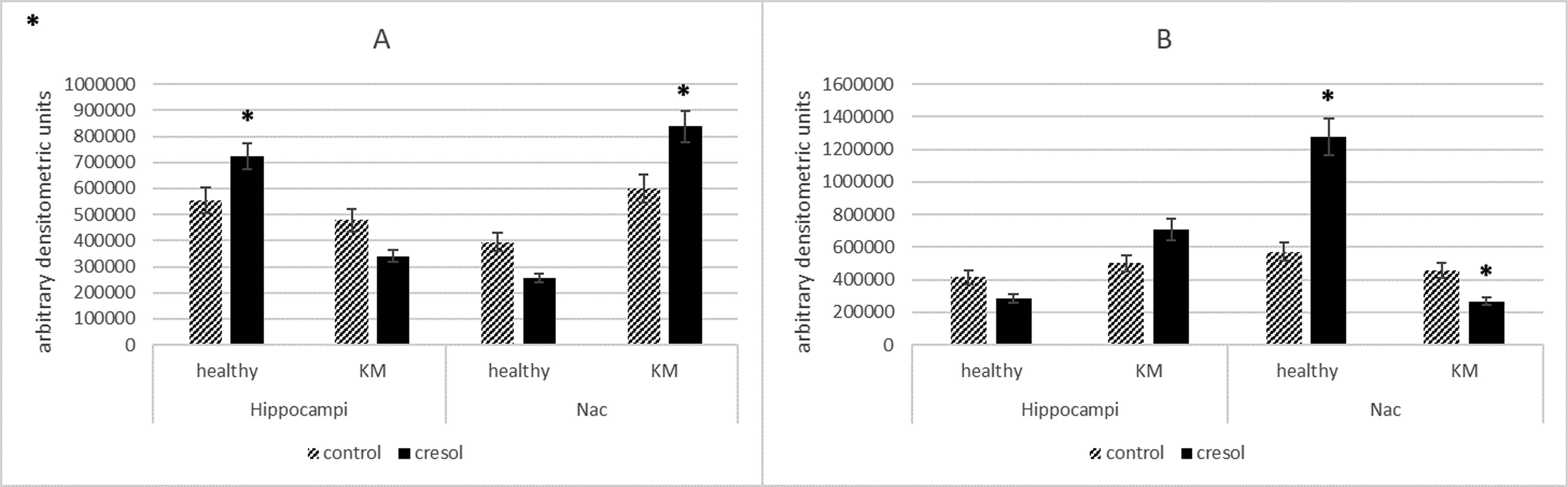
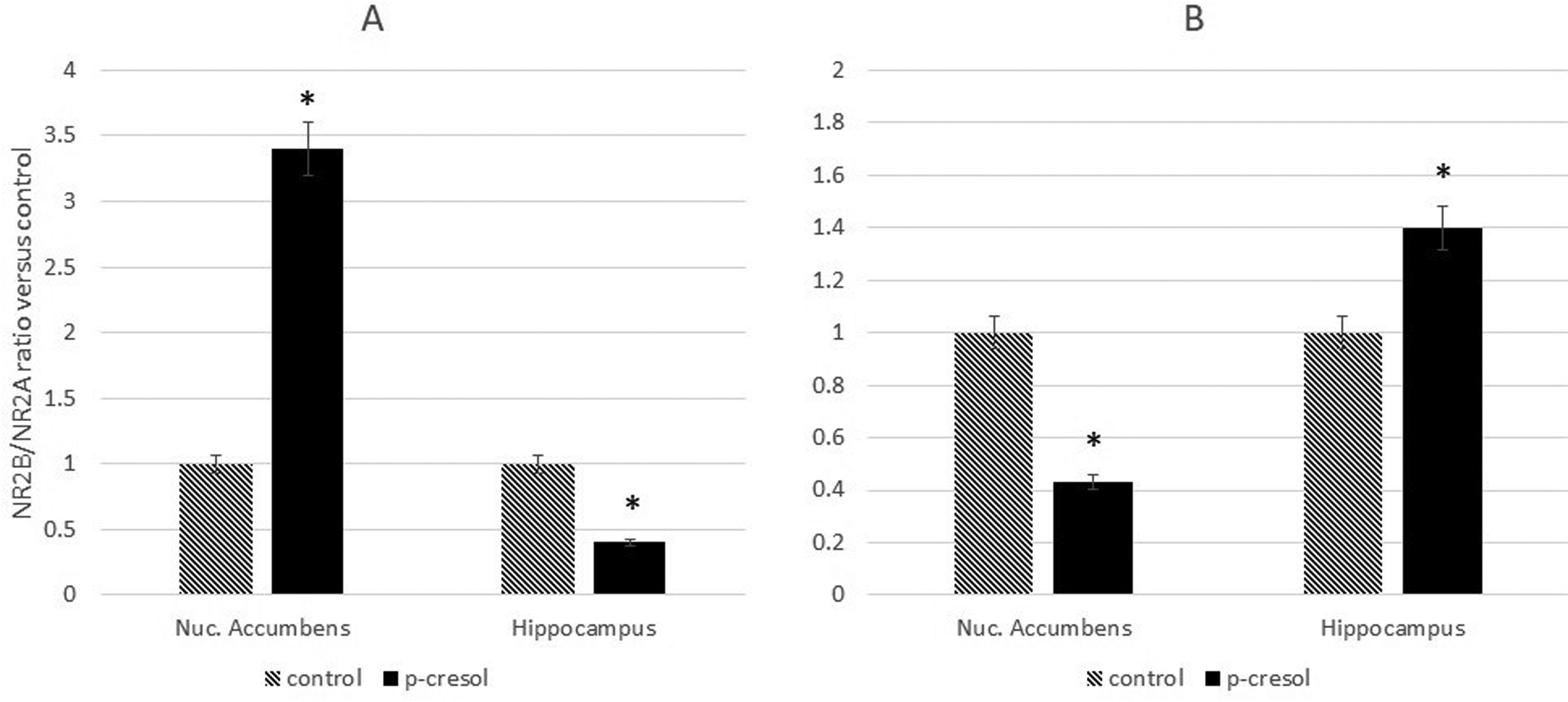
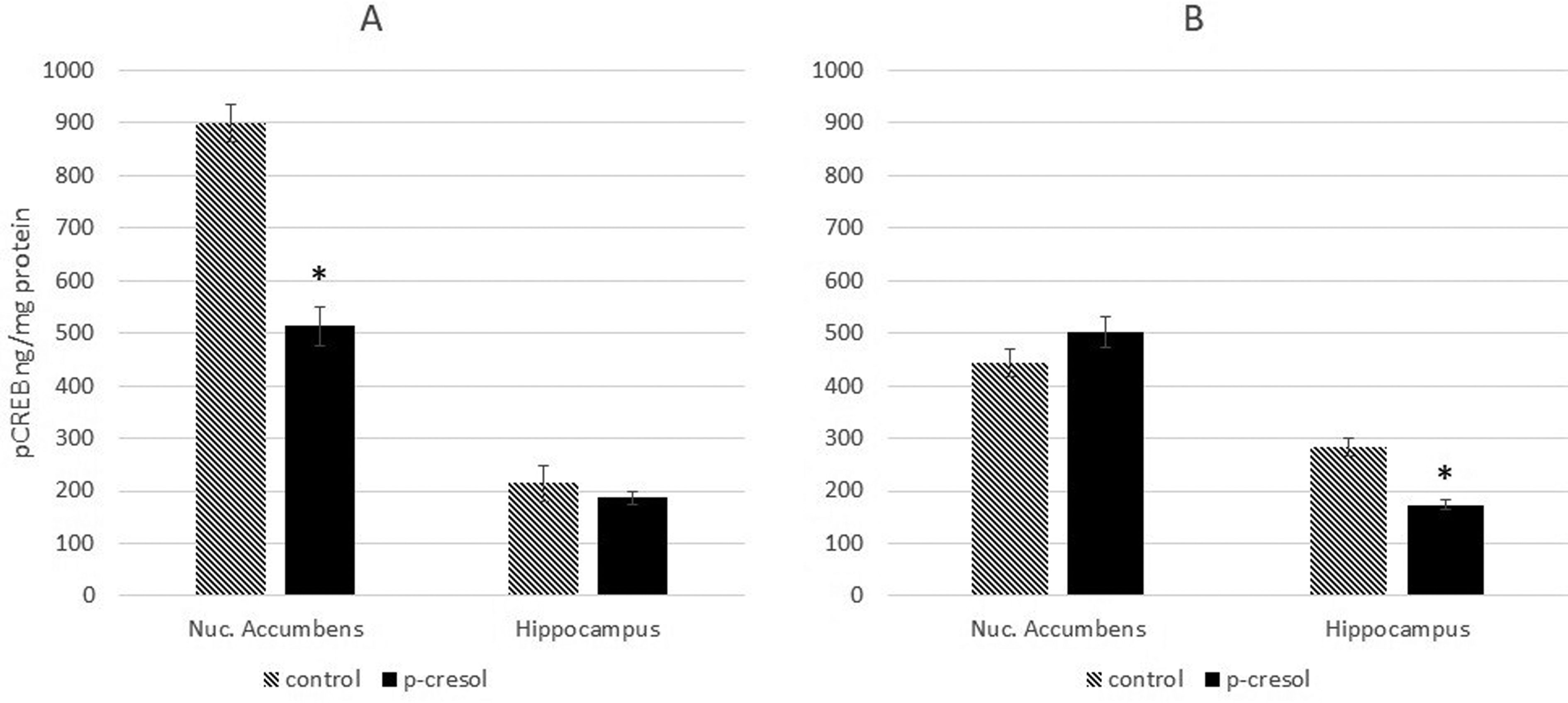
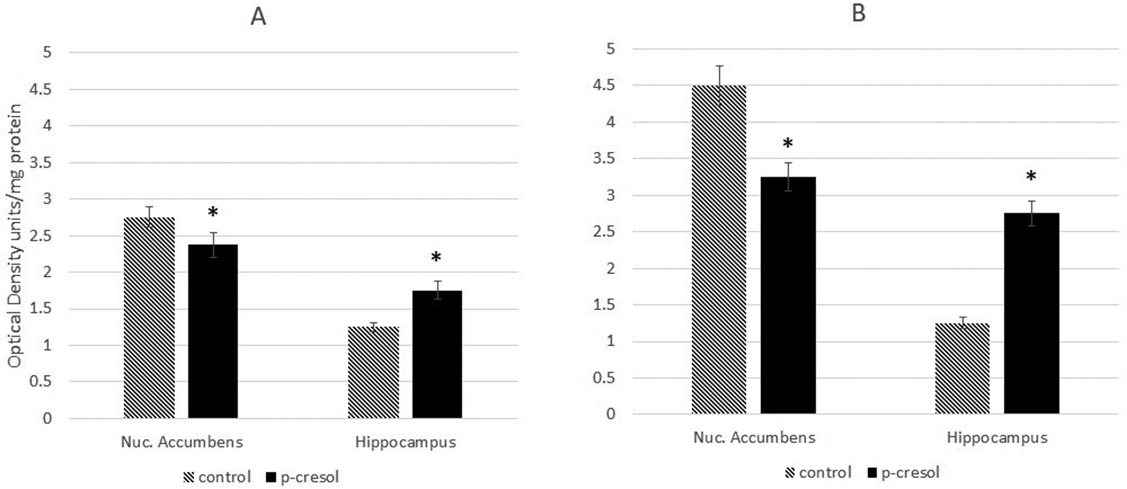
Mislocalization and abnormal expression of N-methyl-D-aspartate glutamate receptor (NMDAR) subunits is observed in several brain disorders and pathological conditions. Recently, we have shown that intraperitoneal injection of the gut neurotoxin p-cresol induces autism-like behavior and accelerates seizure reactions in healthy and epilepsy-prone rats, respectively. In this study, we evaluated the expression of GLUN2B and GLUN2A NMDAR subunits, and assessed the activity of cAMP-response element binding protein (CREB) and Rac1 in the hippocampi and nucleus accumbens of healthy and epilepsy-prone rats following p-cresol administration. We have found that subchronic intraperitoneal injection of p-cresol induced differential expression of GLUN2B and GLUN2A between the two brain regions, and altered the GLUN2B/GLUN2A ratio, in rats in both groups. Moreover, p-cresol impaired CREB phosphorylation in both brain structures and stimulated Rac activity in the hippocampus. These data indicate that p-cresol differently modulates the expression of NMDAR subunits in the nucleus accumbens and hippocampi of healthy and epilepsy-prone rats. We propose that these differences are due to the specificity of interactions between dopaminergic and glutamatergic pathways in these structures.
Citation: Tevzadze Gigi, Zhuravliova Elene, Barbakadze Tamar, Shanshiashvili Lali, Dzneladze Davit, Nanobashvili Zaqaria, Lordkipanidze Tamar, Mikeladze David. Gut neurotoxin p-cresol induces differential expression of GLUN2B and GLUN2A subunits of the NMDA receptor in the hippocampus and nucleus accumbens in healthy and audiogenic seizure-prone rats[J]. AIMS Neuroscience, 2020, 7(1): 30-42. doi: 10.3934/Neuroscience.2020003
Mislocalization and abnormal expression of N-methyl-D-aspartate glutamate receptor (NMDAR) subunits is observed in several brain disorders and pathological conditions. Recently, we have shown that intraperitoneal injection of the gut neurotoxin p-cresol induces autism-like behavior and accelerates seizure reactions in healthy and epilepsy-prone rats, respectively. In this study, we evaluated the expression of GLUN2B and GLUN2A NMDAR subunits, and assessed the activity of cAMP-response element binding protein (CREB) and Rac1 in the hippocampi and nucleus accumbens of healthy and epilepsy-prone rats following p-cresol administration. We have found that subchronic intraperitoneal injection of p-cresol induced differential expression of GLUN2B and GLUN2A between the two brain regions, and altered the GLUN2B/GLUN2A ratio, in rats in both groups. Moreover, p-cresol impaired CREB phosphorylation in both brain structures and stimulated Rac activity in the hippocampus. These data indicate that p-cresol differently modulates the expression of NMDAR subunits in the nucleus accumbens and hippocampi of healthy and epilepsy-prone rats. We propose that these differences are due to the specificity of interactions between dopaminergic and glutamatergic pathways in these structures.

| [1] |
Yamamoto H, Hagino Y, Kasai S, et al. (2015) Specific roles of NMDA receptor subunits in mental disorders. Curr Mol Med 15: 193-205. doi: 10.2174/1566524015666150330142807

|
| [2] |
Zhou Q, Sheng M (2013) NMDA receptors in nervous system diseases. Neuropharmacology 74: 69-75. doi: 10.1016/j.neuropharm.2013.03.030
|
| [3] |
Sanz-Clemente A, Nicoll RA, Roche KW (2013) Diversity in NMDA receptor composition: many regulators, many consequences. Neuroscientist 19: 62-75. doi: 10.1177/1073858411435129
|
| [4] | Mao LM, Guo ML, Jin DZ, et al. (2011) Post-translational modification biology of glutamate receptors and drug addiction. Front Neuroanat 5: 19. |
| [5] |
Koster KP, Francesconi W, Berton F, et al. (2019) Developmental NMDA receptor dysregulation in the infantile neuronal ceroid lipofuscinosis mouse model. Elife 8: pii: e40316. doi: 10.7554/eLife.40316
|
| [6] |
Hardingham GE, Bading H (2010) Synaptic versus extrasynaptic NMDA receptor signalling: Implications for neurodegenerative disorders. Nat Rev Neurosci 11: 682-696. doi: 10.1038/nrn2911
|
| [7] |
Monti B, Marri L, Contestabile A (2002) NMDA receptor-dependent CREB activation in survival of cerebellar granule cells during in vivo and in vitro development. Eur J Neurosci 16: 1490-1498. doi: 10.1046/j.1460-9568.2002.02232.x
|
| [8] |
Lee B, Butcher GQ, Hoyt KR, et al. (2005) Activity-dependent neuroprotection and cAMP response element-binding protein (CREB): Kinase coupling, stimulus intensity, and temporal regulation of CREB phosphorylation at serine 133. J Neurosci 25: 1137-1148. doi: 10.1523/JNEUROSCI.4288-04.2005
|
| [9] |
Chen BS, Roche KW (2009) Growth factor-dependent trafficking of cerebellar NMDA receptors via protein kinase B/Akt phosphorylation of NR2C. Neuron 62: 471-478. doi: 10.1016/j.neuron.2009.04.015
|
| [10] |
Duffney LJ, Wei J, Cheng J, et al. (2013) Shank3 deficiency induces NMDA receptor hypofunction via an actin-dependent mechanism. J Neurosci 33: 15767-15778. doi: 10.1523/JNEUROSCI.1175-13.2013
|
| [11] |
Ridley AJ (2006) Rho GTPases and actin dynamics in membrane protrusions and vesicle trafficking. Trends Cell Biol 16: 522-529. doi: 10.1016/j.tcb.2006.08.006
|
| [12] |
Li J, Xing H, Jiang G, et al. (2016) Increased expression of Rac1 in epilepsy patients and animal models. Neurochem Res 41: 836-843. doi: 10.1007/s11064-015-1759-y
|
| [13] |
Liu L, Wong TP, Pozza MF, et al. (2004) Role of NMDA receptor subtypes in governing the direction of hippocampal synaptic plasticity. Science 304: 1021-1024. doi: 10.1126/science.1096615
|
| [14] |
Krapivinsky G, Krapivinsky L, Manasian Y, et al. (2003) The NMDA receptor is coupled to the ERK pathway by a direct interaction between NR2B and RasGRF1. Neuron 40: 775-784. doi: 10.1016/S0896-6273(03)00645-7
|
| [15] |
Liu XY, Chu XP, Mao LM, et al. (2006) Modulation of D2R-NR2B interactions in response to cocaine. Neuron 52: 897-909. doi: 10.1016/j.neuron.2006.10.011
|
| [16] |
Hsiao EY, McBride SW, Hsien S, et al. (2013) Microbiota modulate behavioral and physiological abnormalities associated with neurodevelopmental disorders. Cell 155: 1451-1463. doi: 10.1016/j.cell.2013.11.024
|
| [17] | Thakur AK, Shakya A, Husain GM, et al. (2014) Gut-Microbiota and mental health: current and future perspectives. J Pharmacol Clin Toxicol 2: 1016. |
| [18] |
Persico AM, Napolioni V (2013) Autism genetics. Behav Brain Res 251: 95-112. doi: 10.1016/j.bbr.2013.06.012
|
| [19] |
Nicholson JK, Holmes E, Kinross J, et al. (2012) Host-gut microbiota metabolic interactions. Science 336: 1262-1267. doi: 10.1126/science.1223813
|
| [20] |
Goodhart PJ, DeWolf WE, Kruse LI (1987) Mechanism-based inactivation of dopamine .beta.-hydroxylase by p-cresol and related alkylphenols. Biochemistry 26: 2576-2583. doi: 10.1021/bi00383a025
|
| [21] |
Southan C, DeWolf WE, Kruse LI (1990) Inactivation of dopamine β-hydroxylase by p-cresol: Evidence for a second, minor site of covalent modification at tyrosine 357. Biochim Biophys Acta 1037: 256-258. doi: 10.1016/0167-4838(90)90176-G
|
| [22] |
Pavăl D (2017) A dopamine hypothesis of autism spectrum disorder. Dev Neurosci 39: 355-360. doi: 10.1159/000478725
|
| [23] |
Tevzadze G, Oniani N, Zhuravliova E, et al. (2018) Effects of a gut microbiome toxin, p-cresol, on the indices of social behavior in rats. Neurophysiology 50: 372-377. doi: 10.1007/s11062-019-09764-1
|
| [24] |
Tevzadze G, Nanobashvili Z, Zhuravliova E, et al. (2018) Effects of a gut microbiome toxin, p-Cresol, on the susceptibility to seizures in rats. Neurophysiology 50: 424-427. doi: 10.1007/s11062-019-09774-z
|
| [25] |
Tevzadze G, Zhuravliova E, Meparishvili M, et al. (2019) Effects of a Gut Microbiome Toxin, p-Cresol, on the Contents of the NMDA2B Receptor Subunit in the Nucl. Accumbens of Rats. Neurophysiology 51: 72-76. doi: 10.1007/s11062-019-09795-8
|
| [26] |
Poletaeva II, Surina NM, Kostina ZA, et al. (2017) The Krushinsky-Molodkina rat strain: The study of audiogenic epilepsy for 65 years. Epilepsy Behav 71: 130-141. doi: 10.1016/j.yebeh.2015.04.072
|
| [27] | Ma L, Chung WK (2014) Quantitative analysis of copy number variants based on real-time lightcycler PCR. Curr Protoc Hum Genet 80. |
| [28] |
Ishii A, Hirose S (2017) New genes for epilepsy–autism comorbidity. J Pediatr Neurol 15: 105-114. doi: 10.1055/s-0037-1602822
|
| [29] |
Ryley PR, Albertson AJ, Buckingham SC, et al. (2013) Status epilepticus triggers early and late alterations in brain-derived neurotrophic factor and NMDA glutamate receptor Grin2b DNA methylation levels in the hippocampus. Neuroscience 248: 602-619. doi: 10.1016/j.neuroscience.2013.06.029
|
| [30] |
Paoletti P, Bellone C, Zhou Q (2013) NMDA receptor subunit diversity: Impact on receptor properties, synaptic plasticity and disease. Nat Rev Neurosci 14: 383-400. doi: 10.1038/nrn3504
|
| [31] |
Endele S, Rosenberger G, Geider K, et al. (2010) Mutations in GRIN2A and GRIN2B encoding regulatory subunits of NMDA receptors cause variable neurodevelopmental phenotypes. Nat Genet 42: 1021-1026. doi: 10.1038/ng.677
|
| [32] |
Sebat J, Levy DL, McCarthy SE (2009) Rare structural variants in schizophrenia: one disorder, multiple mutations; one mutation, multiple disorders. Trends Genet 25: 528-535. doi: 10.1016/j.tig.2009.10.004
|
| [33] |
Weiss LA, Shen Y, Korn JM, et al. (2008) Association between microdeletion and microduplication at 16p11.2 and autism. N Engl J Med 358: 667-675. doi: 10.1056/NEJMoa075974
|
| [34] |
Coe BP, Witherspoon K, Rosenfeld JA, et al. (2014) Refining analyses of copy number variation identifies specific genes associated with developmental delay. Nat Genet 46: 1063-1071. doi: 10.1038/ng.3092
|
| [35] |
Gladding CM, Raymond LA (2011) Mechanisms underlying NMDA receptor synaptic/extrasynaptic distribution and function. Mol Cell Neurosci 48: 308-320. doi: 10.1016/j.mcn.2011.05.001
|
| [36] |
Chen M, Lu TJ, Chen XJ, et al. (2008) Differential roles of NMDA receptor subtypes in ischemic neuronal cell death and ischemic tolerance. Stroke 39: 3042-3048. doi: 10.1161/STROKEAHA.108.521898
|
| [37] |
Nakayama AY, Harms MB, Luo L (2000) Small GTPases Rac and Rho in the maintenance of dendritic spines and branches in hippocampal pyramidal neurons. J Neurosci 20: 5329-5338. doi: 10.1523/JNEUROSCI.20-14-05329.2000
|
| [38] |
Kowski AB, Voges J, Heinze HJ, et al. (2015) Nucleus accumbens stimulation in partial epilepsy - A randomized controlled case series. Epilepsia 56: e78-82. doi: 10.1111/epi.12999
|
| [39] |
Fu J, Liu Y, Yang K, et al. (2018) Effect of accumbens nucleus shell lesioning on bitemporal lobe epilepsy in rat model. Folia Neuropathol 56: 346-353. doi: 10.5114/fn.2018.80868
|
| [40] |
Yang CR, Mogenson GJ (1984) Electrophysiological responses of neurones in the nucleus accumbens to hippocampal stimulation and the attenuation of the excitatory responses by the mesolimbic dopaminergic system. Brain Res 324: 69-84. doi: 10.1016/0006-8993(84)90623-1
|
| [41] |
Pennartz CMA, Dolleman-Van der Weel MJ, Lopes da Silva FHL (1992) Differential membrane properties and dopamine effects in the shell and core of the rat nucleus accumbens studied in vitro. Neurosci Lett 136: 109-112. doi: 10.1016/0304-3940(92)90660-Y
|
| [42] |
Gonon F, Sundstrom L (1996) Excitatory effects of dopamine released by impulse flow in the rat nucleus accumbens in vivo. Neuroscience 75: 13-18. doi: 10.1016/0306-4522(96)00320-X
|
| [43] |
Shen H, Moussawi K, Zhou W, et al. (2011) Heroin relapse requires long-term potentiation-like plasticity mediated by NMDA2b-containing receptors. Proc Natl Acad Sci U S A 108: 19407-19412. doi: 10.1073/pnas.1112052108
|
| [44] |
Vega-Villar M, Horvitz JC, Nicola SM (2019) NMDA receptor-dependent plasticity in the nucleus accumbens connects reward-predictive cues to approach responses. Nat Commun 10: 4429. doi: 10.1038/s41467-019-12387-z
|
| [45] |
Ortega-Martínez S (2015) A new perspective on the role of the CREB family of transcription factors in memory consolidation via adult hippocampal neurogenesis. Front Mol Neurosci 8: 46. doi: 10.3389/fnmol.2015.00046
|
| [46] |
Sakamoto K, Karelina K, Obrietan K (2011) CREB: A multifaceted regulator of neuronal plasticity and protection. J Neurochem 116: 1-9. doi: 10.1111/j.1471-4159.2010.07080.x
|
| [47] |
Marie H, Morishita W, Yu X, et al. (2005) Generation of silent synapses by acute in vivo expression of CaMKIV and CREB. Neuron 45: 741-752. doi: 10.1016/j.neuron.2005.01.039
|
| [48] |
Brown TE, Lee BR, Mu P, et al. (2011) A silent synapse-based mechanism for cocaine-induced locomotor sensitization. J Neurosci 31: 8163-8174. doi: 10.1523/JNEUROSCI.0016-11.2011
|
| [49] |
Wayman GA, Impey S, Marks D, et al. (2006) Activity-dependent dendritic arborization mediated by CaM-kinase I activation and enhanced CREB-dependent transcription of Wnt-2. Neuron 50: 897-909. doi: 10.1016/j.neuron.2006.05.008
|
| [50] |
Gao C, Wolf ME (2008) Dopamine receptors regulate NMDA receptor surface expression in prefrontal cortex neurons. J Neurochem 106: 2489-2501. doi: 10.1111/j.1471-4159.2008.05597.x
|
| [51] |
Hu JL, Liu G, Li YC, et al. (2010) Dopamine D1 receptor-mediated NMDA receptor insertion depends on Fyn but not Src kinase pathway in prefrontal cortical neurons. Mol Brain 3: 20. doi: 10.1186/1756-6606-3-20
|
| [52] |
Dudman JT, Eaton ME, Rajadhyaksha A, et al. (2003) Dopamine D1 receptors mediate CREB phosphorylation via phosphorylation of the NMDA receptor at Ser897-NR1. J Neurochem 87: 922-934. doi: 10.1046/j.1471-4159.2003.02067.x
|
| [53] |
Dunah AW, Sirianni AC, Fienberg AA, et al. (2004) Dopamine D1-dependent trafficking of striatal N-Methyl-D-aspartate glutamate receptors requires Fyn protein tyrosine kinase but not DARPP-32. Mol Pharmacol 65: 121-129. doi: 10.1124/mol.65.1.121
|
| [54] |
Chernigovskaya EV, Korotkov AA, Dorofeeva NA, et al. (2019) Delayed audiogenic seizure development in a genetic rat model is associated with overactivation of ERK1/2 and disturbances in glutamatergic signaling. Epilepsy Behav 99: 106494. doi: 10.1016/j.yebeh.2019.106494
|
Figures(4) / Tables(1)

Tevzadze Gigi, Zhuravliova Elene, Barbakadze Tamar, Shanshiashvili Lali, Dzneladze Davit, Nanobashvili Zaqaria, Lordkipanidze Tamar, Mikeladze David. Gut neurotoxin p-cresol induces differential expression of GLUN2B and GLUN2A subunits of the NMDA receptor in the hippocampus and nucleus accumbens in healthy and audiogenic seizure-prone rats[J]. AIMS Neuroscience, 2020, 7(1): 30-42. doi: 10.3934/Neuroscience.2020003


 DownLoad: