Research article Topical Sections

Public perception of climate change in a period of economic crisis in Puerto Rico

  • Received: 30 August 2016 Accepted: 22 January 2017 Published: 08 February 2017
  • In this study, an analysis is performed to measure Puerto Rican citizens’ perceptions of environmental issues and global climate change (GCC). Puerto Rico (PR) is presently facing an economic crisis that began in 2010. In addition, the island has faced many extreme weather events that have greatly impacted the economy, including a dry period that lasted from the beginning of 2014 until the end of 2015 [1]. By replicating a study done in Greece (GR), [2] found that the citizens of PR are more informed about GCC (43%) than Greeks (34%). However, only 29% of Puerto Ricans said they knew about the effects of greenhouse gases, as compared to 44% of Greeks. The citizens of PR (67%) and GR (91%) claim they do not have confidence that their respective governments and agencies will be able to face GCC. In both cases, they have a higher confidence level in environmental organizations than in the scientific community. In PR, 89% of the citizens are concerned about the degradation of natural resources. At 64%, this concern is also significant in the case of GR, although the figure has decreased from previous years. Both countries place more responsibility in the hands of those in government and industry to take action on GCC. That being said, the people of GR have made it known they are willing to perform individual actions as well.

    Citation: María Santos-Corrada, Rafael Méndez-Tejeda. Public perception of climate change in a period of economic crisis in Puerto Rico[J]. AIMS Environmental Science, 2017, 4(1): 83-93. doi: 10.3934/environsci.2017.1.83
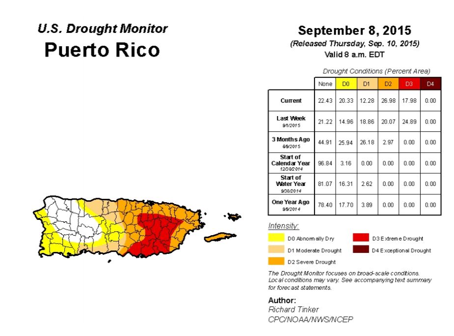
    Related Papers:

    [1] Britta Muster, Alexander Rapp, M. Cristina Cardoso . Systematic analysis of DNA damage induction and DNA repair pathway activation by continuous wave visible light laser micro-irradiation. AIMS Genetics, 2017, 4(1): 47-68. doi: 10.3934/genet.2017.1.47
    [2] Mrinalini Tiwari, Suhel Parvez, Paban K Agrawala . Role of some epigenetic factors in DNA damage response pathway. AIMS Genetics, 2017, 4(1): 69-83. doi: 10.3934/genet.2017.1.69
    [3] William M. Scovell . Nucleosome dynamics: HMGB1 facilitates nucleosome restructuring and collaborates in estrogen-responsive gene expression. AIMS Genetics, 2016, 3(4): 252-279. doi: 10.3934/genet.2016.4.252
    [4] Harem Othman Smail . The epigenetics of diabetes, obesity, overweight and cardiovascular disease. AIMS Genetics, 2019, 6(3): 36-45. doi: 10.3934/genet.2019.3.36
    [5] Huong Thi Thu Phung, Hoa Luong Hieu Nguyen, Dung Hoang Nguyen . The possible function of Flp1 in homologous recombination repair in Saccharomyces cerevisiae. AIMS Genetics, 2018, 5(2): 161-176. doi: 10.3934/genet.2018.2.161
    [6] Fabrizio Gentile, Alessia Arcaro, Stefania Pizzimenti, Martina Daga, Giovanni Paolo Cetrangolo, Chiara Dianzani, Alessio Lepore, Maria Graf, Paul R. J. Ames, Giuseppina Barrera . DNA damage by lipid peroxidation products: implications in cancer, inflammation and autoimmunity. AIMS Genetics, 2017, 4(2): 103-137. doi: 10.3934/genet.2017.2.103
    [7] Michael T. Fasullo, Mingzeng Sun . Both RAD5-dependent and independent pathways are involved in DNA damage-associated sister chromatid exchange in budding yeast. AIMS Genetics, 2017, 4(2): 84-102. doi: 10.3934/genet.2017.2.84
    [8] Mi Young Son, Paul Hasty . Homologous recombination defects and how they affect replication fork maintenance. AIMS Genetics, 2018, 5(4): 192-211. doi: 10.3934/genet.2018.4.192
    [9] Achal Rastogi, Xin Lin, Bérangère Lombard, Damarys Loew, Leïla Tirichine . Probing the evolutionary history of epigenetic mechanisms: what can we learn from marine diatoms. AIMS Genetics, 2015, 2(3): 173-191. doi: 10.3934/genet.2015.3.173
    [10] Asaad M Mahmood, Jim M Dunwell . Evidence for novel epigenetic marks within plants. AIMS Genetics, 2019, 6(4): 70-87. doi: 10.3934/genet.2019.4.70
  • In this study, an analysis is performed to measure Puerto Rican citizens’ perceptions of environmental issues and global climate change (GCC). Puerto Rico (PR) is presently facing an economic crisis that began in 2010. In addition, the island has faced many extreme weather events that have greatly impacted the economy, including a dry period that lasted from the beginning of 2014 until the end of 2015 [1]. By replicating a study done in Greece (GR), [2] found that the citizens of PR are more informed about GCC (43%) than Greeks (34%). However, only 29% of Puerto Ricans said they knew about the effects of greenhouse gases, as compared to 44% of Greeks. The citizens of PR (67%) and GR (91%) claim they do not have confidence that their respective governments and agencies will be able to face GCC. In both cases, they have a higher confidence level in environmental organizations than in the scientific community. In PR, 89% of the citizens are concerned about the degradation of natural resources. At 64%, this concern is also significant in the case of GR, although the figure has decreased from previous years. Both countries place more responsibility in the hands of those in government and industry to take action on GCC. That being said, the people of GR have made it known they are willing to perform individual actions as well.


    1. Introduction

    Eukaryotic genome is composed of nucleosomes that consist of 145-148 bp DNA segments wrapped around the histone octamer in 1.65-1.7 superhelical coils. Nucleosomal organization limits DNA accessibility to various proteins, including protein complexes involved in DNA repair [1]. Various protein complexes, including ATP-dependent chromatin remodelers and PARP-1 protein reorganize chromatin, making it more accessible to other DNA-interacting proteins.

    PARP-1 is an abundant multi-domain protein, localized in cell nuclei of higher eukaryotes, with a range of diversity functions, playing role in DNA repair [2,3], chromatin organization and transcription [4]. One of the crucial roles of the protein in a cell is detection of DNA damages through its DNA-binding zinc-finger domains that recognize single- and double-strand DNA breaks [5]. A variety of factors (e.g., ionizing radiation) cause genome damage making double-strand breaks in DNA, which can lead to mutations. PARP-1 binding to a DNA strand breakinduces a conformational change in the protein [6,7], which results in its DNA-dependent activation and poly(ADP)-ribosylation (pADP-r) of the target proteins (including automodification of PARP-1) using NAD+ as a substrate. Core histones [8] and linker histone H1 [9] are among the targets for pADP-r. Some direct inhibitors of PARP-1 enzymatic activity are important anticancer compounds. Thus anticancer compound olaparib interferes with essential nuclear processes in various tumors and causes cell death due to synthetic lethality [10,11].

    Although PARP-1 can bind to intact nucleosomes and to a variety of nucleosome substrates through double-strand break in nucleosomal DNA with different affinities [12], it is unknown whether it affects the structure of the nucleosome core. Using a single-particle Forster resonance energy transfer (spFRET) approach [13,14,15,16], we report that PARP-1 causes a considerable nucleosome unfolding in vitro that can be almost completely reversed by its automodification.


    2. Materials and Methods


    2.1. Protein purification and DNA templates

    Human recombinant PARP-1 was expressed in E.coli and purified as described [17].

    Fluorescently labeled DNA templates used for nucleosome assembly were synthesized by PCR using modified nucleosome-positioning sequence s603-42 [18] as a template. The following oligonucleotides were used to introduce fluorescent labels in nucleosomal DNA:

    for nucleosomes N13/91: forward - 5'-ACCCCAGGGACTTGAAGTAATAAGGACGGAGGGCCT#CTTTCAACATCGAT-3' (Т# refers to a nucleotide with a Cy3 label), reverse - 5'-CAAGCGACACCGGCACTGGGCCCGGTTCGCGCTCCCTCCTTCCGTGTGTTGTCGT*CTCT-3' (T* refers to a nucleotide with a Cy5 label). For nucleosomes N35/112: forward - 5'-AAGCGACACCGGCACTGGGCCCGGTTCGCGCT#CCCGCCTTCCGTGTGTTGTCGTCTCTCGGGCGT-3', reverse - 5'-ACCCCAGGGACTTGAAGTAATAAGGACGGAGGGCCTCTTTCAACATCGATGCACGGT*GGTTAG; for N57/135: forward - 5'- ACACCGGCACTGGGCCCGGTTCGCGCTCCCTCCTTCCGTGTGTTGTCGTCTCTCGGGCGTCTAAGTACGCT#TAGGC-3', reverse - 5'-ACCCCAGGGACTT*GAAGTAATAAG-3'.


    2.2. Nucleosome assembly and purification

    Nucleosomes were assembled using chicken donor chromatin without linker histone by salt dialysis as described [19]. The mononucleosomes were then gel-purified as described [20]. In-gel FRET analysis was performed using a Typhoon PhosphorImager. Fluorescence was excited in gel at 532 nm wavelength and recorded at 570-610 nm (for Cy3) and 650-700 nm (for Cy5) spectral regions.


    2.3. spFRET measurements

    Fluorescently labeled nucleosomes at 3 nM were incubated with 50 or 100 nM PARP-1 for 20 minutes in a buffer containing 20 mM Tris-HCl pH 7.9, 5 mM MgCl2, 150 mM KCl and 0.15 mM ZnCl2 at +25 °C in siliconized tubes. To induce poly(ADP)-ribosylation, nucleosomes were incubated with 50 nM PARP-1 for 20 min and further incubated with 2 or 4 µM NAD+ for 15 min. spFRET analysis was performed for 15 min using facilities and settings described previously [13]. spFRET measurements were repeated in at least two independent experiments. In each experiment, data from 700 to 7000 single nucleosomes were analyzed. Preservation of structures of nucleosomes and PARP-1-nucleosome complexes during the analysis was further verified by comparing the results of two consequent measurements.

    Efficiency of FRET and its changes were characterized by calculating proximity ratio (EPR) for each single nucleosome:

    EPR = (I5  0.19 × I3)/(I5 + 0.81 × I3) (1)

    where I5 and I3 are measured fluorescence intensities of Cy5 and Cy3, respectively, and factors 0.19 and 0.81 provide correction for the contribution of Cy3 fluorescence in the Cy5 detection channel (spectral cross-talk). EPR values calculated for nucleosome samplings were presented as frequency distribution histograms and fitted by two Gaussians. Goodness of the fit (R2) varied from 0.84 to 0.99.


    3. Results


    3.1. The experimental approach for analysis of PARP-1-dependent changes in nucleosome structure

    To study the effect of PARP-1 on the nucleosomal structure, spFRET microscopy experiments were conducted using three mononucleosomal templates; each nucleosome was labeled with a single pair of Cy3 and Cy5 fluorophores (Figure 1A). These labels were introduced in DNA based on known crystal structure of a nucleosome [21] to obtain efficient FRET between them in assembled nucleosomes without interfering with DNA structure or contacts between the DNA and core histones [13,14,15,16]. Labels were positioned into different parts of nucleosomal DNA: at +13 (Cy3) and +91 (Cy5) base pairs, relatively to the entry of linker DNA into nucleosome core (referred to as N 13/91), at positions +35 and +112 (N 57/135) and at +57 and +135 (N 57/135). In the assembled nucleosome, these positions are localized near the entry point of DNA into the nucleosome, near a contact between H2A-H2B histone dimers and close to the exit of DNA from nucleosome, respectively (Figure 1A).

    Nucleosome assembly was carried out using chicken chromatin as a donor of core histones and a short DNA fragment containing nucleosome positioning sequence 603 [22] and additional terminal 20 bp linker. This linker provided a DNA end ("dsDNA break") for PARP-1 binding to the nucleosome (Figure 1A); PARP-1 cannot bind to the other DNA end localized at the nucleosomal boundary [12,23]. Quality of the assembly was estimated by native PAGE; the expected changes of the FRET signal in the nucleosomes as compared with histone-free DNA were observed in the gel (Figure 1B). Single-particle FRET measurements were conducted using gel-purified nucleosomes in solution under microscope. The Cy3 label was excited with 514.5 nm laser wavelength in single nucleosomes or complexes when they diffused freely through a small focal volume [13] (Figure 1C), and fluorescence intensities of both Cy3 (donor) and Cy5 (acceptor) dyes were measured. Proximity of the labels in single nucleosomes was characterized by calculating of so-called proximity ratio (EPR), and the frequency distribution of EPR was plotted for each nucleosome sample. Absolute distances between labels were not calculated because of insufficient data about quantum yields and an instrumental factor.

    Figure 1. The experimental approach for analysis of PARP-1-dependent changes in nucleosome structure. A. Three types of mononucleosomes containing the single pair of Cy3 and Cy5 dyes in different positions on the nucleosomal DNA (the positions of Cy3 and Cy5 are shown by green and red circles, respectively). B. PAGE and in-gel FRET analysis of assembled nucleosomes and DNA template. Distributions of Cy3 and Cy5 fluorescence in a gel at a Cy3 excitation are shown in green and red, respectively. Yellow color (superposition of green and red colors) indicates a considerable FRET efficiency. C. Experimental approach. spFRET from nucleosomes was measured in the absence or presence of PARP-1 and subsequent addition of NAD+.

    3.2. PARP-1 induces structural changes in nucleosomal DNA

    In agreement with the previously published data [16], spFRET analysis revealed two populations of N 13/91 nucleosomes (Figure 2A, Table S1). A peak with the EPR maximum at 0.69 corresponds to a major fraction of compact nucleosomes, while a peak with the EPR maximum at 0.01 likely indicates the presence of a minor fraction of nucleosomes with partially unwrapped DNA. After addition of 50 nM PARP-1, the distribution of nucleosomes by EPR is changed (Figure 2A) indicating formation of PARP-1-nucleosome complexes. These complexes are characterized by EPR with a maximum at 0.31, while a shoulder in a higher EPR region corresponds to PARP-1-free, more compact intact nucleosomes. Incomplete PARP-1 binding is observed most likely because the dissociation constant for PARP-1 complexes with similar nucleosome constructs is 85 nM [12]. In agreement with this proposal, a high EPR shoulder disappears after increasing concentration of PARP-1 to 100 nM (Figure 2A). The low-EPR peak at 0.01 is also diminished in the presence of PARP-1, suggesting that spontaneous DNA uncoiling from the octamer is diminished in the complex.

    Figure 2. PARP-1 induces structural changes in nucleosomal DNA. A. spFRET analysis ofPARP-1 binding to N 13/91 nucleosomes. Typical frequency distributions of EPR are shown for N 13/91 nucleosomes before and after addition of 50 or 100 nM of PARP-1 (for quantitative and statistical data see Table S1). B. spFRET analysis of +13/+91-labeled DNA before and after addition of 50 or 100 nM of PARP-1.

    Domination of the single Gaussian peak in the frequency distribution of EPR indicates formation of a single uniform population of nucleosome complexes with PARP-1. A shift of EPR maximum from 0.69 to 0.31 shows that PARP-1 binding causes structural changes in nucleosomal DNA near the entrance of DNA into nucleosome, namely in the region, where DNA interacts with the H2A-H2B dimer (position +13) and H4-H2B interface (position +91). To evaluate a possibility that PARP-1 disrupts nucleosomes and forms complexes with histone-free DNA, the same experiments were conducted with DNA template used for nucleosome assembly (Figure 2B). In more extended histone-free DNA, the Cy3 and Cy5 labels are positioned far from each other and no FRET occurs (maximum of EPR is 0.01, Figure 2B, Table S1). The frequency distribution of EPR, which is observed for the histone-free DNA, is minimally affected by PARP-1 (Figure 2B).

    In summary, binding PARP-1 to the N 13/91 nucleosome causes considerable structural changes in nucleosomal DNA that are accompanied by an increase in the distance between the labels introduced near the entrance of DNA into a nucleosome, indicating that gyres of nucleosomal DNA are coming apart and, at the same time, by diminished spontaneous DNA uncoiling from the octamer in the complex, suggesting that the DNA end has a lower mobility in the PARP-1-nucleosome complex. These observations, taken together, suggest that PARP-1 causes a mobility of the end of nucleosomal DNA, but, at the same time, induces uncoiling of nucleosomal DNA together with histones.


    3.3. PARP-1 induces similar structural changes in different regions of nucleosomal DNA

    To evaluate how PARP-1 affects other parts of nucleosomal DNA, N 35/112 and N 57/135 nucleosomes were analyzed (Figure 3). The N 13/91 (Figure 2A) and N 35/112 nucleosomes (Figure 3A) are characterized by similar frequency distributions of EPR (see Table S1 for the statistical data). Although labels in the nucleosomes N 35/112 were positioned far from the extending DNA end (PARP-1 target), FRET between theses labels was significantly affected by PARP-1 binding. The maximum of the main EPR peak was shifted from 0.63 to 0.37, indicating to reorganization of nucleosomal DNA structure near the interface between the H2A/H2B dimers and H3/H4 tetramers (position +35) and/or H3/H4 tetramers (position +112) that resulted in the increase in the inter-label distance.

    Figure 3. PARP-1 induces similar structural changes in different regions of nucleosomal DNA. spFRET analysis of PARP-1 binding to N 35/112 (A) and N 57/135 nucleosomes (B). Typical frequency distributions of EPR are shown for nucleosomes before and after incubation in the presence of 50 or 100 nM of PARP-1 (for quantitative and statistical data see Table S1).

    N 57/135 nucleosomes were also characterized by a bimodal distribution of EPR (Figure 3B and Table S1). In the presence of 100 nM PARP-1, a broad EPR distribution was formed with a maximum at 0.43. This broadening could be explained by an increased mobility of nucleosomal DNA localized near the position +135 in the PARP-1-nucleosome complex.

    For every combination of the labels, the shift of the main peak from higher to intermediate EPR values was observed. In the case of N 13/91 and N 35/112 the shift was accompanied by a significant decrease in the height of the low-EPR peak (Figures 2 and 3). Taken together, the data indicate that PARP-1 can partially and similarly uncoil different regions of nucleosomal DNA, and, at the same time, can restrict mobilities of the +13, +35 and +112 regions of nucleosomal DNA.


    3.4. PARP-1-induced changes in nucleosome structure are reversed after PARP-1 automodification

    To elucidate how activation of the enzymatic activity of PARP-1 affects the structure of the PARP-1-nucleosome complex, pre-formed PARP-1 complexes with N 13/91 nucleosomes were incubated in the presence of different concentrations of NAD+. DNA-bound PARP-1 is activated, auto-poly(ADP)-ribosylated in the presence of NAD+ and loses its capability to interact with damaged DNA and nucleosomes [23,24]. Therefore, it was expected that nucleosomal EPR distribution would be recovered in the presence of NAD+. spFRET analysis revealed that incubation of the PARP-1-nucleosome complex in the presence of 2 or 4 µM NAD+ results in a progressive, stepwise shift of the mean value of EPR peak from 0.31 to 0.43 or to 0.62, respectively (Figure 4 and Table S1). In the presence of 4 µM NAD+, the main EPR maximum (0.62) approaches the value, which is a characteristic of free nucleosomes (0.69), suggesting that nucleosome structure is almost completely recovered. Since the principal NAD+-dependent reaction is PARP-1 automodification [25], the data indicate that after partial automodification (i.e., at 2 µM NAD+) PARP-1 remains bound to nucleosomes and forms a discrete intermediate PARP-1-nucleosome complex.

    Figure 4. PARP-1-induced changes in nucleosome structure are reversed after PARP-1 automodification. spFRET analysis of PARP-1 automodification in the complex with nucleosomes N 13/91 after addition of NAD+. Typical frequency distributions of EPR are shown (for quantitative and statistical data see Table S1).

    Incomplete reversal of the nucleosomal EPR distributions at 4 µM NAD+ is likely explained by incomplete automodification of PARP-1 that remains bound to the nucleosome. Alternatively, poly(ADP)-ribosylation of core histones [8] prevents complete recovery of the nucleosomal structure.


    4. Discussion

    Our spFRET experiments suggest that PARP-1 binds to a nucleosome and induces disturbance of different regions of nucleosomal DNA: near the entrance/exit of DNA into/from a nucleosome, and in the region positioned ~35 bp from the boundaries of nucleosomal DNA (Figures 2 and 3). This uncoiling of nucleosomal DNA is accompanied by a reduced mobilities of the +13, +35 and +112 regions of nucleosomal DNA (Figures 2 and 3). PARP-1 automodification (self-PARylation) is accompanied by formation of an intermediate complex, and eventually leads to nearly complete recovery of the initial structure of nucleosome (Figure 4). Thus spFRET is a sensitive method for analysis of PARP-1-induced changes in chromatin structure that could also be used for analysis of PARP-1 inhibition by various compounds.

    PARP-1 binds to nucleosomes having one linker DNA with an exposed double-strand break with stoichiometry of one PARP-1 molecule per nucleosome [12]. Rearrangements in the enzyme structure after binding to a double-strand DNA break [7] make HD subdomain unstable, resulting in activation of the catalytic center of PARP-1 [26,27]. If PARP-1 is bound in the vicinity of a nucleosome, activated PARP-1 can also induce a considerable, partial and reversible disturbance of nucleosomal DNA structure (Figure 5). Similar, although less pronounced changes of nucleosomal structure have been observed after acetylation of core histones and DNA methylation in a nucleosome [28,29]. Much more dramatic uncoiling of nucleosomal DNA together with the associated core histones was observed in the complex between yFACT and a nucleosome [16]. It is possible that yFACT and PARP-1 induce conformational changes in nucleosomal DNA of similar nature, but different magnitude.

    Nucleosome structure can be considerably changed during various processes, such as transcription [30,31,32] and protein binding to nucleosomal DNA [33]. These conformational changes include: (i) DNA unwrapping from an intact octamer; (ii) DNA unwrapping accompanied by opening of the (H2A-H2B) dimer/(H3-H4)2 tetramer interface; (iii) DNA unwrapping with complete octamer disassembly and (iv) the unwrapping involving opening of the (H3-H4)2 tetramer [34,35,36]. Since different regions of nucleosomal DNA are uncoiled in the PARP-1-nucleosome complex to a similar degree (Figure 5), the global change in the nucleosome structure involving structural changes in the entire histone octamer likely occurs. The nature and extent of these conformational changes in nucleosome structure remain to be determined.

    Figure 5. The model of PARP-1-induced conformational changes in nucleosomal DNA in the vicinity of double-strand DNA break. DNA is tightly wrapped around histone octamer, but there are fluctuations in the structure of the double helix, especially in an area near DNA entering or exiting the nucleosome core. Unmodified PARP-1 is inactive and its domains (shown by different colors) are connected together by flexible linkers [26]. When PARP-1 binds to a DNA damage or available DNA end in the vicinity of a nucleosome, it forms a compact structure [7], which induces activation of PARP-1 and partial unwrapping of nucleosomal DNA. This nucleosome unfolding is accompanied by a reduced mobilities of the +13, +35 and +112 regions of nucleosomal DNA and possibly by destabilization of the intranucleosomal interactions between core histones. In the presence of NAD+ PARP-1 is automodified and released from the nucleosome; PARP-1 release is accompanied by spontaneous recoiling of nucleosomal DNA. Yellow circles indicate positions of the pairs of fluorescent dyes on nucleosomal DNA.

    Detection and repair of double-strand breaks in cells require multi-step chromatin remodeling. Thus p400/Tip60 chromatin remodeler induces exchange of histones H2A to H2A.Z onto nucleosomes at the break, which is important for downstream acetylation of H4 histone and maintenance of less compact chromatin structure in the break region [37]. PARP-1-induced DNA uncoiling could facilitate the histone exchange and/or displacement during the chromatin remodeling, before PARP-1 leaves the DNA break after automodification. It is also possible that the stable PARP-1-nucleosome complexes could be formed at transcription start sites [4].


    5. Conclusions

    Nucleosome structure can be considerably and reversibly unfolded after PARP-1 binding. These changes include transient and partial uncoiling of nucleosomal DNA along its entire length. These PARP-1-dependent changes in nucleosome structure are nearly completely reversed after PARP-1 eviction due to its auto-poly(ADP)-ribosylation.


    Acknowledgments

    We thank Daria Gaykalova for help with designing the fluorescent probes. This work was supported by NIH grants GM58650 to V.M.S. and GM087282 to J.M.P., and by the Federal Target Program "Research and developments in the priority directions of the scientific-technological complex of the Russian Federation for 2014-2020" (Agreement of the Ministry of Education of the Russian Federation No 14.604.21.0063, RFMEFI60414X0063).


    Conflict of Interest

    The authors declare no competing financial interests.


    [1] The national drougt mitigation center, available from: http://droughtmonitor.unl.edu/Home/StateDroughtMonitor.aspx?PR
    [2] Papoulis D, Kaika D, Bampatsou C, et al. (2015) Public Perception of Climate Change in a Period of Economic Crisis. Climate 3: 715-726. doi: 10.3390/cli3030715
    [3] Symon Carolyn (2005) Artic Climate Impact Assessment. Cambridge University Press. 1042p.
    [4] Pielke Sr RA (2005) Land Use and Climate Change. Science 310: 1625-1626. doi: 10.1126/science.1120529
    [5] PRCCC. Puerto Rico's State of the Climate 2010-2013 Assessing Puerto Rico's Social-Ecological Vulnerabilities in a Changing Climate, 2013. Available from: http://pr-ccc.org/download/PR%20State%20of%20the%20Climate-FINAL_ENE2015.pdf
    [6] Rafael MT, María SC, Sergio OM, et al. (2015) Environmental and Economic Impact of Forest Fires in Puerto Rico 2013-2014. Open J Forestry 5: 353. doi: 10.4236/ojf.2015.54030
    [7] Atiles-Osoria JM (2014) Environmental Colonialism, Criminalization and Resistance: Puerto Rican Mobilizations for Environmental Justice in the 21st Century. RCCS Annual Review. A selection from the Portuguese journal Revista Crítica de Ciências Sociais.
    [8] Chisari O, Galiani S, Miller S (2013) Optimal Climate Change Adaptation and Mitigation in Environmentally Small Economies. IDB Working Paper No. 417. Inter-American Development Bank, Washington, DC.
    [9] Censo de Puerto Rico 2010, 2012. Available from: https://www.census.gov/prod/cen2010/cph-2-53sp.pdf.
    [10] Pagan-Trinidad I, Statistical Analyses of Spatial and Temporal Storm Rainfall Characteristics in Puerto Rico; Project No. 374103 Final Technical Report; United States Department of Interior: Mayaguez, PR, USA, 1984.
    [11] Torres-Valcárcel A, Harbor J, González-Aviles C, et al. (2014) Impacts of Urban Development on Precipitation in the Tropical Maritime Climate of Puerto Rico. Climate 2: 47-77. doi: 10.3390/cli2020047
    [12] Méndez-Lázaro PA, Nieves-Santiago A, Miranda-Bermúdez J (2014) Trends in Total Rainfall, Heavy Rain Events, and Number of Dry Days in San Juan, Puerto Rico, 1955-2009. Ecol Soc 2: 50.
    [13] Rigau-Perez JG, Clark GG, Gubler DJ, et al. (1998) Dengue and Dengue Hemorrhagic Fever. Lancet 352: 971-977. doi: 10.1016/S0140-6736(97)12483-7
    [14] Rigau-Perez JG, Ayala-Lopez A, Garcia-Rivera EJ, et al. (2002) The Reappearance of Dengue-3 and a Subsequent Dengue-4 and Dengue-1 Epidemic in Puerto Rico in 1998. ASTMH 67: 355-362.
    [15] Méndez-Lázaro P, Muller-Karger FE, Otis D, et al. (2014). Assessing Climate Variability Effects on Dengue Incidence in San Juan, Puerto Rico. Int J Environ Res Public Health 11: 9409-9428. doi: 10.3390/ijerph110909409
    [16] CDC. Zika in Puerto Rico a 'challenge and crisis'. Centers for Disease Control and Prevention (CDC). Available from: http://www.cdc.gov/.
    [17] Armstrong RA, Singh H, Torres J (2002) Benthic Survey of Insular Slope Coral Reefs using the SeaBed AUV. Backscatter 13: 22-25.
    [18] Ballantine DL, Appeldoorn RS, Yoshioka P, et al. (2008) Biology and ecology of Puerto Rican coral reefs. Coral Reefs of the USA, Springer Netherlands 2008: 375-406.
    [19] Hernández-Delgado EA (2000) Effects of Anthropogenic Stress Gradients in the Structure of Coral Reef Epibenthic and Fish Communities. Ph.D. Dissertation, Department of Biology, University of Puerto Rico, San Juan, 330 p.
    [20] Irizarry-Soto E, Weil E (2009) Spatial and Temporal Variability in Juvenile Coral Densities, Survivorship and Recruitment in La Parguera, Southwestern Puerto Rico. Caribb J Sci 45: 269-281. doi: 10.18475/cjos.v45i2.a14
    [21] Rafael MT, María SC, Sergio OM, et al. (2015) Environmental and Economic Impact of Forest Fires in Puerto Rico 2013-2014. Open J Forestry 5: 353.
    [22] PRCCC. Puerto Rico's State of the Climate 2010-2013: Assessing Puerto Rico's Social-Ecological Vulnerabilities in a Changing Climate. (2013). Available from: http://pr-ccc.org/download/PR%20State%20of%20the%20Climate-FINAL_ENE2015.pdf
    [23] Cabrera, Nahir, and Barreto, Maritza (2013) A Study of Beach Profile Changes at Selected Beaches on the North Coast of Puerto Rico (2009-2013). Geography Department, University of Puerto Rico, Río Piedras Campus Graduate Student, Graduate School of Planning, University Puerto Rico, Río Piedras Campus. Available from: http://cara.uprm.edu/?q=node/64/
    [24] Van Beusekom AE, González G, Rivera MM (2014) Short-term Precipitation and Temperature Trends along an Elevation Gradient in Northeastern Puerto Rico. Earth Interactions 19: 1-33.
    [25] Carter M, Elsner J (1997) A Statistical Method for Forecasting Rainfall Over Puerto Rico. Wea Forecasting 12: 515–525.
    [26] García-Martinó AR, Warner GS, Scatena FN, et al. (1996). Rainfall, Runoff and Elevation Relationships in the Luquillo Mountains of Puerto Rico. Caribb J Sci 32: 413-424.
    [27] Larsen MC, Simon A (1993) A Rainfall Intensity-Duration Threshold for Landslides in a Humid-Tropical Environment, Puerto Rico. Geogr Ann 75A: 13-23.
    [28] Johansson MA, Cummings DAT, Glass GE (2009) Multiyear Climate Variability and Dengue-El Niño Southern Oscillation, Weather, and Dengue Incidence in Puerto Rico, Mexico, and Thailand: A Longitudinal Data Analysis. PLoS Med 6: e1000168. doi: 10.1371/journal.pmed.1000168
    [29] Jury MR (2011) Caribbean Hurricanes: Interaction of Easterly and Westerly Waves. Theor Appl Climatol 106: 117-126. doi: 10.1007/s00704-011-0424-x
    [30] Jury MR, Rios-Berrios R, Garcia E (2011) Caribbean Hurricanes: Changes of Intensity and Track Prediction. Theor Appl Climatol 107: 297-311.
    [31] Donnelly JP, Woodruff JD (2007) Intense Hurricane Activity over the Past 5,000 Years. Nature 447: 465-468. doi: 10.1038/nature05834
    [32] Chen AA, Taylor MA (2002) Investigating the Link between Early Season Caribbean Rainfall and the El Niño +1 year. Int J Climatology 22: 87-106. doi: 10.1002/joc.711
    [33] Chen AA, Roy A, McTavish J, et al. (1997) Using SST Anomalies to Predict Flood and Drought Conditions for the Caribbean. Center for Ocean-Land Atmosphere Studies.
    [34] Elsner JB, Kossin JP, Jagger TH (2008) The increasing intensity of the strongest tropical cyclones. Nature 455: 92-95. doi: 10.1038/nature07234
    [35] Mann ME, Emanuel KA (2006) Atlantic Hurricane Trends Linked to Climate Change. EOS, Transactions American Geophysical Union 87: 233-241.
    [36] NOAA. Available from: http://www.nhc.noaa.gov/data/publications.php#NOAA.
    [37] Impacto Económico del Huracán Georges en Puerto Rico. Available from: http://www.jp.gobierno.pr/Portal_JP/Portals/0/Publicaciones/PublicacionesHistoricasOnline/Impacto%20Econ%C3%B3mico%20del%20Hurac%C3%A1n%20Georges%20de%20PR%20-%20Abril%201999-2.pdf.
    [38] National hurricane center, Hurricanes in history. Avaible from: http://www.nhc.noaa.gov/outreach/history/
    [39] Bueno R, Herzfeld C, Stanton AE, et al. (2008) The Caribbean and climate change. Available from: https://www.sei-international.org/mediamanager/documents/Publications/Climate/inaction-caribbean-es-eng.pdf.
    [40] Gros D, Puerto Rico and Greece: A Tale of Two Defaults in a Monetary Union. Centre for European Policy Studies, 2015. Available from: https://www.ceps.eu/system/files/HLB5_DG_PuertoRico_0.pdfGren.
    [41] Greene D, Puerto Rico's Battered Economy: The Greece of the Caribbean? 2013. Available from: http://www.npr.org/2013/02/06/171071377/puerto-ricos-battered-economy-the-greece-of-the-caribbean
    [42] United Nations Millennium Development Goals Indicators. Available from: http://datos.bancomundial.org/indicador/NY.GDP.PCAP.CD.
    [43] EPA. Environmental Protection Agency and the Municipality. Available from: https://www.epa.gov/newsreleases/municipality.
  • This article has been cited by:

    1. N. V. Malyuchenko, D. O. Koshkina, A. N. Korovina, N. S. Gerasimova, M. P. Kirpichnikov, V. M. Studitsky, A. V. Feofanov, The Effect of Gossypol on the Structure of Nucleosomes, 2020, 75, 0096-3925, 142, 10.3103/S0096392520030050
    2. Anastasiia L. Sivkina, Maria G. Karlova, Maria E. Valieva, Laura L. McCullough, Timothy Formosa, Alexey K. Shaytan, Alexey V. Feofanov, Mikhail P. Kirpichnikov, Olga S. Sokolova, Vasily M. Studitsky, Electron microscopy analysis of ATP-independent nucleosome unfolding by FACT, 2022, 5, 2399-3642, 10.1038/s42003-021-02948-8
    3. Dmitry Nilov, Natalya Maluchenko, Tatyana Kurgina, Sergey Pushkarev, Alexandra Lys, Mikhail Kutuzov, Nadezhda Gerasimova, Alexey Feofanov, Vytas Švedas, Olga Lavrik, Vasily M. Studitsky, Molecular Mechanisms of PARP-1 Inhibitor 7-Methylguanine, 2020, 21, 1422-0067, 2159, 10.3390/ijms21062159
    4. Natalya Maluchenko, Darya Koshkina, Anna Korovina, Vasily Studitsky, Alexey Feofanov, Interactions of PARP1 Inhibitors with PARP1-Nucleosome Complexes, 2022, 11, 2073-4409, 3343, 10.3390/cells11213343
    5. Grigoriy A Armeev, Anna K Gribkova, Iunona Pospelova, Galina A Komarova, Alexey K Shaytan, Linking chromatin composition and structural dynamics at the nucleosome level, 2019, 56, 0959440X, 46, 10.1016/j.sbi.2018.11.006
    6. Natalya Maluchenko, Dmitry Nilov, Alexey Feofanov, Alexandra Lys, Mikhail Kutuzov, Nadezhda Gerasimova, Vasily Studitsky, 7-Methylguanine Traps PARP-1 on Nucleosomes: spFRET Microscopy Study, 2019, 25, 1431-9276, 1282, 10.1017/S1431927619007141
    7. M. M. Kutuzov, E. A. Belousova, T. A. Kurgina, A. A. Ukraintsev, I. A. Vasil’eva, S. N. Khodyreva, O. I. Lavrik, The contribution of PARP1, PARP2 and poly(ADP-ribosyl)ation to base excision repair in the nucleosomal context, 2021, 11, 2045-2322, 10.1038/s41598-021-84351-1
    8. Yi Qiao, Yuhan Luo, Naiyun Long, Yi Xing, Jing Tu, Single-Molecular Förster Resonance Energy Transfer Measurement on Structures and Interactions of Biomolecules, 2021, 12, 2072-666X, 492, 10.3390/mi12050492
    9. Giovanna De Matteis, Anna Reale, Francesco Grandoni, Mirella L. Meyer-Ficca, Maria Carmela Scatà, Ralph G. Meyer, Luca Buttazzoni, Bianca Moioli, Assessment of Poly(ADP-ribose) Polymerase1 (PARP1) expression and activity in cells purified from blood and milk of dairy cattle, 2018, 202, 01652427, 102, 10.1016/j.vetimm.2018.06.013
    10. Natalya V. Maluchenko, Dmitry K. Nilov, Sergey V. Pushkarev, Elena Y. Kotova, Nadezhda S. Gerasimova, Mikhail P. Kirpichnikov, Marie-France Langelier, John M. Pascal, Md. Sohail Akhtar, Alexey V. Feofanov, Vasily M. Studitsky, Mechanisms of Nucleosome Reorganization by PARP1, 2021, 22, 1422-0067, 12127, 10.3390/ijms222212127
    11. M. M. Kutuzov, E. A. Belousova, E. S. Ilina, O. I. Lavrik, 2020, Chapter 4, 978-3-030-41282-1, 47, 10.1007/978-3-030-41283-8_4
    12. Tatyana V. Andreeva, Natalya V. Maluchenko, Anastasiia L. Sivkina, Oleg V. Chertkov, Maria E. Valieva, Elena Y. Kotova, Mikhail P. Kirpichnikov, Vasily M. Studitsky, Alexey V. Feofanov, Na+ and K+ Ions Differently Affect Nucleosome Structure, Stability, and Interactions with Proteins, 2022, 28, 1431-9276, 243, 10.1017/S1431927621013751
    13. Ekaterina A. Belousova, Olga I. Lavrik, The Role of PARP1 and PAR in ATP-Independent Nucleosome Reorganisation during the DNA Damage Response, 2022, 14, 2073-4425, 112, 10.3390/genes14010112
    14. A. V. Lyubitelev, V. M. Studitsky, A. V. Feofanov, M. P. Kirpichnikov, Effect of sodium and potassium ions on conformation of linker parts of nucleosomes, 2017, 72, 0096-3925, 146, 10.3103/S0096392517030075
    15. Elena Y. Kotova, Fu-Kai Hsieh, Han-Wen Chang, Natalia V. Maluchenko, Marie-France Langelier, John M. Pascal, Donal S. Luse, Alexey V. Feofanov, Vasily M. Studitsky, Human PARP1 Facilitates Transcription through a Nucleosome and Histone Displacement by Pol II In Vitro, 2022, 23, 1422-0067, 7107, 10.3390/ijms23137107
    16. N. V. Malyuchenko, E. Yu. Kotova, M. P. Kirpichnikov, V. M. Studitsky, A. V. Feofanov, PARP1 Binding to DNA Breaks and Hairpins Alters Nucleosome Structure, 2019, 74, 0096-3925, 158, 10.3103/S0096392519030076
    17. Deepti Sharma, Louis De Falco, Sivaraman Padavattan, Chang Rao, Susana Geifman-Shochat, Chuan-Fa Liu, Curt A. Davey, PARP1 exhibits enhanced association and catalytic efficiency with γH2A.X-nucleosome, 2019, 10, 2041-1723, 10.1038/s41467-019-13641-0
    18. O. V. Chertkov, M. E. Valieva, N. V. Malyuchenko, A. V. Feofanov, Analysis of Nucleosome Structure in Polyacrylamide Gel by the Förster Resonance Energy Transfer Method, 2017, 72, 0096-3925, 196, 10.3103/S0096392517040034
    19. N. V. Maluchenko, D. S. Sultanov, E. Yu. Kotova, M. P. Kirpichnikov, V. M. Studitsky, A. V. Feofanov, Histone Tails Promote PARP1-Dependent Structural Rearrangements in Nucleosomes, 2019, 489, 1607-6729, 377, 10.1134/S1607672919060061
    20. Tatiana Andreeva, Natalya Maluchenko, Oleg Chertkov, Vasily Studitsky, Alexey Feofanov, Mikhail Kirpichnikov, Length of DNA Linker Affects Nucleosomal DNA Structure, 2020, 26, 1431-9276, 1390, 10.1017/S1431927620017948
    21. Alexander Ukraintsev, Mikhail Kutuzov, Ekaterina Belousova, Marie Joyeau, Victor Golyshev, Alexander Lomzov, Olga Lavrik, PARP3 Affects Nucleosome Compaction Regulation, 2023, 24, 1422-0067, 9042, 10.3390/ijms24109042
    22. Maria E. Stefanova, Olesya I. Volokh, Oleg V. Chertkov, Grigory A. Armeev, Alexey K. Shaytan, Alexey V. Feofanov, Mikhail P. Kirpichnikov, Olga S. Sokolova, Vasily M. Studitsky, Structure and Dynamics of Compact Dinucleosomes: Analysis by Electron Microscopy and spFRET, 2023, 24, 1422-0067, 12127, 10.3390/ijms241512127
    23. Luis Gregory Zamalloa, Margaret M Pruitt, Nicole M Hermance, Himabindu Gali, Rachel L Flynn, Amity L Manning, RB loss sensitizes cells to replication-associated DNA damage after PARP inhibition by trapping, 2023, 6, 2575-1077, e202302067, 10.26508/lsa.202302067
    24. Safwen Ghediri, Parvathy A. P. Sarma, Vinnarasi Saravanan, Corinne Abbadie, Ralf Blossey, Fabrizio Cleri, Mechanisms of DNA Damage Recognition by UDG and PARP1 in the Nucleosome, 2025, 15, 2218-273X, 649, 10.3390/biom15050649
    25. Natalya Maluchenko, Alexandra Saulina, Olga Geraskina, Elena Kotova, Anna Korovina, Grigoriy Armeev, Mikhail Kirpichnikov, Alexey Feofanov, Vasily Studitsky, Poly(ADP-ribose)polymerase 2 is zinc-dependent enzyme and nucleosome reorganizer, 2025, 82, 1420-9071, 10.1007/s00018-025-05785-8
  • Reader Comments
  • © 2017 the Author(s), licensee AIMS Press. This is an open access article distributed under the terms of the Creative Commons Attribution License (http://creativecommons.org/licenses/by/4.0)
通讯作者: 陈斌, bchen63@163.com
  • 1. 

    沈阳化工大学材料科学与工程学院 沈阳 110142

  1. 本站搜索
  2. 百度学术搜索
  3. 万方数据库搜索
  4. CNKI搜索

Metrics

Article views(7746) PDF downloads(1077) Cited by(4)

Article outline

Figures and Tables

Figures(1)  /  Tables(1)

Other Articles By Authors

/

DownLoad:  Full-Size Img  PowerPoint
Return
Return

Catalog