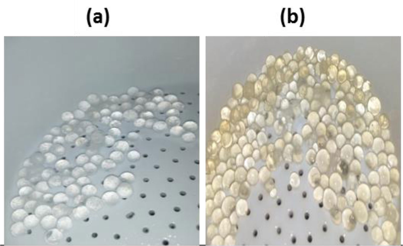
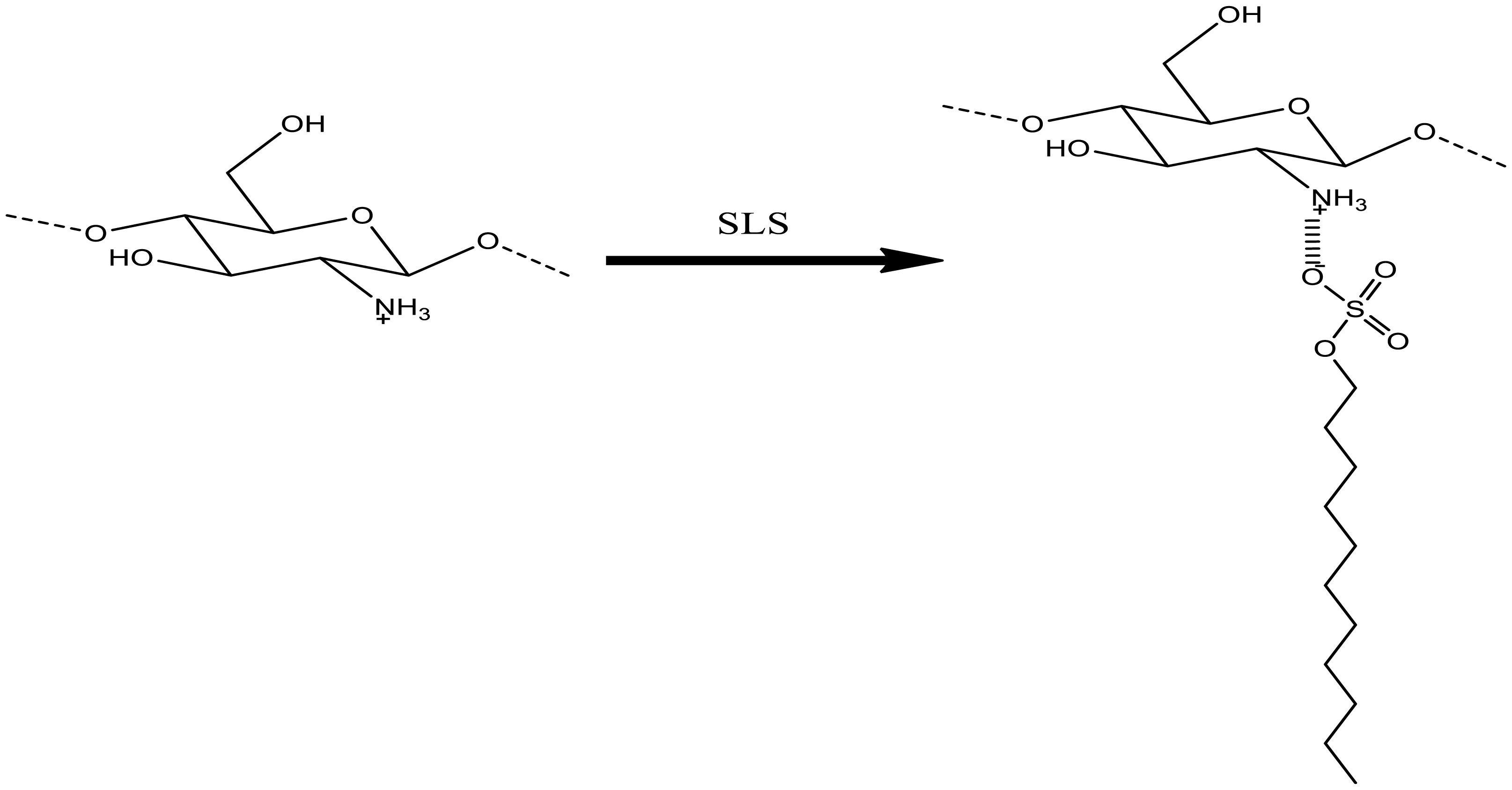
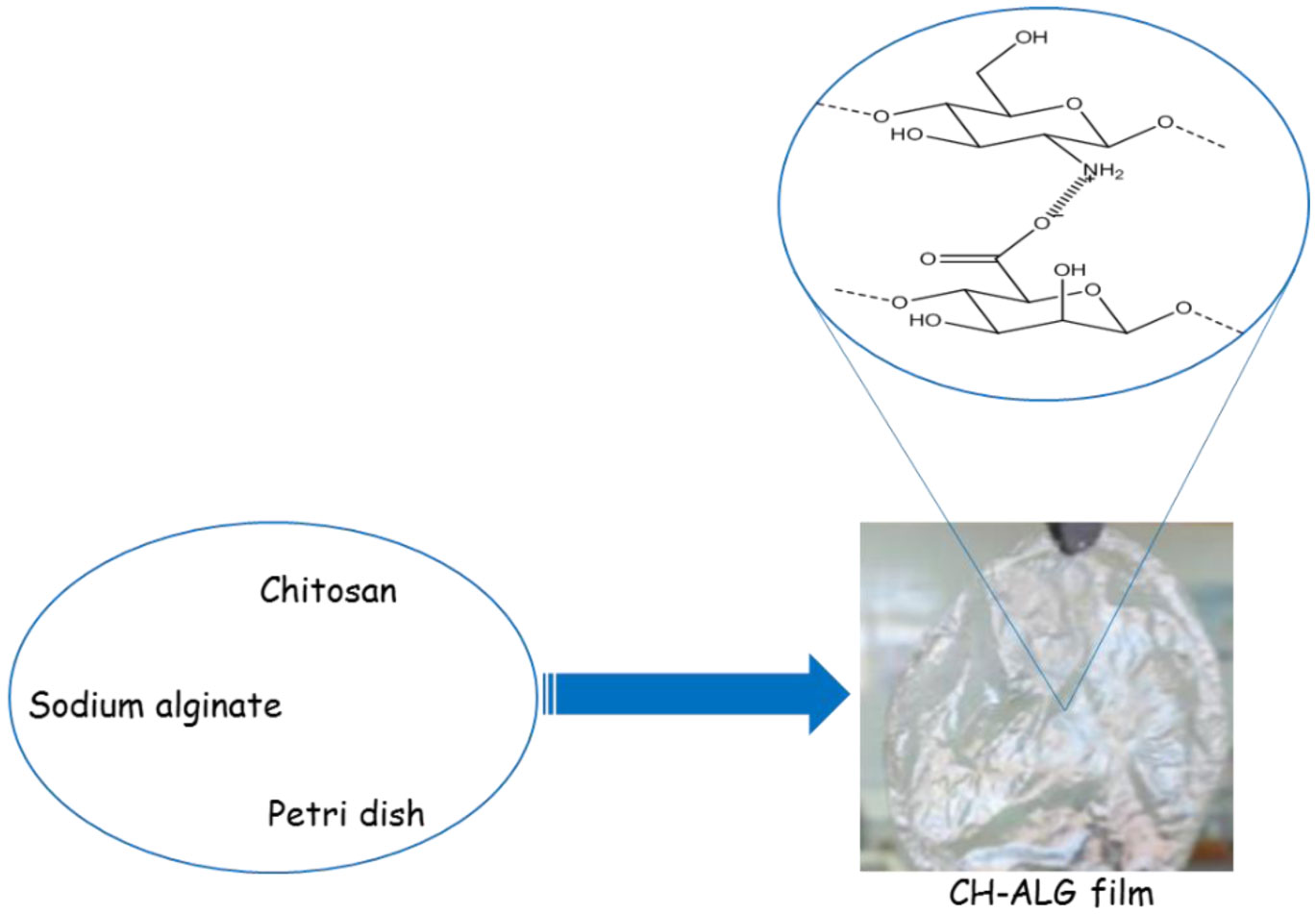
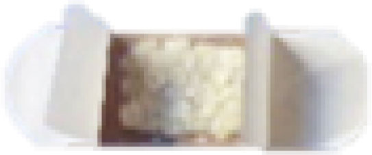
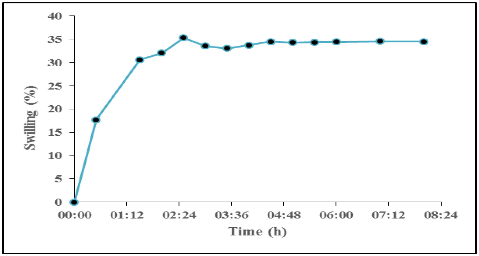
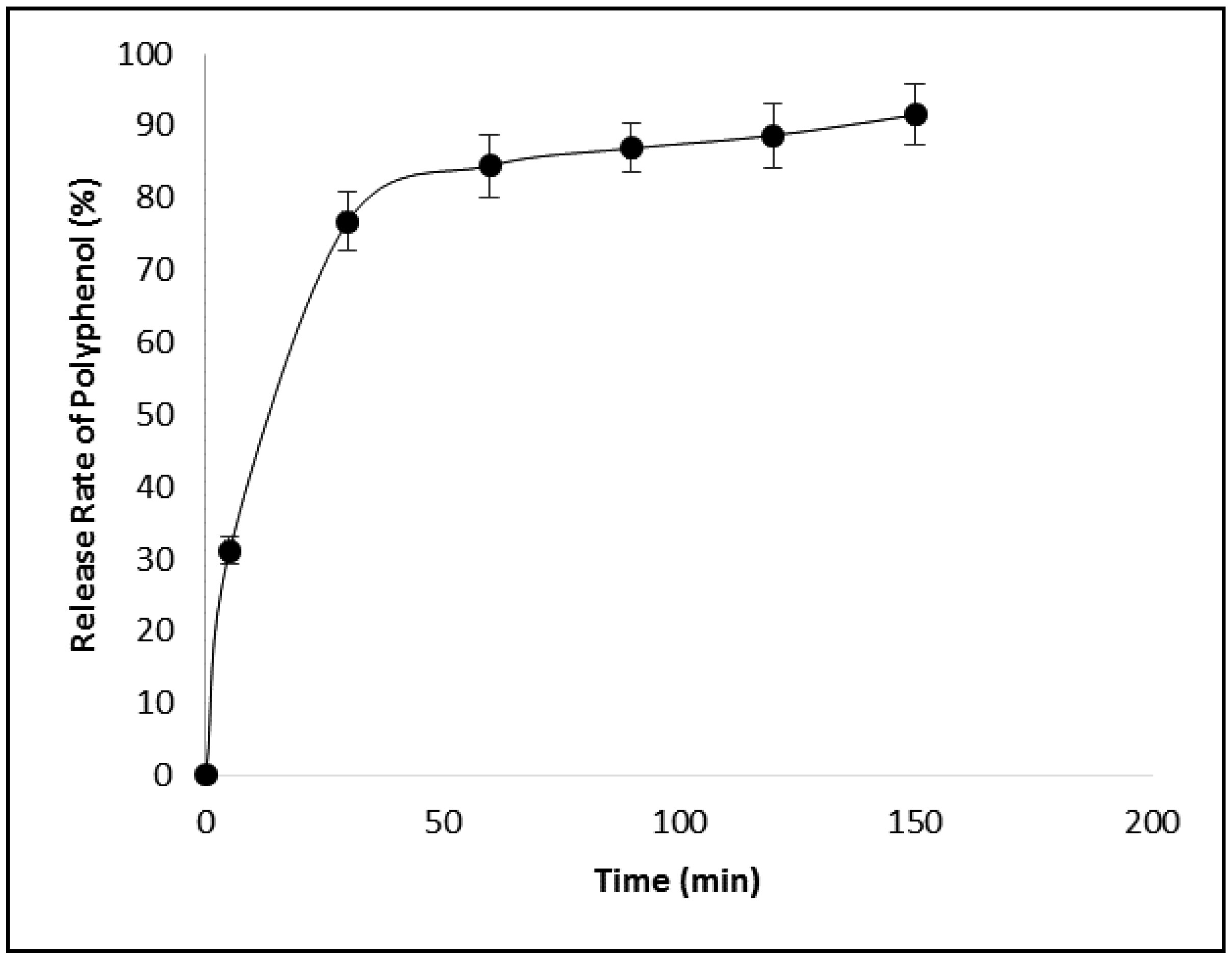
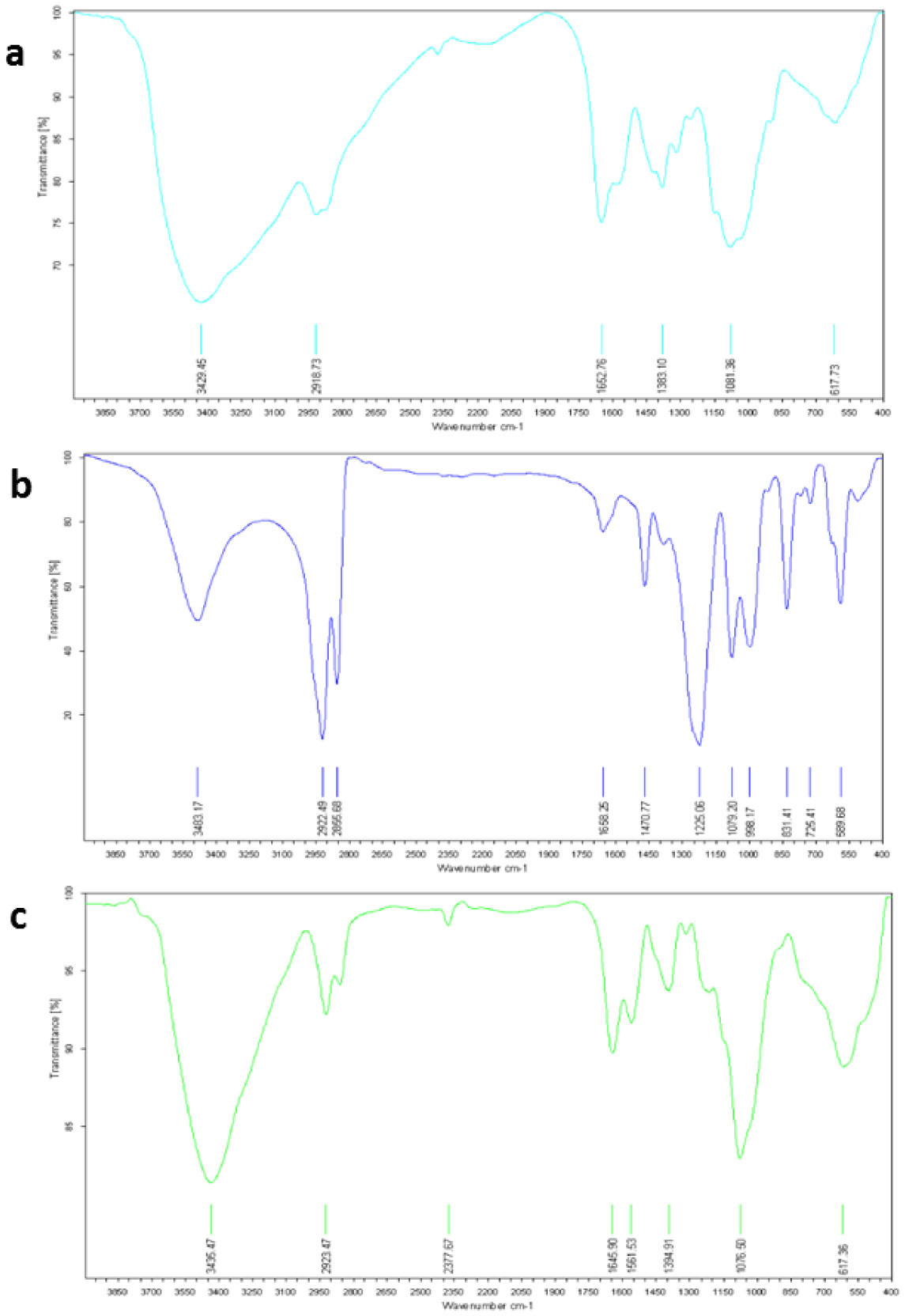
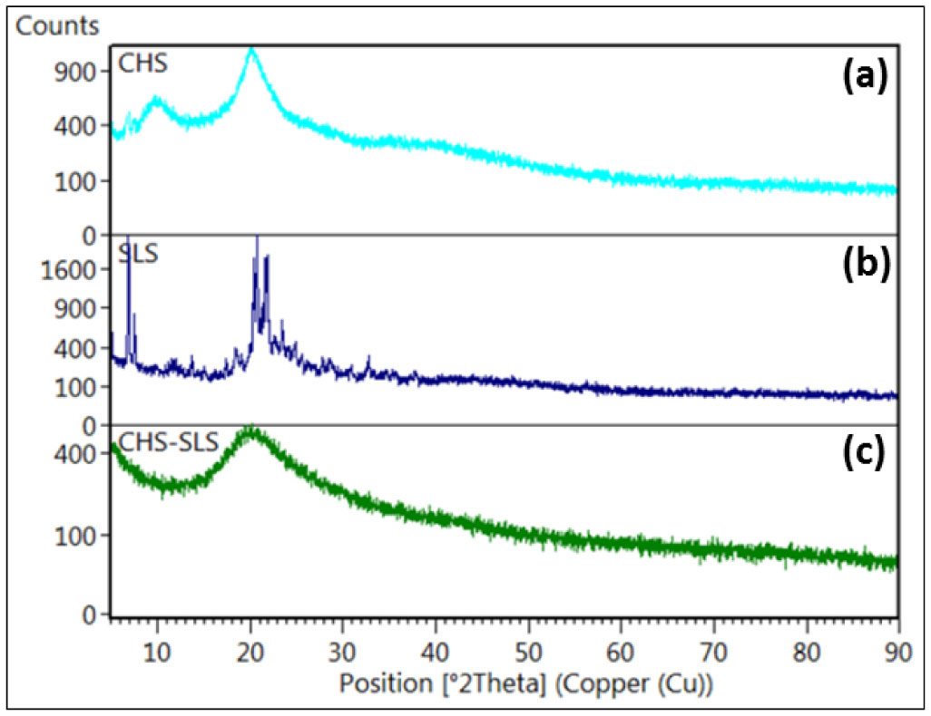
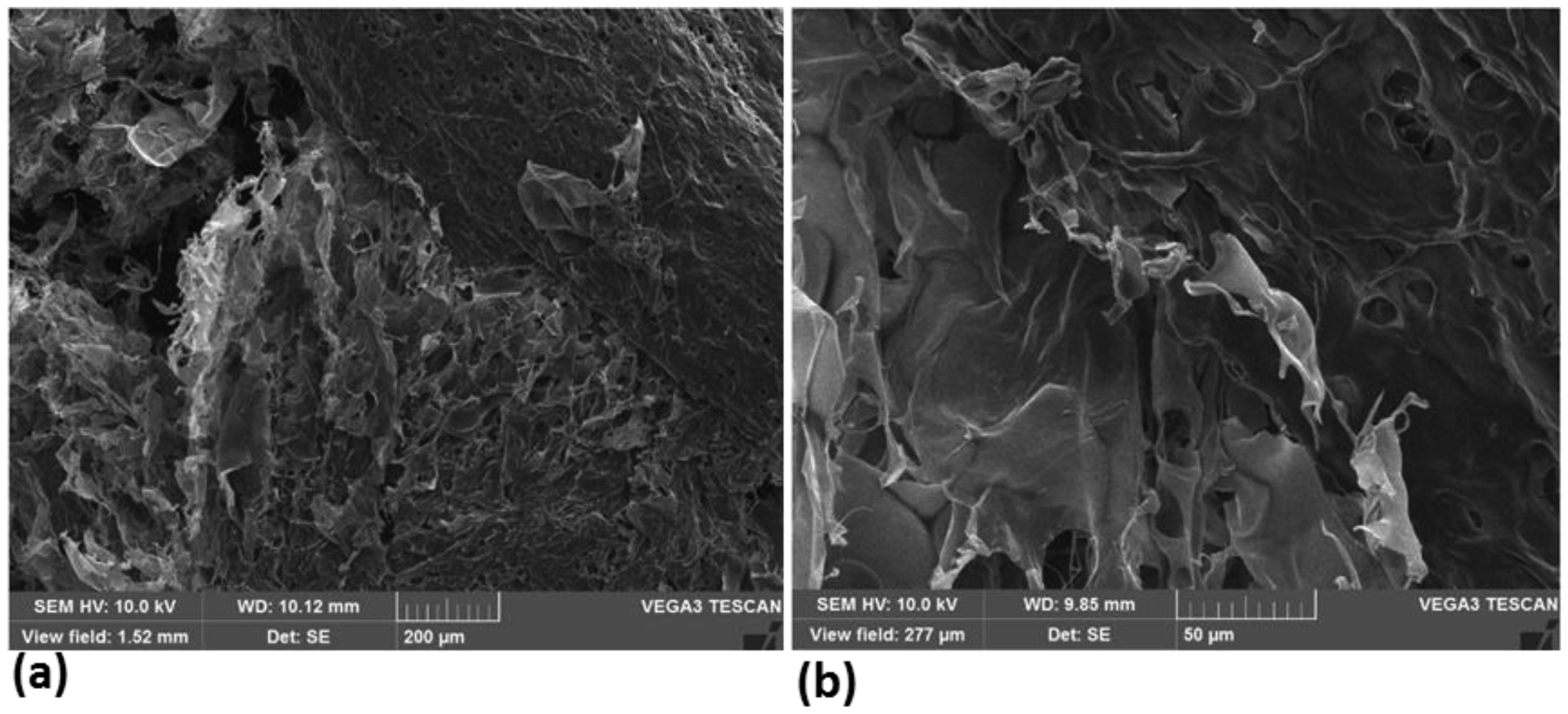
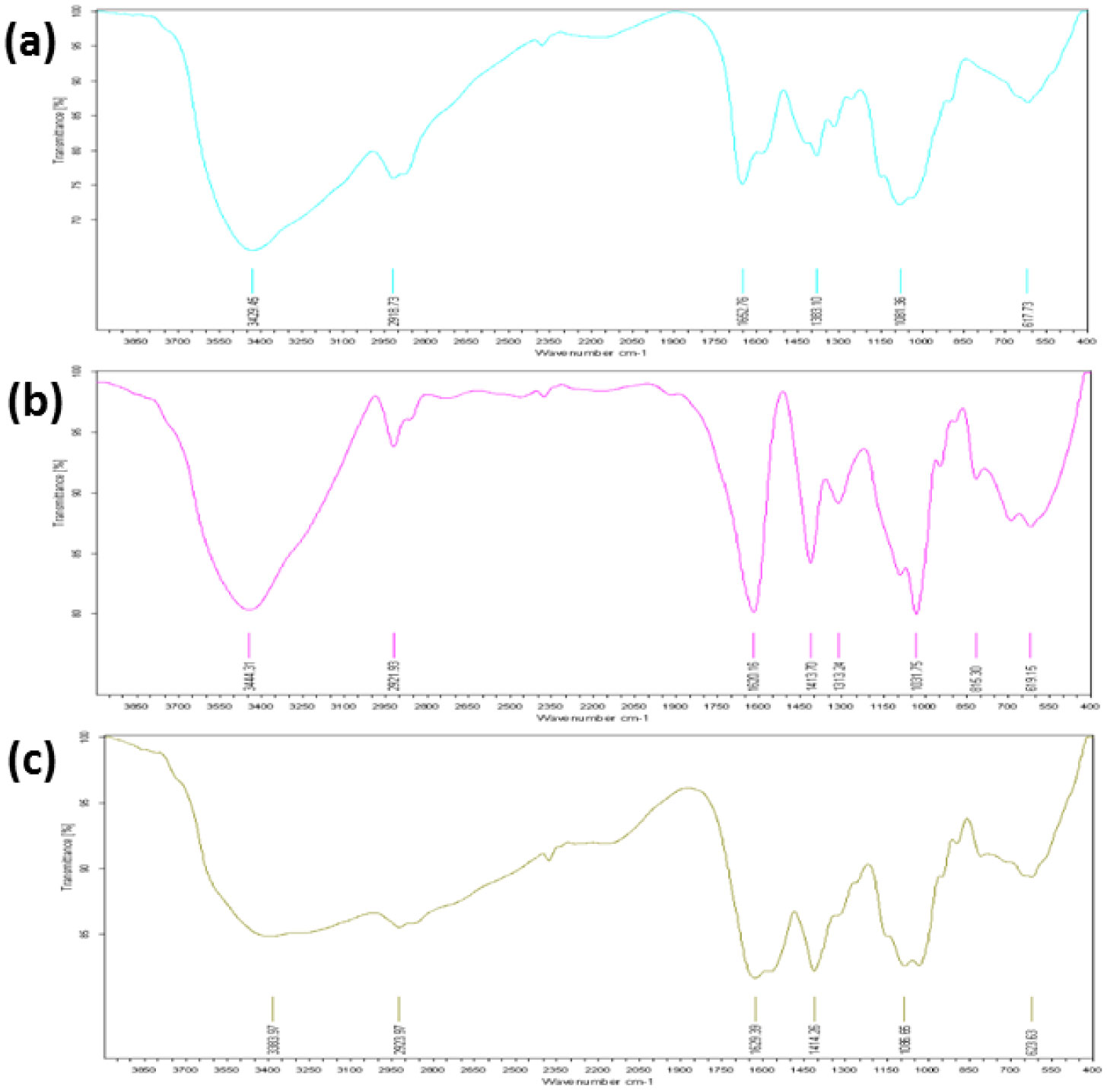
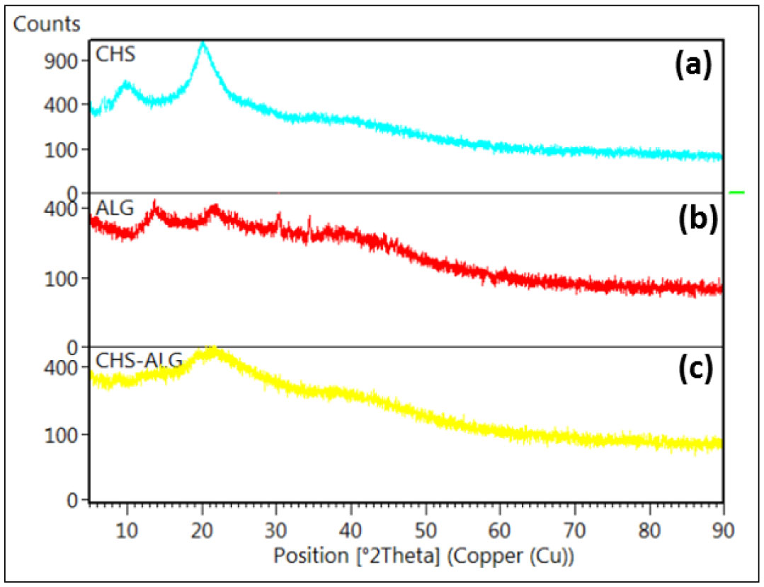
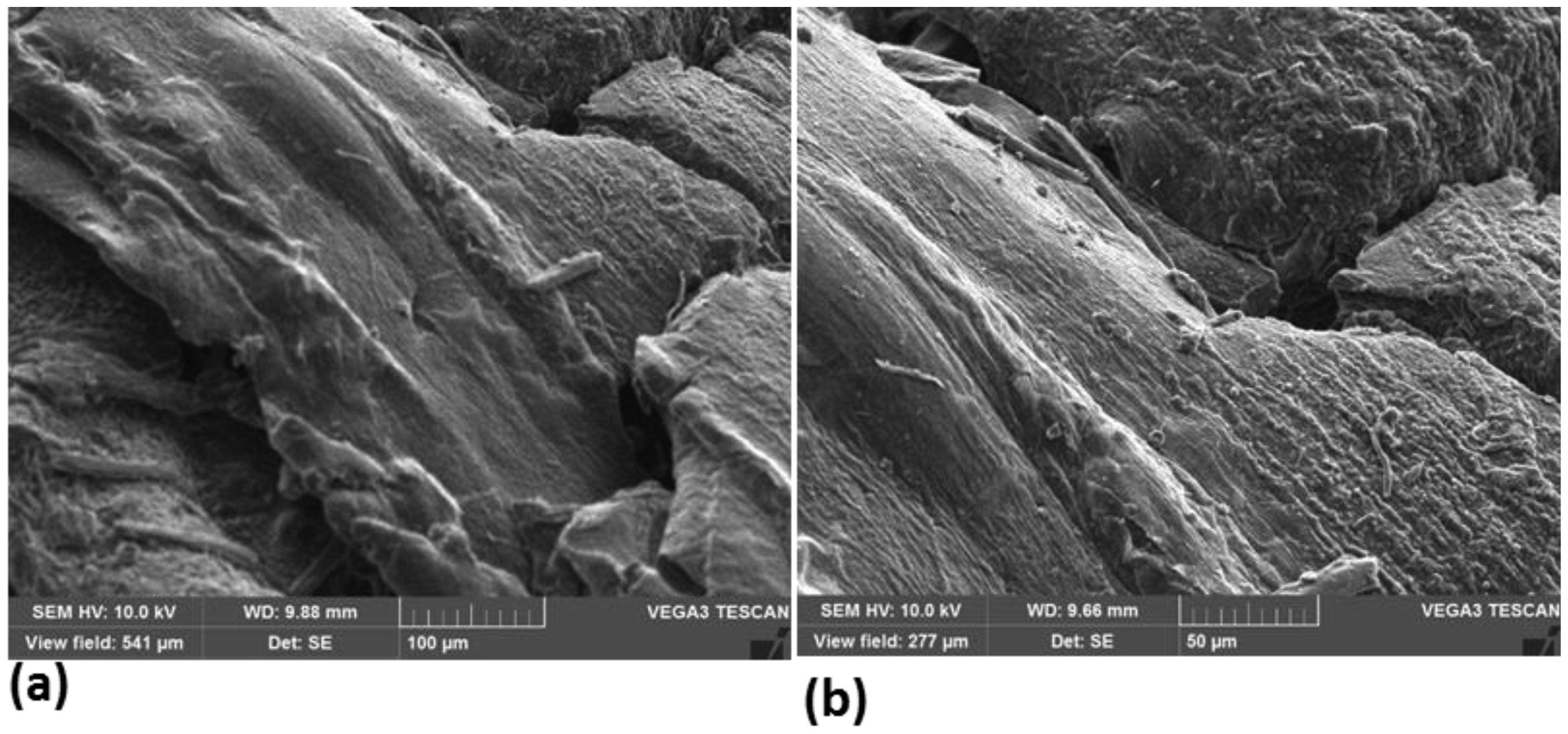
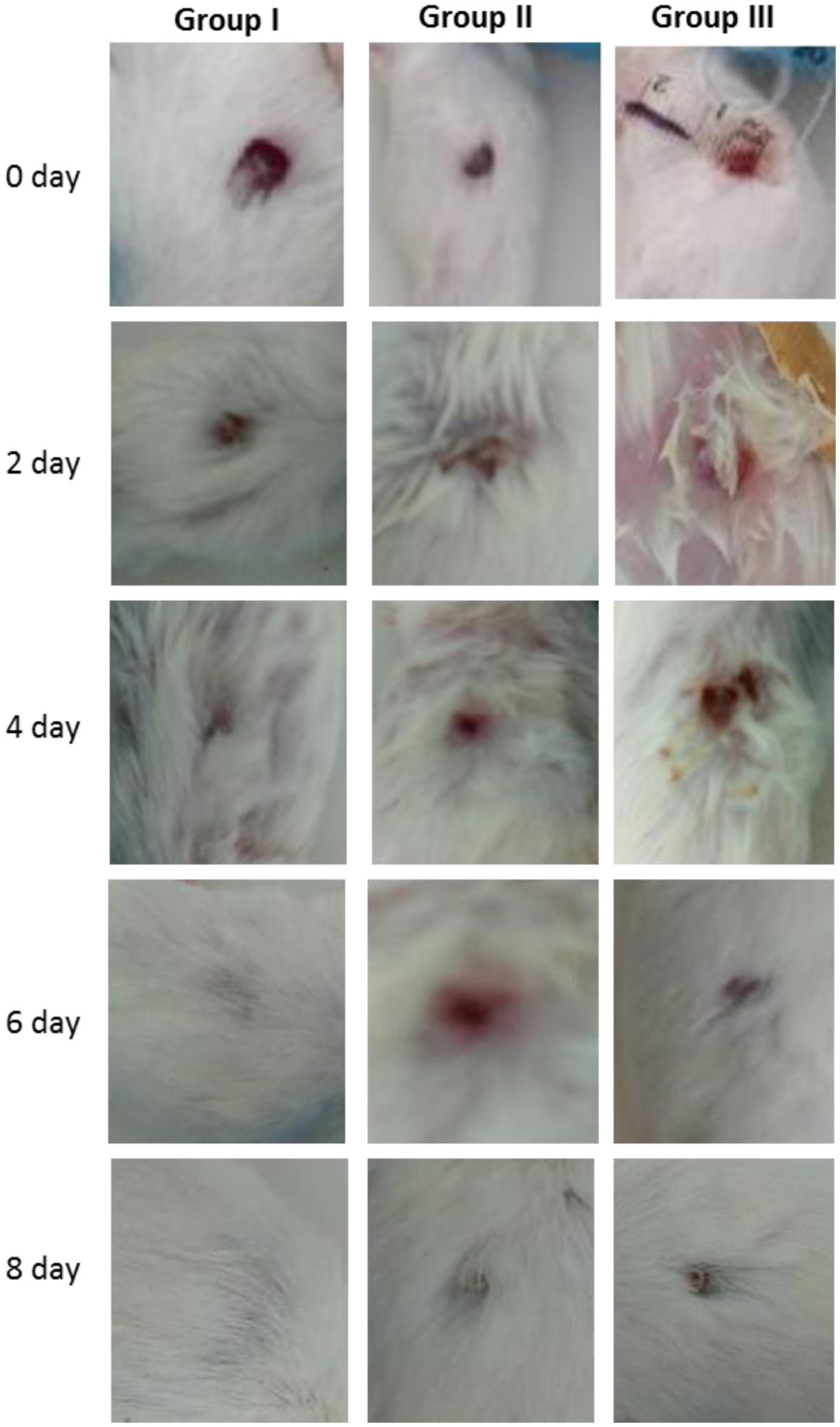
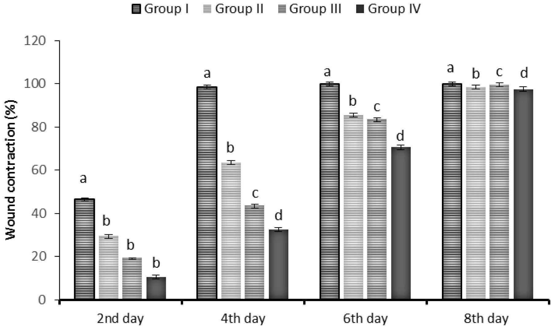
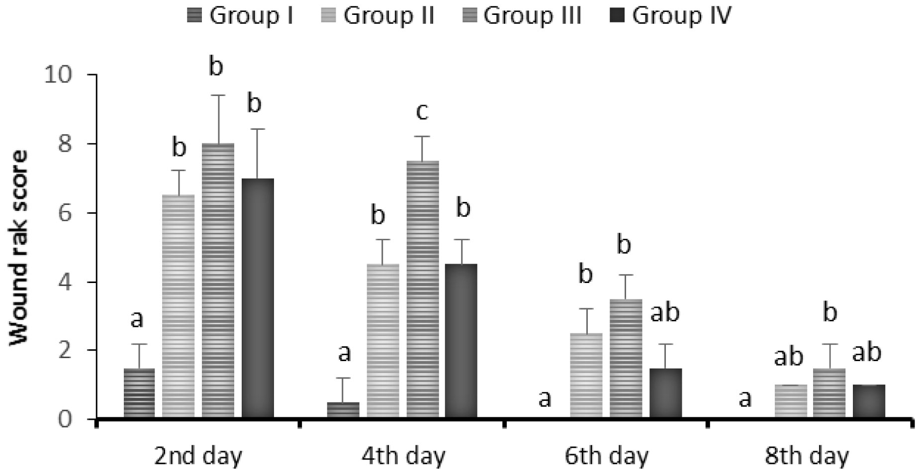
This study aimed to formulate sodium lauryl sulfate cross-linked chitosan beads and sodium alginate-chitosan films for designing a dressing that would shorten the healing time of skin wounds. Teucrium polium extract-loaded chitosan-sodium lauryl sulfate beads (CH-SLS) and chitosan-alginate (CH-ALG) films were prepared and characterized by using Fourier transform infrared spectroscopy (FT-IR), X-ray diffraction (XRD) analysis, and scanning electron microscopy (SEM). The swelling properties of the CH-SLS beads were also analyzed in a water solution. The obtained Teucrium polium extract-loaded CH-SLS beads and CH-ALG films (TBF) were further incorporated into the commercial adhesive dressing. This TBF wound dressing was then investigated for evaluation of its wound healing potential in the mice using the excision wound model. Healing was assessed by the macroscopic appearance and the rate of wound contraction during 8 days. On day 4, the TBF-treated wounds exhibited 98% reduction in the wound area when they were compared with healing ointment, elastic adhesive dressing, and untreated wounds which were exhibited 63%, 43%, and 32%, respectively. Furthermore, the application of TBF dressing reduced skin wound rank scores and increased the percentage of wounds contraction. These results demonstrate that TBF dressing improves considerably the healing rate and the macroscopic wound appearance at a short delay and this application may have therapeutic benefits in wound healing.
Citation: Mariem Kharroubi, Fatima Bellali, Abdelhafid Karrat, Mohamed Bouchdoug, Abderrahim Jaouad. Preparation of Teucrium polium extract-loaded chitosan-sodium lauryl sulfate beads and chitosan-alginate films for wound dressing application[J]. AIMS Public Health, 2021, 8(4): 754-775. doi: 10.3934/publichealth.2021059
This study aimed to formulate sodium lauryl sulfate cross-linked chitosan beads and sodium alginate-chitosan films for designing a dressing that would shorten the healing time of skin wounds. Teucrium polium extract-loaded chitosan-sodium lauryl sulfate beads (CH-SLS) and chitosan-alginate (CH-ALG) films were prepared and characterized by using Fourier transform infrared spectroscopy (FT-IR), X-ray diffraction (XRD) analysis, and scanning electron microscopy (SEM). The swelling properties of the CH-SLS beads were also analyzed in a water solution. The obtained Teucrium polium extract-loaded CH-SLS beads and CH-ALG films (TBF) were further incorporated into the commercial adhesive dressing. This TBF wound dressing was then investigated for evaluation of its wound healing potential in the mice using the excision wound model. Healing was assessed by the macroscopic appearance and the rate of wound contraction during 8 days. On day 4, the TBF-treated wounds exhibited 98% reduction in the wound area when they were compared with healing ointment, elastic adhesive dressing, and untreated wounds which were exhibited 63%, 43%, and 32%, respectively. Furthermore, the application of TBF dressing reduced skin wound rank scores and increased the percentage of wounds contraction. These results demonstrate that TBF dressing improves considerably the healing rate and the macroscopic wound appearance at a short delay and this application may have therapeutic benefits in wound healing.

| [1] |
Hananeh WM, Bani Ismail Z, Alshehabat MA, et al. (2015) Review of animal models used to study effects of bee products on wound healing: findings and applications. Bull Vet Inst Pulawy 59: 425-431. doi: 10.1515/bvip-2015-0062

|
| [2] |
Mayet N, Choonara YE, Kumar P, et al. (2014) A comprehensive review of advanced biopolymeric wound healing systems. J Pharm Sci 103: 2211-2230. doi: 10.1002/jps.24068
|
| [3] |
Muzzarelli RAA (2009) Chitins and chitosans for the repair of wounded skin, nerve, cartilage and bone. Carbohydr Polym 76: 167-182. doi: 10.1016/j.carbpol.2008.11.002
|
| [4] |
Domard A (2011) A perspective on 30 years research on chitin and chitosan. Carbohydr Polym 84: 696-703. doi: 10.1016/j.carbpol.2010.04.083
|
| [5] | Barbucci R (2010) Hydrogels: Biological properties and applications. SSBM . |
| [6] |
Chen SH, Tsao CT, Chang CH, et al. (2013) Assessment of reinforced poly (ethylene glycol) chitosan hydrogels as dressings in a mouse skin wound defect model. Mater Sci Eng C Mater Biol Appl 33: 2584-2594. doi: 10.1016/j.msec.2013.02.031
|
| [7] |
Montembault A, Tahiri K, Korwin-Zmijowska C, et al. (2006) A material decoy of biological media based on chitosan physical hydrogels: application to cartilage tissue engineering. Biochimie 88: 551-564. doi: 10.1016/j.biochi.2006.03.002
|
| [8] |
Ladet SG, Tahiri K, Montembault AS, et al. (2011) Multi-membrane chitosan hydrogels as chondrocytic cell bioreactors. Biomaterials 32: 5354-5364. doi: 10.1016/j.biomaterials.2011.04.012
|
| [9] |
Peluso G, Petillo O, Ranieri M, et al. (1994) Chitosan-mediated stimulation of macrophage function. Biomaterials 15: 1215-1220. doi: 10.1016/0142-9612(94)90272-0
|
| [10] |
Ueno H, Mori T, Fujinaga T (2001) Topical formulations and wound healing applications of chitosan. Adv Drug Deliv Rev 52: 105-115. doi: 10.1016/S0169-409X(01)00189-2
|
| [11] |
Mi FL, Shyu SS, Wu YB, et al. (2001) Fabrication and characterization of a sponge-like asymmetric chitosan membrane as a wound dressing. Biomaterials 22: 165-173. doi: 10.1016/S0142-9612(00)00167-8
|
| [12] |
Jarry C, Chaput C, Chenite A, et al. (2001) Effects of steam sterilization on thermogelling chitosan-based gels. J Biomed Mater Res 58: 127-135. doi: 10.1002/1097-4636(2001)58:1<127::AID-JBM190>3.0.CO;2-G
|
| [13] |
Pandit AP, Koyate KR, Kedar AS, et al. (2019) Spongy wound dressing of pectin/carboxymethyl tamarind seed polysaccharide loaded with moxifloxacin beads for effective wound heal. Int J Biol Macromol 140: 1106-1115. doi: 10.1016/j.ijbiomac.2019.08.202
|
| [14] | Perrine M, Morgane EB, Marie-Christine M, et al. (2007) Hémostase: quels pansements choisir?: Hemostasis: which are the most appropriate dressings? Le Pharm Hosp 42: 193-199. |
| [15] |
Thomas S (2000) Alginate dressings in surgery and wound management—Part 1. J Wound Care 9: 56-60. doi: 10.12968/jowc.2000.9.2.26338
|
| [16] |
Otterlei M, Ostgaard K, Skjåk-Bræk G, et al. (1991) Induction of cytokine production from human monocytes stimulated with alginate. J Immunother 10: 286-291. doi: 10.1097/00002371-199108000-00007
|
| [17] |
Zimmermann U, Klöck G, Federlin K, et al. (1992) Production of mitogen-contamination free alginates with variable ratios of mannuronic acid to guluronic acid by free flow electrophoresis. Electrophoresis 13: 269-274. doi: 10.1002/elps.1150130156
|
| [18] |
Klöck G, Frank H, Houben R, et al. (1994) Production of purified alginates suitable for use in immunoisolated transplantation. Appl Microbiol Biotechnol 40: 638-643. doi: 10.1007/BF00173321
|
| [19] |
Venkatesan J, Lowe B, Anil S, et al. (2015) Seaweed polysaccharides and their potential biomedical applications. Starch-Stärke 67: 381-390. doi: 10.1002/star.201400127
|
| [20] | Tariq M, Ageel AM, Al-Yahya MA, et al. (1989) Anti-inflammatory activity of Teucrium polium. Int J Tissue React 11: 185-188. |
| [21] | Belmekki N, Bendimerad N, Bekhechi C (2013) Chemical analysis and antimicrobial activity of Teucrium polium L. essential oil from Western Algeria. J Med Plants Res 7: 897-902. |
| [22] |
Elmasri WA, Hegazy MEF, Aziz M, et al. (2014) Biofilm blocking sesquiterpenes from Teucrium polium. Phytochemistry 103: 107-113. doi: 10.1016/j.phytochem.2014.03.029
|
| [23] |
Sharififar F, Dehghn-Nudeh G, Mirtajaldini M (2009) Major flavonoids with antioxidant activity from Teucrium polium L. Food Chem 112: 885-888. doi: 10.1016/j.foodchem.2008.06.064
|
| [24] | Ansari R, Sahinfard N, Namjoo A, et al. (2013) Ameliorative property of Teucrium polium on second degree burn. J HerbMed Pharmacol 2: 9-11. |
| [25] | Jamshid M, Jafar N, Bahram M, et al. (2018) The healing effect of combined hydroalcoholic extract of Teocurium polium and the seed hull of Quercus brantii on burn wounds in rats. Int J Med Res Health 5: 232-237. |
| [26] | Hammoudi R, Khenfer S, Medjouel M, et al. (2017) Optimization of extraction conditions for phenolic compounds from Salvia chudaei. Leban Sci J 18: 234. |
| [27] |
Zannou O, Koca I (2020) Optimization and stabilization of the antioxidant properties from Alkanet (Alkanna tinctoria) with natural deep eutectic solvents. Arab J Chem 13: 6437-6450. doi: 10.1016/j.arabjc.2020.06.002
|
| [28] |
Belscak-Cvitanovic A, Komes D, Karlovic S, et al. (2015) Improving the controlled delivery formulations of caffeine in alginate hydrogel beads combined with pectin, carrageenan, chitosan and psyllium. Food Chem 167: 378-386. doi: 10.1016/j.foodchem.2014.07.011
|
| [29] |
Arriola NDA, de Medeiros PM, Prudencio ES, et al. (2016) Encapsulation of aqueous leaf extract of Stevia rebaudiana Bertoni with sodium alginate and its impact on phenolic content. Food Biosci 13: 32-40. doi: 10.1016/j.fbio.2015.12.001
|
| [30] |
Martucci JF, Ruseckaite RA (2009) Biodegradation of three-layer laminate films based on gelatin under indoor soil conditions. Polym Degrad Stab 94: 1307-1313. doi: 10.1016/j.polymdegradstab.2009.03.018
|
| [31] |
Qiu C, Coutinho P, Frank S, et al. (2003) Targeting connexin43 expression accelerates the rate of wound repair. Curr Biol 13: 1697-1703. doi: 10.1016/j.cub.2003.09.007
|
| [32] |
El-Gibaly I, Meki AMA, Abdel-Ghaffar SK (2003) Novel B melatonin-loaded chitosan microcapsules: in vitro characterization and antiapoptosis efficacy for aflatoxin B1-induced apoptosis in rat liver. Int J Pharm 260: 5-22. doi: 10.1016/S0378-5173(03)00149-2
|
| [33] |
Muniyandy S, Yi LM, Santhagunam A, et al. (2020) Chitosan-sodium lauryl sulfate/Eudragit S100 beads loaded with 5-fluorouracil: Influence of solvent and duration of crosslinking the crosslinking on physicochemical properties. Mater Res Express 7: 115402. doi: 10.1088/2053-1591/abc90f
|
| [34] |
Chen P, Zhang WA, Luo W, et al. (2004) Synthesis of superabsorbent polymers by irradiation and their applications in agriculture. J Appl Polym Sci 93: 1748-1755. doi: 10.1002/app.20612
|
| [35] |
Gundogan N, Okay O, Oppermann W (2004) Swelling, elasticity and spatial inhomogeneity of poly (N, N-dimethylacrylamide) hydrogels formed at various polymer concentrations. Macromol Chem Phys 205: 814-823. doi: 10.1002/macp.200300174
|
| [36] |
Salmerón-González E, García-Vilariño E, Ruiz-Cases A, et al. (2018) Absorption capacity of wound dressings: a comparative experimental study. Plast Surg Nurs 38: 73-75. doi: 10.1097/PSN.0000000000000218
|
| [37] |
Li Q, Duan M, Hou D, et al. (2021) Fabrication and characterization of Ca (II)-alginate-based beads combined with different polysaccharides as vehicles for delivery, release and storage of tea polyphenols. Food Hydrocoll 112: 106274. doi: 10.1016/j.foodhyd.2020.106274
|
| [38] |
Yuvarani I, Kumar SS, Venkatesan J, et al. (2012) Preparation and characterization of curcumin coated chitosan-alginate blend for wound dressing application. J Biomater Tissue Eng 2: 54-60. doi: 10.1166/jbt.2012.1037
|
| [39] | Peretz S, Spiroiu F, Anghel DF, et al. (2013) Chitosan microparticulate systems prepared by polymer-surfactant interaction. Rev Roum Chim 58: 275-281. |
| [40] |
Berthold A, Cremer K, Kreuter J (1996) Preparation and characterization of chitosan microspheres as drug carrier for prednisolone sodium phosphate as model for anti-inflammatory drugs. J Control Release 39: 17-25. doi: 10.1016/0168-3659(95)00129-8
|
| [41] |
Al-Remawi MMA (2012) Properties of chitosan nanoparticles formed using sulfate anions as crosslinking bridges. Am J Applied Sci 9: 1091. doi: 10.3844/ajassp.2012.1091.1100
|
| [42] |
Elsayed A, Al-Remawi M, Qinna N, et al. (2011) Chitosan-sodium lauryl sulfate nanoparticles as a carrier system for the in vivo delivery of oral insulin. AAPS PharmSciTech 12: 958-964. doi: 10.1208/s12249-011-9647-5
|
| [43] |
Sankararamakrishnan N, Sanghi R (2006) Preparation and characterization of a novel xanthated chitosan. Carbohydr Polym 66: 160-167. doi: 10.1016/j.carbpol.2006.02.035
|
| [44] |
Wang W, Lu H, Liu Y, et al. (2014) Sodium dodecyl sulfate/epoxy composite: water-induced shape memory effect and its mechanism. J Mater Chem 2: 5441-5449. doi: 10.1039/c3ta15204a
|
| [45] |
Paës G, Chabbert B (2012) Characterization of arabinoxylan/cellulose nanocrystals gels to investigate fluorescent probes mobility in bioinspired models of plant secondary cell wall. Biomacromolecules 13: 206-214. doi: 10.1021/bm201475a
|
| [46] |
Moura MJ, Faneca H, Lima MP, et al. (2011) In situ forming chitosan hydrogels prepared via ionic/covalent co-cross-linking. Biomacromolecules 12: 3275-3284. doi: 10.1021/bm200731x
|
| [47] |
Yan X, Khor E, Lim LY (2000) PEC films prepared from chitosan-alginate coacervates. Chem Pharm Bull 48: 941-946. doi: 10.1248/cpb.48.941
|
| [48] |
Baysal K, Aroguz AZ, Adiguzel Z, et al. (2013) Chitosan/alginate crosslinked hydrogels: Preparation, characterization and application for cell growth purposes. Int J Biol Macromol 59: 342-348. doi: 10.1016/j.ijbiomac.2013.04.073
|
| [49] |
Pallela R, Venkatesan J, Janapala VR, et al. (2012) Biophysicochemical evaluation of chitosan-hydroxyapatite-marine sponge collagen composite for bone tissue engineering. J Biomed Mater Res 100: 486-495. doi: 10.1002/jbm.a.33292
|
| [50] |
Cheng M, Deng J, Yang F, et al. (2003) Study on physical properties and nerve cell affinity of composite films from chitosan and gelatin solutions. Biomaterials 24: 2871-2880. doi: 10.1016/S0142-9612(03)00117-0
|
| [51] |
Li Z, Zhang M (2005) Chitosan-alginate as scaffolding material for cartilage tissue engineering. J Biomed Mater Res A 75: 485-493. doi: 10.1002/jbm.a.30449
|
| [52] |
Li Z, Ramay HR, Hauch KD, et al. (2005) Chitosan-alginate hybrid scaffolds for bone tissue engineering. Biomaterials 26: 3919-3928. doi: 10.1016/j.biomaterials.2004.09.062
|
| [53] | Wang G, Wang X, Huang L (2017) Feasibility of chitosan-alginate (Chi-Alg) hydrogel used as scaffold for neural tissue engineering: a pilot study in vitro. Biotechnol Biotec Eq 31: 766-773. |
| [54] |
Khodja AN, Mahlous M, Tahtat D, et al. (2013) Evaluation of healing activity of PVA/chitosan hydrogels on deep second degree burn: pharmacological and toxicological tests. Burns 39: 98-104. doi: 10.1016/j.burns.2012.05.021
|
| [55] |
Rudyardjo DI, Wijayanto S (2017) The synthesis and characterization of hydrogel chitosan-alginate with the addition of plasticizer lauric acid for wound dressing application. J Phys Conf Ser 853: 012042. doi: 10.1088/1742-6596/853/1/012042
|
| [56] | Halim AS, Keong LC, Zainol, et al. (2012) Chitosan-based systems for biopharmaceuticals: delivery, targeting and polymer therapeutics Hoboken: John Wiley & Sons, Chapter 4: p. 57. |
| [57] |
Rachmawati N, Triwibowo R, Widianto R (2015) Mechanical properties and biodegradability of acid-soluble chitosan-starch based film. Squalen Bull Mar Fish Postharvest Biotechnol 10: 1-7. doi: 10.15578/squalen.v10i1.132
|
| [58] |
Kumar PS, Raj NM, Praveen G, et al. (2013) In vitro and in vivo evaluation of microporous chitosan hydrogel/nanofibrin composite bandage for skin tissue regeneration. Tissue Eng Part A 19: 380-392. doi: 10.1089/ten.tea.2012.0376
|
| [59] |
Boucher F, Château J, Ferry T, et al. (2017) Diagnostic de l'infection d'une plaie chronique et principes de traitement. Revue Francophone Cicatrisation 1: 15-22. doi: 10.1016/S2468-9114(17)30343-2
|
| [60] |
Slotosch CM, Kampf G, Löffler H (2007) Effects of disinfectants and detergents on skin irritation. Contact Dermatitis 57: 235-241. doi: 10.1111/j.1600-0536.2007.01200.x
|
| [61] |
Autore G, Capasoo F, Fuso RD, et al. (1984) Antipyretic and antibacterial actions of Teucrium polium. Pharmacol Res Commun 16: 21-29. doi: 10.1016/S0031-6989(84)80101-0
|
| [62] |
Menichini F, Conforti F, Rigano D, et al. (2009) Phytochemical composition, anti-inflammatory and antitumour activities of four Teucrium essential oils from Greece. Food Chem 115: 679-686. doi: 10.1016/j.foodchem.2008.12.067
|
| [63] |
Guimarães I, Baptista-Silva S, Pintado M, et al. (2021) Polyphenols: a promising avenue in therapeutic solutions for wound care. Appl Sci 11: 1230. doi: 10.3390/app11031230
|
| [64] |
Kaparekar PS, Poddar N, Anandasadagopa SK (2021) Fabrication and characterization of Chrysin—a plant polyphenol loaded alginate-chitosan composite for wound healing application. Colloids Surf B Biointerfaces 206: 111922. doi: 10.1016/j.colsurfb.2021.111922
|
| [65] | Alizadeh AM, Sohanaki H, Khaniki M, et al. (2011) The effect of teucrium polium honey on the wound healing and tensile strength in rat. Iran J Basic Med Sci 14: 499-505. |
Figures(15) / Tables(3)

Mariem Kharroubi, Fatima Bellali, Abdelhafid Karrat, Mohamed Bouchdoug, Abderrahim Jaouad. Preparation of Teucrium polium extract-loaded chitosan-sodium lauryl sulfate beads and chitosan-alginate films for wound dressing application[J]. AIMS Public Health, 2021, 8(4): 754-775. doi: 10.3934/publichealth.2021059


 DownLoad: