Loading [Contrib]/a11y/accessibility-menu.js
Communication

Assessing the utility and limitations of high throughput virtual screening

  • Received: 24 March 2016 Accepted: 15 May 2016 Published: 25 January 2016
  • Due to low cost, speed, and unmatched ability to explore large numbers of compounds, high throughput virtual screening and molecular docking engines have become widely utilized by computational scientists. It is generally accepted that docking engines, such as AutoDock, produce reliable qualitative results for ligand-macromolecular receptor binding, and molecular docking results are commonly reported in literature in the absence of complementary wet lab experimental data. In this investigation, three variants of the sixteen amino acid peptide, α-conotoxin MII, were docked to a homology model of the a3β2-nicotinic acetylcholine receptor. DockoMatic version 2.0 was used to perform a virtual screen of each peptide ligand to the receptor for ten docking trials consisting of 100 AutoDock cycles per trial. The results were analyzed for both variation in the calculated binding energy obtained from AutoDock, and the orientation of bound peptide within the receptor. The results show that, while no clear correlation exists between consistent ligand binding pose and the calculated binding energy, AutoDock is able to determine a consistent positioning of bound peptide in the majority of trials when at least ten trials were evaluated.

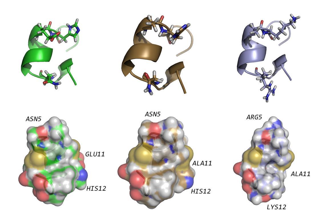
    Citation: Paul Daniel Phillips, Timothy Andersen, Owen M. McDougal. Assessing the utility and limitations of high throughput virtual screening[J]. AIMS Molecular Science, 2016, 3(2): 238-245. doi: 10.3934/molsci.2016.2.238

    Related Papers:

    [1] Yuri Pevzner, Daniel N. Santiago, Jacqueline L. von Salm, Rainer S. Metcalf, Kenyon G. Daniel, Laurent Calcul, H. Lee Woodcock, Bill J. Baker, Wayne C. Guida, Wesley H. Brooks . Virtual target screening to rapidly identify potential protein targets of natural products in drug discovery. AIMS Molecular Science, 2014, 1(2): 81-98. doi: 10.3934/molsci.2014.2.81
    [2] Uday Chintapula, Samir M Iqbal, Young-Tae Kim . A compendium of single cell analysis in aging and disease. AIMS Molecular Science, 2020, 7(1): 49-69. doi: 10.3934/molsci.2020004
    [3] Abdulrahman Mahmoud Dogara, Ateeq Ahmed Al-Zahrani, Sarwan W. Bradosty, Saber W. Hamad, Aisha Abdullahi Mahmud, Hussain D. Almalki, Mustapha Abdullahi, Abubakar Abdullahi Lema, Hasan Nudin Nur Fatihah . In-vitro biological activity and in-silico studies of some volatile phytochemicals from the ethanol extract of Eugenia uniflora. AIMS Molecular Science, 2024, 11(3): 303-321. doi: 10.3934/molsci.2024018
    [4] Mohd Shukri Abd Shukor, Mohd Yunus Abd Shukor . Molecular docking and dynamics studies show: Phytochemicals from Papaya leaves extracts as potential inhibitors of SARS–CoV–2 proteins targets and TNF–alpha and alpha thrombin human targets for combating COVID-19. AIMS Molecular Science, 2023, 10(3): 213-262. doi: 10.3934/molsci.2023015
    [5] Jean Emmanuel Mbosso Teinkela, Philippe Belle Ebanda Kedi, Jean Baptiste Hzounda Fokou, Michelle Isaacs, Lisette Pulchérie Yoyo Ngando, Gaelle Wea Tchepnou, Hassan Oumarou, Xavier Siwe Noundou . In vitro anti-trypanosomal activity of crude extract and fractions of Trichoscypha acuminata stem bark, Spathodea campanulata flowers, and Ficus elastica lianas on Trypanosoma brucei brucei. AIMS Molecular Science, 2024, 11(1): 63-71. doi: 10.3934/molsci.2024005
    [6] Souravh Bais, Renu Kumari, Nirmal Dongre, Aakash Singh Panwar . Insights into neurometabolic diseases. AIMS Molecular Science, 2024, 11(2): 116-139. doi: 10.3934/molsci.2024008
    [7] Marcus Vinícius Dias-Souza, Arthur Azevedo Perpétuo, Gabriel Souza dos Santos, Luiz Felipe Carreiro Machado, Renan Martins dos Santos . Natural products in drug discovery: meeting the urgency for new antimicrobials for human and veterinary use. AIMS Molecular Science, 2023, 10(1): 11-21. doi: 10.3934/molsci.2023002
    [8] Bhuvana Selvaraj, Ganesan Subramanian, Senthil Kumar Ramanathan, Sangeetha Soundararajan, Shettu Narayanasamy . Studies on molecular spectrum of beta thalassemia among residents of Chennai. AIMS Molecular Science, 2022, 9(3): 107-135. doi: 10.3934/molsci.2022007
    [9] Dimitra Beroukas, Maurice Selhorst, Stuart M. Pitson, Dusan Matusica, Ian L. Gibbins, Michaela Kress, Rainer V. Haberberger . Sphingosine kinase 1 in murine dorsal root ganglia. AIMS Molecular Science, 2015, 1(1): 22-33. doi: 10.3934/molsci.2015.1.22
    [10] Bilal Ahmad Mir, P. F. Rahaman, Arif Ahmad . Viral load and interaction of HPV oncoprotein E6 and E7 with host cellular markers in the progression of cervical cancer. AIMS Molecular Science, 2021, 8(3): 184-192. doi: 10.3934/molsci.2021014
  • Due to low cost, speed, and unmatched ability to explore large numbers of compounds, high throughput virtual screening and molecular docking engines have become widely utilized by computational scientists. It is generally accepted that docking engines, such as AutoDock, produce reliable qualitative results for ligand-macromolecular receptor binding, and molecular docking results are commonly reported in literature in the absence of complementary wet lab experimental data. In this investigation, three variants of the sixteen amino acid peptide, α-conotoxin MII, were docked to a homology model of the a3β2-nicotinic acetylcholine receptor. DockoMatic version 2.0 was used to perform a virtual screen of each peptide ligand to the receptor for ten docking trials consisting of 100 AutoDock cycles per trial. The results were analyzed for both variation in the calculated binding energy obtained from AutoDock, and the orientation of bound peptide within the receptor. The results show that, while no clear correlation exists between consistent ligand binding pose and the calculated binding energy, AutoDock is able to determine a consistent positioning of bound peptide in the majority of trials when at least ten trials were evaluated.


    1. Introduction

    High throughput virtual screening (HTVS) is a widely used technique for determining the binding affinity of a large number of ligands to a target receptor 1,2,3. The output of HTVS provides a qualitative comparison of a ligand library, which is to say the ligands can be ranked from best to worst based on receptor binding affinity. The speed and accuracy of HTVS is dependent on how well the binding pocket of the receptor has been characterized. Commonly, the binding pocket for HTVS docking experiments is determined from the crystal structure of a biomacromolecular receptor with bound ligand.

    Jacob et al. (2011) provided a step-by-step description of the process of initiating a docking experiment beginning with a pdb file for a ligand-receptor complex 4. Briefly, the authors obtained the protein database pdb file for Aplysia californica-acetylcholine binding protein (Ac-AChBP) (accession number 2BR8) containing a-conotoxin (a-CTx) PnIA[A10L:D14K] bound to the ligand binding domain. The Cartesian coordinate information specifying each atom of the ligand was removed from the pdb file of the ligand-receptor complex leaving the remaining pdb file consisting of only apoenzyme information. AutoDock Tools was then used to generate a grid parameter file encompassing this binding pocket, allowing for HTVS docking experiments to determine how new a-CTx ligands bind to the Ac-AChBP. HTVS experiments have been used to discover DNA gyrase inhibitors, improve knowledge of dihydrodipicolinate reductase binding paradigms, discover STAT3 inhibitors, optimizing JAK2 inhibitors, kinase inhibitors, G-quadruplex ligands to demonstrate inert group 9 metals inhibiting protein-protein interfaces, and others 5,6,7,8,9,10,11,12.

    In this study, a-CTx peptide ligands were docked to a homology model of the a3β2-nicotinic acetylcholine receptor (nAChR) using DockoMatic version 2.0 software suite 13. The HTVS was conducted using a-CTxMII as a ligand because experimental data supports a very high affinity of the peptide for the a3β2-nAChR isoform (IC50 = 0.5 nM) 14. The sequence for a-CTxMII is GCCSNPVCHLEHSNLC with disulfide bonds connecting Cys2-Cys8 and Cys3-Cys16. The peptide has a well-established a-helix originated at Pro6 and ending at His12. Coordinate files have been deposited in the Research Collaboratory for Structural Bioinformatics (RCSB) 15 representing a-CTxMII from two separate groups; in this investigation we used the original work submitted by Shon et al. 16 rather than Hill et al. 17. The structure generated by Shon et al. was based on traditional aqueous solution conditions for NMR experiments in contrast to the further refinement in structure gained by Hill et al. as a result of solvent condition manipulation to reduce polarity by addition of either acetonitrile or trifluoroethanol. In their work, Hill et al. acknowledge the global fold of the peptide remains the same as that reported by Shon et al. For our purposes, either system would have sufficed as a starting point for computational studies. The structure of a-CTxMII contains electropositive amino acids at either end of the helix (Asn5 and His12) that have been proposed to be significant for ligand to receptor interaction 18,19. Two additional analogs of a-CTxMII were also evaluated, a-CTxMII[E11A] and a-CTxMII[N5R:E11A:H12K].

    Conotoxins are small, cysteine-rich peptides found in the venom of snails belonging to the genus Conus. a-CTxs selectively bind and inhibit nAChRs leading to a great deal of research into their use as analgesics and treatments for Parkinson’s disease 20,21,22. α-CTxMII from the venom of Conus magus, is a well-studied molecular probe for α3β2-nAChRs. The single point mutant a-CTxMII[E11A] was determined to shift binding selectivity from α3β2-nAChR isoforms to preferential binding of α6β2β3-containing nAChRs 23. The triple mutant, a-CTxMII[N5R:E11A:H12K], was designed to have positively charged amino acids at both ends of the α-helix, which was hypothesized to enhance receptor binding affinity while still retaining the E11A selectivity for α6β2β3-containing nAChRs 19. Figure 1 shows the structures of the three peptides, where the top structures indicate the larger size of the triple mutant (right), which measures 16 Å from end-to-end of the α-helix, in comparison to either a-CTxMII (left) or a-CTxMII[E11A] (middle), which are both on the order of 11 Å from end-to-end of the α-helix. The bottom set of structures shows the electrostatic topography of each peptide for visualization of the removal of negative charge (red) in position 11 for the a-CTxMII[E11A] mutant, and the enhancement of positive charge (blue) character for the triple mutant.

    Figure 1.Top, from left to right: distance between amino acid positions 5 and 12 of a-CTxMII (~11 Å), a-CTxMII[E11A] (~11 Å), and a-CTxMII[N5R:E11A:H12K] (~16 Å). Bottom, from left to right: electrostatic topography of a-CTxMII, a-CTxMII[E11A], and a-CTxMII[N5R:E11A:H12K] showing the decrease in negative charge (red) and increase in positive charge (blue) with each successive amino acid mutation

    The aim of this study was to identify the reproducibility and limitations of HTVS for each ligand individually and for the three ligands compared to one another for binding to a homology model of the a3β2-nAChR isoform.


    2. Materials and Methods

    DockoMatic is a comprehensive open source computational application with a graphical user interface (GUI) designed to facilitate HTVS; the docking engine used by DockoMatic is AutoDock v 4.2 24. DockoMatic was installed and made accessible on a high performance computing (HPC) cluster consisting of 32 AMD processors per node with 16 nodes available.

    DockoMatic was then used to generate mutant analogs of a-CTxMII beginning with the pdb file (accession number 1M2C) originating from nuclear magnetic resonance spectroscopy structure elucidation 16. The two mutations to a-CTxMII, a-CTxMII[E11A] and a-CTxMII[N5R:E11A:H12K], were generated using the Treepack program, subordinate to the DockoMatic GUI 4. These three peptides were docked to the α3β2-nAChR for a total of ten trials each. Every trial consisted of 100 AutoDock cycles, resulting in 1000 docking trials per peptide 25.


    3. Results and Discussion

    The results from HTVS provide a qualitative ranking of ligands using a binding energy in kcal/mol as the unit of measurement. Analysis of the lowest free energy for binding (DG) resultant from the ten trials for each of the three peptides showed significant variation (Table 1).

    Table 1.Lowest free energy of binding value for a-CTxMII, a-CTxMII[E11A], and a-CTxMII[N5R:E11A:H12K] docking to a homology model for the a3β2-nAChR over 10 trials
    Conotoxin trial1 trial2 trial3 trial4 trial5 trial6 trial7 trial8 trial9 trial10
    MII −14.96 −16.23 −15.32 −13.78 −16.27 −15.39 −14.53 −14.14 −15.32 −14.69
    E11A −15.98 −17.58 −16.22 −16.25 −16.26 −15.92 −17.58 −16.61 −7.14 −16.55
    Triple −17.3 −3.9 −8.21 −9.02 −16.95 −17.4 −17.34 −17.74 −7.43 −17.92
     | Show Table
    DownLoad: CSV

    The average free energy of binding for the most favorable bound ligand, resultant from 100 docking cycles over all ten trials, for a-CTxMII was −15.063 ± 0.816 kcal/mol, a-CTxMII[E11A] was −15.069 ± 3.034 kcal/mol, and a-CTxMII[N5R:E11A:H12K] was −13.321 ± 5.483 kcal/mol (Table 2). From these data, one could conclude that more favorable interaction with the receptor is achieved for a-CTxMII and a-CTxMII[E11A] as compared to a-CTxMII[N5R:E11A:H12K], but the large standard deviation for the triple mutant indicated docking reproducibility was a problem and more stringent analysis of the results was required. We speculate that the triple mutant is subject to greater variability in docking result for two reasons: (1) the peptide is 5 Å larger than either a-CTxMII or a-CTxMII[E11A] resulting in greater difficulty to bind to the same receptor binding site, and (2) the triple mutant has greater charge character at either end of the peptide, lending itself to a larger number of binding opportunities that may constitute local energy wells rather than global energy minimum binding interactions.

    Table 2.Correlation between average energy for docked ligand-receptor complex and binding pose similarity across ten docking trials consisting of 100 cycles per trial
    Peptide Superimposed Docked Peptide Average ΔG (kcal/mol) Standard Deviation Root Mean Square Deviation (Å)
    MII 1, 6, 8, 9, 10 −15.063 ±0.816 0.510
    E11A 1, 3-8, 10 −15.609 ±3.034 0.526
    Triple 1, 4, 5, 7, 8, 10 −13.321 ±5.483 3.459
     | Show Table
    DownLoad: CSV

    From the data provided in Table 2, it can be seen that the root mean square deviation (RMSD) between similar binding pose peptides for a-CTxMII and a-CTxMII[E11A] were roughly comparable and within specifications that are considered generally acceptable (e.g. ≤ 0.5 Å). These data are in marked contrast to the RMSD for the triple mutant, which is much higher (3.459 Å), indicating greater variability in ligand binding pose between docking trials. It should be noted that trial 2 for the triple mutant was omitted from calculation of average free energy of binding because it was well outside of the standard deviation for calculated free energy, and the bound structure did not converge on a common binding site as was observed for the other ligand structures. As stated previously, the larger size and charge character of the triple mutant is suspected to affect convergence across AutoDock trials. Although AutoDock was the docking engine used in this investigation, we suspect other docking engines will be subject to the same limitations. The reason for this is because HTVS protocols limit the number of rotatable bonds (e.g. AutoDock default value = 32) allowed per docking trial to achieve efficient computational processing. To remove the rotatable bond limitation, requires more computationally intensive investigation which then restricts the number of screening experiments that can be run.

    Protein data base files for the thirty most energetically favorable ligand binding poses (top results over ten trials for the three peptides) were visualized to identify whether inconsistency in binding energy correlated to significant differences in bound ligand positioning. Figure 2 shows the ligand-receptor complexes where the image to the left represents all thirty peptide binding poses for the three ligands and the image to the right provides a view of reproducible docking pose that is conserved for nineteen of the docked structures derived from HTVS.

    Figure 2.HTVS results for all thirty docking trials (left); nineteen of the thirty trials identified a single conserved binding pose (right)

    Close visual inspection of the nineteen ligands with conserved binding pose to the receptor was performed to identify that five of the peptides are a-CTxMII, eight are a-CTxMII[E11A], and the remaining six ligands are a-CTxMII[N5R:E11A:H12K] (Figure 3). In order to limit computational time, AutoDock generates these binding poses in an efficient process by limiting the number of rotatable bonds to 32 for the ligand, and not allowing dynamic motion for the receptor 26. This computational docking method is limited in its ability to optimize side chain interactions between the ligand and the receptor for large systems like those explored in this exercise. As a result, the calculated binding energy for peptide ligands is largely variable because of side chain immobility inherent to docking engines, such as AutoDock.

    The conclusion from this investigation is that HTVS should be performed over multiple trials, followed by visualization of the docking results to identify commonalities in binding pose. The qualitative ranking of ligands in this exercise did not extend between ligands. Thus, it was only through visual inspection of ligand binding pose and validation of binding pose across a minimum of ten trials, that the reproducibility of results could be assessed. To achieve ten trials for each ligand-receptor docking, the authors used DockoMatic, drawing on the docking engine AutoDock. Alternatively, AutoDock Vina, which also takes advantage of parallel processing across computing clusters, could have been used for efficient docking 27. Regardless of the docking engine, it is our finding that manual inspection of results and visual comparison of binding pose across trials is required for confident binding pose identification. When data are available in the RCSB, visual inspection of docking results should be validated by comparison to ligand-receptor structure obtained experimentally by x-ray crystallography. This study suggests that results obtained through HTVS should segue into energy calculation using molecular dynamics simulations where ligand and receptor dynamics are permitted. HTVS can be a valuable tool to achieve a starting point for rigorous and time-intensive molecular dynamics simulation experiments or other more thorough methodologies 28.

    Figure 3.Five of the nineteen peptides with consistent binding pose represent a-CTxMII (left), eight of them are a-CTxMII[E11A] (middle), and the remaining six ligands are a-CTxMII[N5R:E11A:H12K] (right)

    Acknowledgements

    Molecular graphics and analyses were performed with the UCSF Chimera package. Chimera was developed by the Resource for Biocomputing, Visualization, and Informatics at the University of California, San Francisco (supported by NIGMS P41-GM103311). Visualization and structure analysis was also performed with the PyMOL Molecular Graphics System, Version 1.8 Schrödinger, LLC. This material is based in part upon work supported by the National Science Foundation under Grant No. 1229709


    Conflict of interest

    The authors declare no conflicts of interest in the reporting of this study.


    [1] Deng W, Verlinde C (2008) Evaluation of Different Virtual Screening Programs for Docking in a Charged Binding Pocket. J Chem Inf Model 48: 2010-2020. doi: 10.1021/ci800154w
    [2] Klebe G (2008) Virtual ligand screening: strategies, perspectives and limitations. Drug Discov Today 11: 580-594. doi: 10.1016/j.drudis.2006.05.012
    [3] Lavecchia A, Cerchia C (2016) In silico methods to address polypharmacology: current status, applications and future perspectives. Drug Discov Today 21: 288-298. doi: 10.1016/j.drudis.2015.12.007
    [4] Jacob RB, Bullock CW, Andersen T, et al. (2011) DockoMatic: Automated Peptide Analog Creation for High Throughput Virtual Screening. J Comput Chem 32: 2936-2941. doi: 10.1002/jcc.21864
    [5] Jorgensen WL (2004) The many roles of computation in drug discovery. Science 303: 1813-1818. doi: 10.1126/science.1096361
    [6] Clark DE (2008) What has virtual screening ever done for drug discovery? Expert Opin Drug Discov 3: 841-851. doi: 10.1517/17460441.3.8.841
    [7] Liu LJ, Leung KH, Chan DSH, et al. (2014) Identification of a natural product-like STAT3 dimerization inhibitor by structure-based virtual screening. Cell Death Dis 5: 96-104. doi: 10.1038/cddis.2014.250
    [8] Ma DL, Chan DSH, Lee P, et al. (2011) Molecular modeling of drug-DNA interactions: Virtual screening to structure-based design. Biochimie 93: 1252-1266. doi: 10.1016/j.biochi.2011.04.002
    [9] Brenk R, Schipani A, James D, et al. (2008) Lessons learnt from assembling screening libraries for drug discovery for neglected diseases. Chemmedchem 3: 435-444. doi: 10.1002/cmdc.200700139
    [10] Ma DL, Chan DSH, Wei G, et al. (2014) Virtual screening and optimization of Type II inhibitors of JAK2 from a natural product library. Chem Commun 50: 13885-13888. doi: 10.1039/C4CC04498C
    [11] Ma DL, Lai TS, Chan FY, et al. (2008) Discovery of a drug-like G-quadruplex binding ligand by high-throughput docking. Chemmedchem 3: 881-884. doi: 10.1002/cmdc.200700342
    [12] Ma DL, Chan DSH, Leung CH (2014) Group 9 Organometallic Compounds for Therapeutic and Bioanalytical Applications. Acc Chem Res 47: 3614-3631. doi: 10.1021/ar500310z
    [13] Sambasivarao SV, Roberts J, Bharadwaj VS, et al. (2014) Acetylcholine Promotes Binding of alpha-Conotoxin MII at alpha(3)beta(2) Nicotinic Acetylcholine Receptors. Chembiochem 15: 413-424. doi: 10.1002/cbic.201300577
    [14] Cartier GE, Yoshikami DJ, Gray WR, et al. (1996) A new alpha-conotoxin which targets alpha 3 beta 2 nicotinic acetylcholine receptors. J Biol Chem 271: 7522-7528. doi: 10.1074/jbc.271.13.7522
    [15] Berman HM, Westbrook J, Feng Z, et al. (2000) The Protein Data Bank. Nucleic Acids Res 28: 235-242. doi: 10.1093/nar/28.1.235
    [16] Shon KJ, Koerber SC, Rivier JE, et al. (1997) Three-dimensional solution structure of alpha-conotoxin MII, an alpha(3)beta(2) neuronal nicotinic acetylcholine receptor-targeted ligand. Biochemistry 36: 15693-15700. doi: 10.1021/bi971443r
    [17] Hill JM, Oomen CJ, Miranda LP, et al. (1998) Three-dimensional solution structure of alpha-conotoxin MII by NMR spectroscopy: Effects of solution environment on helicity. Biochemistry 37: 15621-15630. doi: 10.1021/bi981535w
    [18] Turner M, Eidemiller S, Martin B, et al. (2009) Structural basis for α-conotoxin potency and selectivity. Bioorg Med Chem 17: 5894-5899. doi: 10.1016/j.bmc.2009.07.005
    [19] McDougal OM, Granum DM, Swartz M, et al. (2013) pKa Determination of Histidine Residues in α-Conotoxin MII Peptides by 1H NMR and Constant pH Molecular Dynamics Simulation. J Physic Chem B 117: 2653-2661. doi: 10.1021/jp3117227
    [20] Armishaw CJ (2010) Synthetic alpha-Conotoxin Mutants as Probes for Studying Nicotinic Acetylcholine Receptors and in the Development of Novel Drug Leads. Toxins 2: 1471-1499. doi: 10.3390/toxins2061471
    [21] Azam L, McIntosh JM (2009) Alpha-conotoxins as pharmacological probes of nicotinic acetylcholine receptors. Acta Pharmacol Sin 30: 771-783. doi: 10.1038/aps.2009.47
    [22] Bordia T, Grady SR, McIntosh JM, et al. (2007) Nigrostriatal damage preferentially decreases a subpopulation of α6β2 nAChRs in mouse, monkey, and Parkinson's disease striatum. Mol Pharmacol 72: 52-61. doi: 10.1124/mol.107.035998
    [23] McIntosh JM, Azam L, Staheli S, et al. (2004) Analogs of α-Conotoxin MII Are Selective for α6-Containing Nicotinic Acetylcholine Receptors. Mol Pharmacol 65: 944-952. doi: 10.1124/mol.65.4.944
    [24] Bullock C, Cornia N, Jacob R, et al. (2013) DockoMatic 2.0: High Throughput Inverse Virtual Screening and Homology Modeling. J Chem Inf Model 53: 2161-2170. doi: 10.1021/ci400047w
    [25] McDougal OM, Cornia N, Sambasivarao SV, et al. (2014) Homology Modeling and Molecular Docking for the Science Curriculum. Biochem Mol Biol Educ 42: 179-182. doi: 10.1002/bmb.20767
    [26] Cavasotto CN, Singh N (2008) Docking and high throughput docking: Successes and the challenge of protein flexibility. Curr Comput-Aided Drug Des 4: 221-234. doi: 10.2174/157340908785747474
    [27] Trott O, Olson AJ (2010) Software News and Update AutoDock Vina: Improving the Speed and Accuracy of Docking with a New Scoring Function, Efficient Optimization, and Multithreading. J Comput Chem 31: 455-461. doi: 10.1002/jcc.21334
    [28] Kufareva I, Handel TM, Abagyan R (2015) Experiment-guided Molecular Modeling of Protein-Protein Complexes Involving GPCRs. Methods Mol Biol 1335: 295-311. doi: 10.1007/978-1-4939-2914-6_19
  • This article has been cited by:

    1. Nitesh Mani Tripathi, Anupam Bandyopadhyay, High throughput virtual screening (HTVS) of peptide library: Technological advancement in ligand discovery, 2022, 243, 02235234, 114766, 10.1016/j.ejmech.2022.114766
    2. Marian Vincenzi, Flavia Anna Mercurio, Marilisa Leone, Virtual Screening of Peptide Libraries: The Search for Peptide-Based Therapeutics Using Computational Tools, 2024, 25, 1422-0067, 1798, 10.3390/ijms25031798
    3. Clement Agoni, Raúl Fernández-Díaz, Patrick Brendan Timmons, Alessandro Adelfio, Hansel Gómez, Denis C. Shields, Molecular Modelling in Bioactive Peptide Discovery and Characterisation, 2025, 15, 2218-273X, 524, 10.3390/biom15040524
  • Reader Comments
  • © 2016 the Author(s), licensee AIMS Press. This is an open access article distributed under the terms of the Creative Commons Attribution License (http://creativecommons.org/licenses/by/4.0)
通讯作者: 陈斌, bchen63@163.com
  • 1. 

    沈阳化工大学材料科学与工程学院 沈阳 110142

  1. 本站搜索
  2. 百度学术搜索
  3. 万方数据库搜索
  4. CNKI搜索

Metrics

Article views(5806) PDF downloads(881) Cited by(3)

Article outline

Figures and Tables

Figures(3)  /  Tables(2)

/

DownLoad:  Full-Size Img  PowerPoint
Return
Return

Catalog