Citation: Neha Sinha, Mark A. Seeley, Daniel S. Horwitz, Hemil Maniar, Andrea H. Seeley. Pediatric Orthogenomics: The Latest Trends and Controversies[J]. AIMS Medical Science, 2017, 4(2): 192-216. doi: 10.3934/medsci.2017.2.192
[1] | Payal A. Shah, John Goldberg . Novel Approaches to Pediatric Cancer: Immunotherapy. AIMS Medical Science, 2015, 2(2): 104-117. doi: 10.3934/medsci.2015.2.104 |
[2] | Ryan D. Davidson, Rebecca Kramer, Sarah Fleet . Satisfaction of telehealth implementation in a pediatric feeding clinic. AIMS Medical Science, 2024, 11(2): 124-136. doi: 10.3934/medsci.2024011 |
[3] | Salma M. AlDallal . Quick glance at Fanconi anemia and BRCA2/FANCD1. AIMS Medical Science, 2019, 6(4): 326-336. doi: 10.3934/medsci.2019.4.326 |
[4] | Maria Wołos, Jan Stachurski . The role of epinephrine in anaphylaxis at all stages of management in pediatric populations. AIMS Medical Science, 2021, 8(2): 80-86. doi: 10.3934/medsci.2021009 |
[5] | Jayson M. Stoffman . Adult learning principles in the development of an academic half day session. AIMS Medical Science, 2024, 11(1): 25-33. doi: 10.3934/medsci.2024002 |
[6] | Auwal Idris Kabuga, Yusuf Ashiru Hassan, Muhammad Ibrahim Getso . Acute infection with measles virus predisposes to mastoiditis with concomitant facial paralysis and neck abscess: A minireview of pathomechanism and diagnostic approach. AIMS Medical Science, 2020, 7(4): 269-277. doi: 10.3934/medsci.2020016 |
[7] | Juliet A Harvey, Sebastien FM Chastin, Dawn A Skelton . What happened to my legs when I broke my arm?. AIMS Medical Science, 2018, 5(3): 252-258. doi: 10.3934/medsci.2018.3.252 |
[8] | Klaus Greier, Clemens Drenowatz, Carla Greier, Gerhard Ruedl, Herbert Riechelmann . Sitting time in different contexts in Austrian adolescents and association with weight status. AIMS Medical Science, 2024, 11(2): 157-169. doi: 10.3934/medsci.2024013 |
[9] | Klaus Greier, Clemens Drenowatz, Carla Greier, Elisabeth Haas, Markus Posch, Gerhard Ruedl, Herbert Riechelmann . Correlates of sedentary behaviors in Austrian children and adolescents. AIMS Medical Science, 2023, 10(4): 291-303. doi: 10.3934/medsci.2023022 |
[10] | Todd T. Nowlen, Luis F. Goncalves, Susan S. Park, Arash A. Sabati . Adolescent Kawasaki Disease patient with coronary calcifications, severe 4-vessel stenosis, and a normal stress MRI. AIMS Medical Science, 2025, 12(1): 28-37. doi: 10.3934/medsci.2025003 |
The twentieth and twenty first centuries have laid the ground work for the Genomics Age [1,2].Perhaps the most defining moment was the discovery of the structural mechanism that supported the DNA (deoxyribonucleic acid) theory of inheritance by Watson and Crick in 1953 where the following was stated, "It has not escaped our notice that the specific pairing we have postulated immediately suggests a possible copying mechanism for the genetic material [3]." The conceptualization of the polymerase chain reaction (PCR) by Karry Mullis led to whole libraries of DNA fragments being constructed [4].The Herculean task of sequencing the entire human genome was conceived in 1990, and to date is one of the largest and most complex scientific collaborations ever undertaken [5]. Knowledge of the human genome has impacted every area of the medical field. Medical disciplines such as oncology and cardiology are advanced in their use of genomics but implementation has been slower in other specialties such as orthopaedics.
Reviews published in the early 2000s focused on the discipline of orthogenomics [6,7,8]. Puzas et al defined an orthopaedic genome as a constellation of thousands of genes that are directly involved in the genesis and maintenance of the musculoskeletal system [7]. In their article they foresaw the possibility of having a genetic score-like a credit score-that would predict the susceptibility to a particular disease. Such ideas have not come to fruition yet.
The application of molecular biology to the clinical realm of pediatric orthopaedics has previously focused on identifying underlying syndromes with a musculoskeletal component. Obtaining a molecular etiology to confirm a diagnosis allows for implementation of appropriate orthopaedic care guidelines and investigation into known non-orthopaedic associated medical conditions that may be pertinent to know prior to a surgical intervention. With advances in genetic technology, however, the medical implications of understanding the genetic make-up of a patient may be greater. Orthopaedic oncology has been the main subspecialty within orthopaedics to utilize molecular genetics for development of a personalized treatment plan. The discovery of various tumor markers has led to better diagnostic, staging and prognostic outcomes [9]. This concept of personalized treatment plans based on molecular genetics should have broader use within orthopaedics. Traditionally, isolated musculoskeletal conditions may not warrant a genetic investigation; however, recent research reveals there may be a role for genetics in pediatric orthopaedics beyond identification of syndromes. The following review summarizes current trends in molecular genomics of common pediatric orthopaedic disorders and alludes to the potential role of genetics in apparently isolated pediatric orthopaedic conditions including scoliosis, talipes equinovarus and congenital limb malformations.
Rapid advances have been made in genomic technologies since the days of banding techniques to visualize genetic material [10,11].Figures 1 and 2 outline the common terms used in genetics along with current analytic techniques.
The first whole genome analytic technique, a karyotype, is a laboratory technique that produces an image of an individual's chromosomes and can identify structural rearrangements or aberrations down to a resolution of 5–10 million base pairs (megabase or Mb) [10]. Introduction of fluorescent in situ hybridization (FISH) increased the resolution at which chromosomal rearrangements could be identified at submicroscopic levels [11]. This technique uses DNA probes that have a fluorescent molecule attached, to detect and locate a specific DNA sequence on a chromosome. The next landmark in the evolution of genetic technologies was the development of comparative genomic hybridization (CGH) which was first introduced in 1992 to study chromosomal abnormalities occurring in solid tumors and other malignancies [12]. Differentially labeled test DNA and normal reference DNA are hybridized simultaneously to normal chromosome spreads and the hybridization is detected with two different fluorochromes. This technique has been refined and array based CGH was developed wherein the patient's DNA is hybridized to a large number of well-characterized probes immobilized on a slide. This remains a useful tool for identifying copy number variants (CNVs) which are regions of chromosomal gain or loss with approximately 100–200 thousand bases (kilobases or kb), that are too small to be detected by karyotype [12,13,14].
Single nucleotide polymorphism (SNPs) refers to variation of a single base pair. The most current form of array based technology incorporates single nucleotide polymorphism (SNP) probes and array CGH into one platform and provides comprehensive clinical testing for both copy number variants CNVs and copy-neutral AOH (absence of heterozygosity) in an efficient manner [15].
If the entire genome was compared to a text book, chromosomal microarrays allow for the detection of missing paragraphs but do not read through the text, letter by letter, assessing for any spelling differences. In order to assess for these changes as small as a single base pair switch, sequencing needs to be performed. The move from Sanger sequencing to next generation sequencing (NGS) has permitted faster and more expansive testing at a lower cost [16]. Due to these benefits, the use of NGS technology has increased dramatically in clinic practice. NGS technology allows for whole exome sequencing and whole genome sequencing. Whole exome sequencing (WES) is the detection of variants in the coding region of approximately 20, 000 genes simultaneously and makes up approximately 1% of the genome, whereas, whole genome sequencing captures the sequence of the entire genetic code that resides in genes and the remaining 99% of non-coding DNA. WES is available clinically and whole genome sequencing remains mainly in the research realm for the moment.
Adolescent idiopathic scoliosis (AIS) is the most common pediatric spinal deformity. It is defined as lateral curvature of the spine greater than 10° with a rotational component and presents in children above 9 years of age [17]. This is differentiated from early onset scoliosis, which occurs at an age less than 9 years and is further divided into infantile (0–3 years) and juvenile idiopathic scoliosis (3–8 years). AIS has a prevalence of 2%–4% [18]. It has also been reported at a significantly higher incidence of 15.8% in first degree relatives of AIS patients [19]. Studies have also shown monozygous twins to have a higher rate of concordance (73%) than dizygous twins (36%) [20]. This higher incidence of AIS within families has led researchers to look for a genetic influence.
Though AIS has been a subject of research for many years, the etiology remains elusive. The past decade has seen a plethora of studies using genetic linkage analysis and candidate gene association analysis to prove the genetic basis of AIS (Figure 3). While the former uses pedigree analysis to track a gene through a family by following the inheritance of a (closely associated) gene or trait, the latter involves the study of genes whose chromosomal locations are associated with a particular disease or phenotype.
Several genes have been reported to be linked to its etiopathogenesis including: estrogen receptors 1 and 2 (ESR1, ESR2) [21,22,23], melatonin receptor 1B (MTNR1B) [24], basonuclin 2 (BNC2) [25], fibrillin 1 and fibrillin 2 (FBN1, FBN2) [26], leptin receptor (LEPR) [27], interleukin-17 RC (IL-17RC) [28], transforming growth factor beta-1 (TGFβ1) [29], matrilin1 (MATRN1) [30,31,32,33,34,35] and tryptophan hydroxylase1(TPH1) [36]. With the increasing discovery of candidate genes, Gorman et al carried out a comprehensive review to summarize genetic associations/linkages with idiopathic scoliosis [37]. Unfortunately, the quality of the association studies may be affected by many factors such as a small sample size, population stratification, racial and ethnic differences, and false positive results [28]. It is now standard practice to replicate these findings in a different population with a larger sample size to validate the results [38].
Genome wide association studies (GWAS) have circumvented the challenges posed by linkage and candidate gene analysis because they measure association between common variations across the entire human genome in the form of SNPs and are used to identify genetic associations with phenotypes; however, they are still susceptible to sample size, variations in population characteristics and statistical issues such as multiple hypothesis testing [39].
Recent research has been focused on delineating the role that LBX1 and GPR126 may play in increasing the susceptibility to AIS [38].
LBX1 (ladybird late, Drosophila homolog of, 1/lady bird-like homeobox) is a homeobox gene and may be implicated in the myogenic and neurogenic etiology of AIS. Its expression during embryogenesis has been shown using mice models. The involvement of LBX1 in spinal cord differentiation and patterning as well as somatosensory signal transduction makes it a strong candidate gene for AIS [40]. A meta-analysis involving Japanese, Han Chinese and Caucasian population confirmed that the SNP rs11190870 is a global AIS susceptibility SNP [41]. This SNP is in a 80kb-LD block and contains 2 genes: LBX1 and FLJ41350. rs11190870 is located in the 3' flanking region of LBX1. This was further validated by Grauers et al using candidate gene analysis and exome sequencing in Scandinavian population [42]. Though scoliosis has not been seen in Lbx1 knockout mice, they have been shown to develop significant muscle loss and cardiac abnormalities along with apoptosis of neurons arising in the dorsal spinal cord [43,44,45,46].
A haplotype refers to a combination of alleles or to a set of SNPs found on the same chromosome. Based on their haplotype analysis Chettier et al [46] concluded that Asian and European populations shared the ancestral haplotype, hap-6, which is associated with sharply increased risk of AIS while a second haplotype, hap-2, seemed to have a protective effect. Individuals carrying both haplotypes showed neither increased nor decreased risk of developing AIS, suggesting that these two have opposing action on the LBX1 locus.
G protein-coupled receptor 126 (GPR126) is another probable susceptible gene, which has been under recent focus. Three SNPs in non-coding regions of GPR126 were found to be strongly associated in the Chinese population [47]. In humans, GPR126 is highly expressed in cartilage. It also has a high expression in the proliferating cartilage of spine in mouse embryos, suggesting its role in spine development [48].
Studies have also linked BNC2 to AIS susceptibility [25,41]. This is a highly conserved protein and belongs to the group of C2H2 zinc finger proteins. In humans BNC2 has been found to be highly expressed in the uterus and the spinal cord, followed by bone and cartilage [25]. BNC2 overexpression in zebra fish embryos has been found to produce axial body curvature associated with abnormal somite formation. In vitro studies have shown that a genomic region containing one of the significantly associated SNPs, rs10738445, has an enhancer activity, which is stronger in the disease associated allele akin to the overexpression in zebrafish. The in vitro and in vivo experiments strongly suggest that increased BNC2 expression predisposes to AIS.
Despite the contribution of the AIS GWASes, no causal variants have been linked to any of the top gene candidates. The GWAS design focuses on common SNPs, most of which do not affect the coding region of the gene. The design is limited in its ability to uncover rare, high impact genetic variations that contribute to development of a disease phenotype [49]. CNVs comprise another type of genetic variation that may play a role in etiologically complex disease like AIS. CNVs account for 13% of the human genome mostly representing variable sized repetitive elements and functionally relevant ones may alter gene copy number, function and regulation leading to recognizable phenotypes [50]. Results of a whole genome study showed that 6% of patients with AIS harbor a clinically important CNV that represent possible risk factors for scoliosis [51].
Whole exome sequencing strategies may help provide a solution to pinpoint these rare variants that segregate within families [49]. HSPG2 encodes for heparin sulfate proteoglycan 2, or perlecan, which is a key component of basement membranes and is strongly associated with musculoskeletal development, both in mouse and in humans [52,53,54]. The known co-localization of perlecan and fibrillin 1 within paraspinal tissues make HSPG2 an excellent candidate gene for IS pathogenesis. Exome sequencing on a multigenerational family with idiopathic scoliosis identified a rare variant in HSPG2 (p.N786S, chromosome 1p36) [55]. Using exome sequencing, Buchan et al identified rare variants in FBN1 and FBN2 (fibrillin1 & 2) as being associated with increased severity of AIS [26]. Of note, pathogenic variants in FBN1 and FBN2 are associated with recognizable genetic conditions, Marfan and Beals syndromes respectively, both of which have severe scoliosis as a key clinical feature [56,57]. Other novel variants have been identified in families with idiopathic scoliosis such as those in POC5 [58] and AKAP2 [59]. Genetic linkage analysis combined with exome sequencing was used to detect a rare missense variant (p.A446T), which results in alterations in the centriolar protein gene POC5 that cosegregated with disease in a family with multiple members with AIS. Other POC5 amino acids alterations (p. A455P and p. A429V) were also identified in families with AIS. Zebrafish models expressing any of the three Poc5 variants resulted in spine deformity providing additional evidence for the role of variants in POC5 in the pathology of AIS [58].
Another study in a Chinese family found a mutation in the AKAP2 gene (leading to amino acid change p. E882A) to co-segregate with AIS phenotype [57]. The AKAP2 gene belongs to the A-kinase anchor protein (AKAP) family. To date, the available information on this gene and its protein is limited. Animal studies have revealed that Akap2 contributes to the development of cartilage explaining how they might contribute to this disorder.
The results of the Bracing in Adolescent Idiopathic Scoliosis Trial (BrAIST) indicate that bracing fails in approximately 15% of patients with AIS [60]. One of the ultimate goals in studying the genetic influence on scoliosis is to try to predict who is going to respond to treatment and who will not so that further management strategies can be pursued. This would require additional studies that focus on genetic variants of responders vs. non-responders since the above studies only focus on predisposition to AIS.
Another problem in AIS is predicting which children who develop a mild curve will progress to scoliosis that requires an intervention—a small percentage of the total. This is a significant issue given that current guidelines recommend serial examinations including radiographs for all children exhibiting a curve. This care is unnecessary for the majority of children who do not show progression after several years of monitoring. A test that could discriminate between children with non-progressive and progressive curves would not only reduce procedures for low risk children, but could signal earlier intervention for children at high risk of progression which could be more effective. Genetics may play an important role in defining the shape of the scoliosis curve as well as the risk of curve progression [38]. In 2010, Ward et al [61] built an algorithm using 53 SNPs to determine risk of spinal curve progression to > 40° for Caucasian patients (ScoliScore, Transgenomics). Initial data supported the discriminatory power of the test, but wider use identified several limitations and there has been variable success with its use [62,63,64]. In 2016, Bohl et al [65] found that ScoliScore predicted final curve better than all traditional methods suggesting that such an approach may be clinically useful. Further research is required into the factors that affect the outcome of brace treatment. Xu et al [66] were the first to report that polymorphisms in estrogen receptor genes and in the TPH1 gene could significantly affect the final outcome of bracing. It is not unreasonable to assume that prior knowledge of the presence of such genetic variations might change the course of treatment in the future.
Limb deficiency or the congenital absence or hypoplasia of long bones/digits vary greatly in their anatomy and their etiology. Despite attempts to standardize descriptions of the malformations, there is inconsistent use of terminology and classification which makes comparisons between studies a daunting task [67]. The range of severity of limb deficiencies is extensive with certain phenotypes being classically linked to a particular etiology. For instance, absence of thumb is strongly associated with Fanconi anemia. Holt-Oram syndrome often presents with hypoplasia of radius and thumb of one or both arms while Thrombocytopenia Absent Radius (TAR) syndrome has absence of the radius with preservation of the thumb [68,69,70,71]. Significant improvements in genetic engineering and genetic manipulation have made it much easier to use model organisms to test the impact of variation in genes yielding discoveries that further our knowledge of the genetics governing limb development. Our current understanding of the molecular basis of human limb development is extrapolated from experiments conducted on animal models. Vertebrate forelimbs and hindlimbs are morphologically similar across species [72]. During the past three-decades molecular analysis of limb bud development in amphibian, chicken and mouse embryos defined the essential functions of most morpho-regulatory genes and pathways [73,74,75]. The limb bud develops along 3 axes: proximal-distal, dorsal-ventral and radial-ulnar/anteroposterior. Each dimension is guided by different group of specialized cells. These are the apical ectodermal ridge (AER) for proximal to distal development, the progress zone (PZ) for dorsal ventral differentiation and the zone of polarizing activity (ZPA) for radial-ulnar differentiation. Several gene families are involved in the spatially and temporally coordinated growth and differentiation of the developing limb bud. The gene products act as signals to activate other downstream genetic pathways. Most genes influence the initiation and patterning of both upper and lower limbs but some genes are differentially expressed in the forelimbs and hindlimbs [72].
Gene families which have been so far implicated in taking part in this intricate pathway are FGF (fibroblast growth factor), SHH (sonic hedgehog), BMP (bone morphogenetic protein), WNT (wingless-type MMTV integration site) and transcription regulator genes like HOX (homeobox) genes, T BOX genes, SALL (Sal like), b HLH (basic helix-loop-helix), LMX (Lim homeobox), DLX (distal less homeobox) [73].
The proximal distal axis is determined by the FGF pathway. There are 22 known FGF genes that have been identified based on sequence homology. Many of the developmental functions of Fgf signaling seem to be conserved between mice and humans [76]. In Fgf10 deficient mice, limb bud is initiated but neither the Apical Ectodermal Ridge (AER) nor the Zone of Polarising Acitvity (ZPA) is formed resulting in complete truncation of forelimbs and hindlimbs [77]. Inactivation of Fgf8 in early limb causes substantial reduction in limb bud size, a delay in Shh expression, misregulation of Fgf4 expression and hypoplasia or aplasia of specific skeletal elements. Inactivation of both Fgf4 and Fgf8 results in complete arrest of limb bud and limb agenesis [78,79]. In humans, mutations of genes encoding FGF receptors have been identified in certain chondrodysplasias, including achondroplasia, and in a few craniosynostoses associated with limb anomalies [77]. Heterozygosity for point mutations in FGFR1 and FGFR2 results in broadening of the radial most digit (thumb), brachydactyly and syndactyly associated with Pfeiffer syndrome. A similar phenotype is seen in Apert syndrome which is caused by two predominant point mutations in FGFR2 [73].
The Hedgehog genes encode proteins which play a key role in the establishment of anteroposterior polarity of limbs. Numerous molecules are involved in the SHH signaling pathway and their anomalies are suspected to play a causal role in human limb malformation. The SHH protein is covalently linked to cholesterol during its maturation process. Serum cholesterol drops in Smith-Lemli Opitz syndrome may be explained by faulty maturation of SHH and subsequent development of an unusual Y-shaped syndactyly of the second and third toes, short proximally placed thumb and post axial polydactyly seen in this syndrome [77].
BMPs belong to the transforming growth factor β (TGFβ) superfamily. Their function which occurs downstream of the SHH pathway is of paramount importance and they are key regulators in determining the anteroposterior axis of limbs. Gremlin1 (Grem1) is its extracellular antagonist and is required to establish and propagate the SHH/GREM1/AER-FGF signaling pathway that takes place between the mesenchyme and AER [80]. Limb Malformations in the form of hypodactyly and fusion of the radius and ulna take place in Grem1 deficient mice [81]. In humans, mutations in the cis regulatory regions GREM1 cause an autosomal recessive condition characterized by synostosis of ulna and radius, hemimelia, syn and hypodactyly along with renal abnormalities [82]. Mutations of genes encoding the BMP antagonist NOGGIN or GDF5 (Growth and differentiation factor) presents with symphalangism, multiple synostosis and brachydactyly [83,84].
The WNT family members are involved in a signaling pathway that regulates many processes in limb development. Variants in WNT7A, which controls patterning of the dorso-ventral and AP axis [72,73] can cause Fuhrmann syndrome which clinically manifests as bowed femurs, aplasia or hypoplasia of the fibula and ulna and syn/hypodactylies or Schinzel phocomelia syndrome which comprises aplasia of the ulna and fibula along with phocomelia and ectrodactyly/hypodactyly [85]. Mutations in genes in the WNT signaling pathway have also been linked to tetra-amelia and Split Hand Foot Malformation syndrome [73].
Transcriptional Regulator genes play a role in limb morphogenesis. There are 39 members in the HOX gene family which are then grouped in 4 clusters. These genes are fundamental to the positioning of the developing wings in the embryos of both vertebrates and invertebrates. Specific HOX genes become upregulated by retinoic acid in areas where the limb buds will develop [72]. Mouse models lacking Hoxa and Hoxd gene clusters show severely truncated limb buds. In humans, nonsense and missense variants of the coding regions of HOXD13 and HOXA13 results in syndactylies and hand foot genital syndrome (HFGS). It presents with malformations of hand (short first metacarpal, distal phalanx of the thumb and middle phalanx of the fifth finger, fusion of retarded maturation of carpus), feet (short first metatarsal and distal phalanx of the great toe) and urogenital anomalies [86,87].
The T Box genes play an important role in establishing the fore and hind limb bud identities [72,88]. There are seven T box genes that are implicated in limb morphogenesis: T, Tbx2, Tbx3, Tbx4, Tbx5, Tbx15 and Tbx18 [89]. T misexpression models in chick models have shown phenotypic changes in skeletal pattern including anterior digit duplications, posterior transformation of anterior digits and thickening of the tibial metatarsal. Similar experiments have also shown that Tbx2 and Tbx3 expression is required for patterning the posterior digits [90]. Tbx3 deficient mice display ulna and digit defects and hindlimbs are severely truncated. This resembles the Ulnar mammary syndrome in humans resulting from mutations in TBX3 and manifesting with a range of abnormalities of the ulnar ray of the upper limb (agenesis or duplication of the fifth finger) and lower limbs (hypoplasia of the fourth and fifth toes) along with apocrine and mammary gland defects [91,92]. In a separate study, the role of Tbx3 as a determinant in axial positioning of the limb was highlighted. When present as a transcriptional repressor in the forelimb it results in a rostral shift in the position of the limb along the main body axis. By contrast, a transcriptional activator form of Tbx3 shifts the limb to more caudal locations [93].
While Tbx4 is expressed in hindlimb, Tbx5 is expressed by the forelimb. Their significance is highlighted in experiments where Tbx4 absence in mouse embryos leads to complete absence of hindlimbs. Delayed inactivation of this gene leads to pelvic anomalies, absence or severe reduction of femur and dysplasias of anterior digits in hindlimbs. A corollary is seen in the pelvic finless zebrafish model. These are a naturally occurring zebrafish strain in which the development of the pelvic fins (the teleost equivalent of hindlimbs) fails. It has now been determined that polymorphisms in Tbx4 are responsible for the specific and complete absence of pelvic fins [94]. In Tbx5 knockout mice there is complete failure of forelimb elements to form. In zebra fish the absence of Tbx5 leads to the loss of pectoral fins [95]. Mutations in Tbx5 is implicated in Holt Oram syndrome [88], where patients present with varied cardiac anomalies along with limb defects of radial ray of the upper limbs (hypoplastic, absent or triphalangeal thumb, agenesis or hypoplasia of the radius).
The SAL-LIKE Zinc Finger transcriptional repressor cluster of genes (SALL1, SALL3, SALL4) are co-expressed in distal limb bud mesenchyme. Experimental studies indicate their participation in various signaling pathways of limb development. Mutations in SALL1 have been identified in Townes Brocks Syndrome in humans which is characterized by anal imperforation and malformations of the ear, hands (preaxial polydactyly, triphalangeal thumb) and urogenital tract [96,97]. Various mutations in SALL4 give rise to a condition called Duane Radial Ray Syndrome. The different limb phenotypes include tri phalangeal thumbs, varying degree of pre-axial polydactylies, reduction or absence of the radius or ulna, phocomelia and shoulder girdle defects [98].
Split-hand foot malformation syndrome (SHFM), also known as ectrodactyly, is a congenital limb defect. Its clinical presentation varies significantly and includes hypoplasia of a single phalanx, aplasia of one or more central digits (classical cleft also known as lobster-claw anomaly) and aplasia of both central and preaxial rays of the autopod amongst others. To date, seven chromosomal loci associated with isolated SHFM have been described, i.e., SHFM1 to 6 and SHFM/SHFLD. Most of the known SHFM loci are associated with chromosomal rearrangements that involve small deletions or duplications of the human genome [99]. Genome Wide Linkage analysis for Split Hand Foot Malformation syndrome with long bone deficiency (SHFD) has identified two novel susceptibility loci on chromosome 1q42.2–q43 and 6q14.1 in a large Arab family [100]. Further genetic heterogeneity for ectrodactyly has been suggested with the presence of an additional SHFM locus in chromosome region 8q21.11–q22.3 [101].
Research into the underlying molecular concepts of errors in this complicated network that leads to limb development funds our knowledge about various limb malformations and may lead to targeted interventional strategies in the future. It is interesting to note that often the same gene is implicated in an isolated limb deformity as well as a syndromic limb deformity. While an isolated limb deformity may have straight forward management, it should prompt a consultation with a geneticist in order to rule out a syndrome that could lead to other treatment recommendations, but also to provide counseling to the family about recurrence, which is of relevance even in isolated limb defects.
Congenital talipes equinovarus (CTEV), also called isolated clubfoot is one of the most common orthopaedic birth defects and is clinically characterized by forefoot adduction, hind foot varus, midfoot cavus and hind foot equines [102]. CTEV occurs in 1 per 700/1000 live births in the United States but the birth prevalence differs substantially among different racial and ethnic groups [103,104,105]. Severe calf muscle hypoplasia is often present and persists even after corrective treatment [106,107].
Both environmental and genetic factors are speculated to play a role in the etiology of CTEV (Figure 4). Environmental influences including maternal smoking [108], maternal diabetes [109] as well as early amniocentesis have been positively correlated to CTEV. With smoking being strongly linked with CTEV, studies were conducted to elucidate the role of genetic variations in smoking metabolism. The chemicals constituting cigarette smoke are metabolized by xenobiotic metabolism and most of these compounds can freely cross the placenta [110]. Fetal concentrations of nicotine have been found to be generally 15% higher than maternal levels [111]. Hect et al examined N-Acetyltransferase genes, NAT1 and NAT2, which are responsible for metabolism of tobacco byproducts [112], and they reported slow acetylation to be a risk factor for CTEV. In a later study, Sommer et al concluded that xenobiotic genes are unlikely to play a major role in clubfoot, but perturbation of this pathway may play a contributory role [113].
Although environmental risk factors likely have a strong influence in CTEV, the higher concordance of CTEV among monozygotic twins (33%) as compared to dizygotic twins (3%) lends impetus to the underlying genetic basis of the pathology, although to a lesser degree than that seen in AIS [114]. This has been substantiated in studies also reporting a higher prevalence among first degree relatives of affected individuals [115,116].
Various genes are thought to play a contributory role in the etiology of CTEV. PITX1 is one such gene. PITX1, paired like homeodomain1, is a transcription factor. A missense variant in PITX1 leads to syndromic clubfoot [117]. Another study provided evidence that PITX1 haploinsufficiency (where only one copy of the gene is functional) may be linked to isolated clubfoot, and closely mimics the lower limb deformity in humans [118].
The T box family genes have also been an area of interest as a potential etiologic cause. Both TBX3 and TBX4 have been shown to be susceptible genes for clubfoot. The TBX3 protein is a transcriptional repressor and mutations are known to affect limb development [119]. TBX4 shares a similar structure with TBX3. It is expressed in the hindlimb and is involved in limb muscle and tendon patterning [120]. The opinion on the role of TBX4 in CTEV is controversial. Alvarado et al concluded that microdeletions/microduplications found in the TBX4 gene are associated with clubfoot [121]. However another study which looked into the role of possible correlation between the TBX4 gene and CTEV reported minimal evidence indicating an association between the two [122]. The contribution of genetic variants in the PITX1 and TBX4 need to be thoroughly investigated to determine a causal role in the etiology of CTEV.
Apoptosis has also been hypothesized to play a significant part in the pathogenesis of CTEV. Apoptosis is the process of programmed cell death where the cell nucleus actively induces its own death. Cysteine dependent aspartate directed proteases (Caspases) belong to the family of cysteine proteases that play an essential role in apoptosis [123]. In 2005, Heck et al reported that a major allele variant in CASP10 is associated with CTEV in simplex white and Hispanic trios [124]. Studies examining the role of mitochondrial apoptotic genes looked into SNPs in each of the genes (CASP3, CASP8, CASP9, CASP10, BID, BCL2 and APAF1) and provided evidence implying correlation with CTEV [125]. It is plausible that apoptosis in the placenta may indirectly result in CTEV. Any imbalance in the process of apoptosis leads to placental dysfunction thereby hindering the supply of nutrients and oxygen to the fetus [126]. This results in fetal blood redistribution leading to decreased amniotic fluid volume, restricting the growth and movement of the fetus [127,128]. All the above has been postulated to affect the development and position of the lower limbs [129].
Many researchers have been successful in inducing clubfoot in mice models using ATRA (all trans-retinoic acid) [102]. The causative role of apoptosis is further strengthened by the significantly higher expression of the pro-apoptotic genes like BAX and caspase 3 at both the mRNA and protein level observed in the ATRA exposed animals. They concluded that their findings not only indicated an association between placental apoptosis and congenital clubfoot, but also reveal that anti apoptotic therapy may reverse the progress of apoptosis in the placenta and recover the function of placenta, which may have some curative effect on CTEV as an adjuvant therapy.
The role of HOX genes in musculoskeletal development during embryogenesis has been mentioned earlier [130,131]. Mutations in both HOXA and HOXD have not directly been associated with clubfoot [132,133]. Although the role of HOX genes is controversial and they may not have a direct effect on development of clubfoot, their interaction with other genes is necessary to disrupt the developmental process.
CTEV has been seen in a number of Mendelian syndromes leading to muscle disorders. Variations in genes underlying these disorders could also have an etiologic role in isolated clubfoot having an effect on muscle contraction and hypoplasia [108]. Weymouth et al studied the association of muscle contractile genes (TNNC2 and TPM1) associated with CTEV [134]. TNNC2 encodes for the Troponin C subunit and is required for initiating muscle contraction in fast twitching skeletal fibers [135]. TPM1 is a member of the tropomyosin family of highly conserved, widely distributed actin binding proteins involved in the contractile system of both striated and smooth muscle cells. Muscle contraction is a highly regulated process and involves TPM1 in the quick easily fatigued fast twitch fibers and TPM2 in the slow, long contracting slow-twitch fibers [136]. In their study, Weymouth et al hypothesized that variation in expression of TPM1 and TPM2 could alter muscle contraction related to changes in binding of regulatory proteins. They found two SNPs in TPM2 had functional activity that significantly differed depending on which variant was present. Decreased TPM2 protein could allow for continuous actin myosin interaction leading to persistent muscle contraction resulting in a contracted foot. On the other hand, increased expression of the promoter activity of TPM2 could lead to actin inhibition limiting muscle contraction and foot movement thereby potentially causing muscle wasting and resulting in muscle hypoplasia. An imbalance in this delicate process may result in CTEV [137].
To date, several genes have been identified but none have solely explained the occurrence of CTEV. Next generation sequencing and study of genetic variants may shed light on the factors involved in CTEV pathogenesis. It is desirable to understand these pathways leading to birth defects like CTEV as they can help with diagnosis and counseling, and integration of genomic risk assessment alongside other clinical investigation may help personalize CTEV treatment [138].
We stand at the brink of an era of personalized medicine. It is easy to foresee a future where clinical decisions and treatments will be based on prior knowledge of the genomic data of a patient. Although WES is available clinically, it is still important to send this test in conjunction with a geneticist. In some cases, WES may not be a substitute for targeted sequencing of suspected genes or gene panels that have been optimized for a particular condition [139]. Moreover, careful interpretation of results by someone familiar with variant classification is imperative [140]. The American College of Medical Genetics and Genomics has recommended reporting back specific variants in clinically actionable genes. The list, which initially contained a set of 56 genes representing 24 disorders, continues to be updated [141]. The field of molecular biology is rapidly advancing and it is the responsibility of the orthopaedic surgeon to stay abreast of the latest innovations and discoveries in this field. The crossover between the two specialties has been a slow but a steady one and a collaborative approach between an orthopaedic surgeon and geneticist can help tailor patient care. Advances in molecular orthopaedics are expected to provide a treating surgeon with greater diagnostic and treatment options which may be individualized to each patient. Genetic characterization of a patient will help indicate the risk of progression of a condition, recurrence and/or response to a treatment modality, recurrence risk for the family and will be instrumental in management decisions. The near future holds promise for the orthopaedic surgeon to expand his/her practice to incorporate preventive strategies for various conditions based on the genetic makeup of the patient.
The authors would like to thank Steven M. Ney for his invaluable help in preparing the manuscript.
Dr. Mark A. Seeley is a paid consultant for orthopaediatrics. The other authors have no conflict of interests related to this topic.
[1] |
Eknoyan G (2006) On the origin of genetics and beginnings of medical genetics of diseases of the kidney. Adv Chronic Kidney Dis 13: 174-177. doi: 10.1053/j.ackd.2006.01.004
![]() |
[2] | Keller EF (2002, Print) The Century of the Gene. Cambridge, MA: Harvard UP, 2002. |
[3] |
Portin P (2014) The birth and development of the DNA theory of inheritance: sixty years since the discovery of the structure of DNA. J Genet 93: 293-302. doi: 10.1007/s12041-014-0337-4
![]() |
[4] |
Mullis KB (1990) The unusual origin of the polymerase chain reaction. Sci Am 262: 56-61, 64-5. doi: 10.1038/scientificamerican0490-56
![]() |
[5] | Sweeney BP (2004) Watson and Crick 50 years on. From double helix to pharmacogenomics. Anaesthesia 59: 150-165. |
[6] | Evans CH, Rosier RN (2005) Molecular biology in orthopaedics: the advent of molecular orthopaedics. J Bone Joint Surg Am 87: 2550-2564. |
[7] | Puzas JE, O'Keefe RJ, Lieberman JR (2002) The orthopaedic genome: what does the future hold and are we ready?. J Bone Joint Surg Am 84-A: 133-141. |
[8] | Bayat A, Barton A, Ollier WE (2004) Dissection of complex genetic disease: implications for orthopaedics. Clin Orthop Relat Res (419): 297-305. |
[9] |
Matzko ME, Bowen TR, Smith WR (2012) Orthogenomics: an update. J Am Acad Orthop Surg 20: 536-546. doi: 10.5435/JAAOS-20-08-536
![]() |
[10] |
Riegel M (2014) Human molecular cytogenetics: From cells to nucleotides. Genet Mol Biol 37: 194-209. doi: 10.1590/S1415-47572014000200006
![]() |
[11] |
Langer-Safer PR, Levine M, Ward DC (1982) Immunological method for mapping genes on Drosophila polytene chromosomes. Proc Natl Acad Sci U S A 79: 4381-4385. doi: 10.1073/pnas.79.14.4381
![]() |
[12] |
Kallioniemi A, Kallioniemi OP, Sudar D, et al. (1992) Comparative genomic hybridization for molecular cytogenetic analysis of solid tumors. Science 258: 818-821. doi: 10.1126/science.1359641
![]() |
[13] |
Pinkel D, Segraves R, Sudar D, et al. (1998) High resolution analysis of DNA copy number variation using comparative genomic hybridization to microarrays. Nat Genet 20: 207-211. doi: 10.1038/2524
![]() |
[14] |
Solinas-Toldo S, Lampel S, Stilgenbauer S, et al. (1997) Matrix-based comparative genomic hybridization: biochips to screen for genomic imbalances. Genes Chromosomes Cancer 20: 399-407. doi: 10.1002/(SICI)1098-2264(199712)20:4<399::AID-GCC12>3.0.CO;2-I
![]() |
[15] |
Wiszniewska J, Bi W, Shaw C, et al. (2014) Combined array CGH plus SNP genome analyses in a single assay for optimized clinical testing. Eur J Hum Genet 22: 79-87. doi: 10.1038/ejhg.2013.77
![]() |
[16] |
Shashi V, McConkie-Rosell A, Rosell B, et al. (2014) The utility of the traditional medical genetics diagnostic evaluation in the context of next-generation sequencing for undiagnosed genetic disorders. Genet Med 16: 176-182. doi: 10.1038/gim.2013.99
![]() |
[17] |
Ogilvie J (2010) Adolescent idiopathic scoliosis and genetic testing. Curr Opin Pediatr 22: 67-70. doi: 10.1097/MOP.0b013e32833419ac
![]() |
[18] | Horne JP, Flannery R, Usman S (2014) Adolescent idiopathic scoliosis: diagnosis and management. Am Fam Physician 89: 193-198. |
[19] |
Riseborough EJ, Wynne-Davies R (1973) A genetic survey of idiopathic scoliosis in Boston, Massachusetts. J Bone Joint Surg Am 55: 974-982. doi: 10.2106/00004623-197355050-00006
![]() |
[20] | Kesling KL, Reinker KA (1997) Scoliosis in twins. A meta-analysis of the literature and report of six cases. Spine (Phila Pa 1976) 22: 2009-2014; |
[21] |
Wu J, Qiu Y, Zhang L, et al. (2006) Association of estrogen receptor gene polymorphisms with susceptibility to adolescent idiopathic scoliosis. Spine (Phila Pa 1976) 31: 1131-1136. doi: 10.1097/01.brs.0000216603.91330.6f
![]() |
[22] |
Chen S, Zhao L, Roffey DM, et al. (2014) Association between the ESR1-351A > G single nucleotide polymorphism (rs9340799) and adolescent idiopathic scoliosis: a systematic review and meta-analysis. Eur Spine J 23: 2586-2593. doi: 10.1007/s00586-014-3481-x
![]() |
[23] | Zhao L, Roffey DM, Chen S (2016) Association between the Estrogen Receptor Beta (ESR2) Rs1256120 Single Nucleotide Polymorphism and Adolescent Idiopathic Scoliosis: A Systematic Review and Meta-Analysis. Spine (Phila Pa 1976): Epub ahead of print. |
[24] | Yang P, Liu H, Lin J, et al. (2015) The Association of rs4753426 Polymorphism in the Melatonin Receptor 1B (MTNR1B) Gene and Susceptibility to Adolescent Idiopathic Scoliosis: A Systematic Review and Meta-analysis. Pain Physician 18: 419-431. |
[25] |
Ogura Y, Kou I, Miura S, et al. (2015) A Functional SNP in BNC2 Is Associated with Adolescent Idiopathic Scoliosis. Am J Hum Genet 97: 337-342. doi: 10.1016/j.ajhg.2015.06.012
![]() |
[26] |
Buchan JG, Alvarado DM, Haller GE, et al. (2014) Rare variants in FBN1 and FBN2 are associated with severe adolescent idiopathic scoliosis. Hum Mol Genet 23: 5271-5282. doi: 10.1093/hmg/ddu224
![]() |
[27] |
Liu Z, Wang F, Xu LL, et al. (2015) Polymorphism of rs2767485 in Leptin Receptor Gene is Associated With the Occurrence of Adolescent Idiopathic Scoliosis. Spine (Phila Pa 1976) 40: 1593-1598. doi: 10.1097/BRS.0000000000001095
![]() |
[28] |
Zhou S, Qiu XS, Zhu ZZ, et al. (2012) A single-nucleotide polymorphism rs708567 in the IL-17RC gene is associated with a susceptibility to and the curve severity of adolescent idiopathic scoliosis in a Chinese Han population: a case-control study. BMC Musculoskelet Disord 13: 181-2474-13-181. doi: 10.1186/1471-2474-13-181
![]() |
[29] |
Ryzhkov II, Borzilov EE, Churnosov MI, et al. (2013) Transforming growth factor beta 1 is a novel susceptibility gene for adolescent idiopathic scoliosis. Spine (Phila Pa 1976) 38: E699-704. doi: 10.1097/BRS.0b013e31828de9e1
![]() |
[30] |
Zhang H, Zhao S, Zhao Z, et al. (2014) The association of rs1149048 polymorphism in matrilin-1(MATN1) gene with adolescent idiopathic scoliosis susceptibility: a meta-analysis. Mol Biol Rep 41: 2543-2549. doi: 10.1007/s11033-014-3112-y
![]() |
[31] |
Bae JW, Cho CH, Min WK, et al. (2012) Associations between matrilin-1 gene polymorphisms and adolescent idiopathic scoliosis curve patterns in a Korean population. Mol Biol Rep 39: 5561-5567. doi: 10.1007/s11033-011-1360-7
![]() |
[32] | Yu Y, Chen ZJ, Qiu Y, et al. (2009) Association between matrilin-1 gene polymorphism and bracing effectiveness in adolescent idiopathic scoliosis girls. Zhonghua Wai Ke Za Zhi 47: 1728-1731. |
[33] | Wang B, Chen ZJ, Qiu Y, et al. (2009) Decreased circulating matrilin-1 levels in adolescent idiopathic scoliosis. Zhonghua Wai Ke Za Zhi 47: 1638-1641. |
[34] | Chen ZJ, Qiu Y, Yu Y, et al. (2009) Association between polymorphism of Matrilin-1 gene (MATN1) with susceptibility to adolescent idiopathic scoliosis. Zhonghua Wai Ke Za Zhi 47: 1332-1335. |
[35] |
Montanaro L, Parisini P, Greggi T, et al. (2006) Evidence of a linkage between matrilin-1 gene (MATN1) and idiopathic scoliosis. Scoliosis 1: 21. doi: 10.1186/1748-7161-1-21
![]() |
[36] |
Wang H, Wu Z, Zhuang Q, et al. (2008) Association study of tryptophan hydroxylase 1 and arylalkylamine N-acetyltransferase polymorphisms with adolescent idiopathic scoliosis in Han Chinese. Spine (Phila Pa 1976) 33: 2199-2203. doi: 10.1097/BRS.0b013e31817c03f9
![]() |
[37] |
Gorman KF, Julien C, Moreau A (2012) The genetic epidemiology of idiopathic scoliosis. Eur Spine J 21: 1905-1919. doi: 10.1007/s00586-012-2389-6
![]() |
[38] | Zhu Z, Xu L, Qiu Y (2015) Current progress in genetic research of adolescent idiopathic scoliosis. Ann Transl Med 3: S19. |
[39] |
Pearson TA, Manolio TA (2008) How to interpret a genome-wide association study. JAMA 299: 1335-1344. doi: 10.1001/jama.299.11.1335
![]() |
[40] |
Chettier R, Nelson L, Ogilvie JW, et al. (2015) Haplotypes at LBX1 have distinct inheritance patterns with opposite effects in adolescent idiopathic scoliosis. PLoS One 10: e0117708. doi: 10.1371/journal.pone.0117708
![]() |
[41] |
Ikegawa S (2016) Genomic study of adolescent idiopathic scoliosis in Japan. Scoliosis Spinal Disord 11: 5-016-0067-x. doi: 10.1186/s13013-016-0067-x
![]() |
[42] |
Grauers A, Wang J, Einarsdottir E, et al. (2015) Candidate gene analysis and exome sequencing confirm LBX1 as a susceptibility gene for idiopathic scoliosis. Spine J 15: 2239-2246. doi: 10.1016/j.spinee.2015.05.013
![]() |
[43] |
Jagla K, Dolle P, Mattei MG, et al. (1995) Mouse Lbx1 and human LBX1 define a novel mammalian homeobox gene family related to the Drosophila lady bird genes. Mech Dev 53: 345-356. doi: 10.1016/0925-4773(95)00450-5
![]() |
[44] | Gross MK, Moran-Rivard L, Velasquez T, et al. (2000) Lbx1 is required for muscle precursor migration along a lateral pathway into the limb. Development 127: 413-424. |
[45] |
Schafer K, Neuhaus P, Kruse J, et al. (2003) The homeobox gene Lbx1 specifies a subpopulation of cardiac neural crest necessary for normal heart development. Circ Res 92: 73-80. doi: 10.1161/01.RES.0000050587.76563.A5
![]() |
[46] |
Gross MK, Dottori M, Goulding M (2002) Lbx1 specifies somatosensory association interneurons in the dorsal spinal cord. Neuron 34: 535-549. doi: 10.1016/S0896-6273(02)00690-6
![]() |
[47] |
Xu JF, Yang GH, Pan XH, et al. (2015) Association of GPR126 gene polymorphism with adolescent idiopathic scoliosis in Chinese populations. Genomics 105: 101-107. doi: 10.1016/j.ygeno.2014.11.009
![]() |
[48] |
Kou I, Takahashi Y, Johnson TA, et al. (2013) Genetic variants in GPR126 are associated with adolescent idiopathic scoliosis. Nat Genet 45: 676-679. doi: 10.1038/ng.2639
![]() |
[49] | Zhao L, Roffey DM, Chen S (2015) Genetics of adolescent idiopathic scoliosis in the post-genome-wide association study era. Ann Transl Med 3: S35. |
[50] |
Stankiewicz P, Lupski JR (2010) Structural variation in the human genome and its role in disease. Annu Rev Med 61: 437-455. doi: 10.1146/annurev-med-100708-204735
![]() |
[51] |
Buchan JG, Alvarado DM, Haller G, et al. (2014) Are copy number variants associated with adolescent idiopathic scoliosis?. Clin Orthop Relat Res 472: 3216-3225. doi: 10.1007/s11999-014-3766-8
![]() |
[52] |
Costell M, Gustafsson E, Aszodi A, et al. (1999) Perlecan maintains the integrity of cartilage and some basement membranes. J Cell Biol 147: 1109-1122. doi: 10.1083/jcb.147.5.1109
![]() |
[53] |
Rodgers KD, Sasaki T, Aszodi A, et al. (2007) Reduced perlecan in mice results in chondrodysplasia resembling Schwartz-Jampel syndrome. Hum Mol Genet 16: 515-528. doi: 10.1093/hmg/ddl484
![]() |
[54] |
Stum M, Davoine CS, Vicart S, et al. (2006) Spectrum of HSPG2 (Perlecan) mutations in patients with Schwartz-Jampel syndrome. Hum Mutat 27: 1082-1091. doi: 10.1002/humu.20388
![]() |
[55] | Baschal EE, Wethey CI, Swindle K, et al. (2014) Exome sequencing identifies a rare HSPG2 variant associated with familial idiopathic scoliosis. G3 (Bethesda) 5: 167-174. |
[56] |
Robinson PN, Godfrey M (2000) The molecular genetics of Marfan syndrome and related microfibrillopathies. J Med Genet 37: 9-25. doi: 10.1136/jmg.37.1.9
![]() |
[57] |
Tuncbilek E, Alanay Y (2006) Congenital contractural arachnodactyly (Beals syndrome). Orphanet J Rare Dis 1: 20. doi: 10.1186/1750-1172-1-20
![]() |
[58] | Patten SA, Margaritte-Jeannin P, Bernard JC, et al. (2015) Functional variants of POC5 identified in patients with idiopathic scoliosis. J Clin Invest 125: 1124-1128. |
[59] |
Li W, Li Y, Zhang L, et al. (2016) AKAP2 identified as a novel gene mutated in a Chinese family with adolescent idiopathic scoliosis. J Med Genet 53: 488-493. doi: 10.1136/jmedgenet-2015-103684
![]() |
[60] |
Weinstein SL, Dolan LA, Wright JG, et al. (2013) Effects of bracing in adolescents with idiopathic scoliosis. N Engl J Med 369: 1512-1521. doi: 10.1056/NEJMoa1307337
![]() |
[61] |
Ward K, Ogilvie JW, Singleton MV, et al. (2010) Validation of DNA-based prognostic testing to predict spinal curve progression in adolescent idiopathic scoliosis. Spine (Phila Pa 1976) 35: E1455-1464. doi: 10.1097/BRS.0b013e3181ed2de1
![]() |
[62] |
Roye BD, Wright ML, Matsumoto H, et al. (2015) An Independent Evaluation of the Validity of a DNA-Based Prognostic Test for Adolescent Idiopathic Scoliosis. J Bone Joint Surg Am 97: 1994-1998. doi: 10.2106/JBJS.O.00217
![]() |
[63] | Lee MC (2015) The Distance from Bench to Bedside: Commentary on an article by Benjamin D. Roye, MD, MPH, et al..: "An Independent Evaluation of the Validity of a DNA-Based Prognostic Test for Adolescent Idiopathic Scoliosis". J Bone Joint Surg Am 97: e79. |
[64] |
Tang QL, Julien C, Eveleigh R, et al. (2015) A replication study for association of 53 single nucleotide polymorphisms in ScoliScore test with adolescent idiopathic scoliosis in French-Canadian population. Spine (Phila Pa 1976) 40: 537-543. doi: 10.1097/BRS.0000000000000807
![]() |
[65] | Bohl DD, Telles CJ, Ruiz FK, et al. (2016) A Genetic Test Predicts Providence Brace Success for Adolescent Idiopathic Scoliosis When Failure Is Defined as Progression to >45 Degrees. Clin Spine Surg 29: E146-50. |
[66] |
Xu L, Qiu X, Sun X, et al. (2011) Potential genetic markers predicting the outcome of brace treatment in patients with adolescent idiopathic scoliosis. Eur Spine J 20: 1757-1764. doi: 10.1007/s00586-011-1874-7
![]() |
[67] |
Lowry RB, Bedard T (2016) Congenital limb deficiency classification and nomenclature: The need for a consensus. Am J Med Genet A 170: 1400-1404. doi: 10.1002/ajmg.a.37608
![]() |
[68] | Gold NB, Westgate MN, Holmes LB (2011) Anatomic and etiological classification of congenital limb deficiencies. Am J Med Genet A 155A: 1225-1235. |
[69] | Auerbach AD, Allen RG (1991) Leukemia and preleukemia in Fanconi anemia patients. A review of the literature and report of the International Fanconi Anemia Registry. Cancer Genet Cytogenet 51: 1-12. |
[70] |
Hurst JA, Hall CM, Baraitser M (1991) The Holt-Oram syndrome. J Med Genet 28: 406-410. doi: 10.1136/jmg.28.6.406
![]() |
[71] |
Hall JG (1987) Thrombocytopenia and absent radius (TAR) syndrome. J Med Genet 24: 79-83. doi: 10.1136/jmg.24.2.79
![]() |
[72] | Barham G, Clarke NM (2008) Genetic regulation of embryological limb development with relation to congenital limb deformity in humans. J Child Orthop 2: 1-9. |
[73] |
Zuniga A, Zeller R, Probst S (2012) The molecular basis of human congenital limb malformations. Wiley Interdiscip Rev Dev Biol 1: 803-822. doi: 10.1002/wdev.59
![]() |
[74] | Wang YH, Keenan SR, Lynn J, et al. (2015) Gremlin1 induces anterior-posterior limb bifurcations in developing Xenopus limbs but does not enhance limb regeneration. Mech Dev 138 Pt 3: 256-267. |
[75] | Amprino R, Bonetti DA (1967) Experimental observations in the development of ectoderm-free mesoderm of the limb bud in chick embryos. Nature 214: 826-827. |
[76] |
Brewer JR, Mazot P, Soriano P (2016) Genetic insights into the mechanisms of Fgf signaling. Genes Dev 30: 751-771. doi: 10.1101/gad.277137.115
![]() |
[77] |
Manouvrier-Hanu S, Holder-Espinasse M, Lyonnet S (1999) Genetics of limb anomalies in humans. Trends Genet 15: 409-417. doi: 10.1016/S0168-9525(99)01823-5
![]() |
[78] |
Sun X, Mariani FV, Martin GR (2002) Functions of FGF signalling from the apical ectodermal ridge in limb development. Nature 418: 501-508. doi: 10.1038/nature00902
![]() |
[79] |
Boulet AM, Moon AM, Arenkiel BR, et al. (2004) The roles of Fgf4 and Fgf8 in limb bud initiation and outgrowth. Dev Biol 273: 361-372. doi: 10.1016/j.ydbio.2004.06.012
![]() |
[80] |
Zeller R, Zuniga A (2007) Shh and Gremlin1 chromosomal landscapes in development and disease. Curr Opin Genet Dev 17: 428-434. doi: 10.1016/j.gde.2007.07.006
![]() |
[81] |
Khokha MK, Hsu D, Brunet LJ, et al. (2003) Gremlin is the BMP antagonist required for maintenance of Shh and Fgf signals during limb patterning. Nat Genet 34: 303-307. doi: 10.1038/ng1178
![]() |
[82] |
Dimitrov BI, Voet T, De Smet L, et al. (2010) Genomic rearrangements of the GREM1-FMN1 locus cause oligosyndactyly, radio-ulnar synostosis, hearing loss, renal defects syndrome and Cenani--Lenz-like non-syndromic oligosyndactyly. J Med Genet 47: 569-574. doi: 10.1136/jmg.2009.073833
![]() |
[83] |
Gong Y, Krakow D, Marcelino J, et al. (1999) Heterozygous mutations in the gene encoding noggin affect human joint morphogenesis. Nat Genet 21: 302-304. doi: 10.1038/6821
![]() |
[84] |
Walsh DW, Godson C, Brazil DP, et al. (2010) Extracellular BMP-antagonist regulation in development and disease: tied up in knots. Trends Cell Biol 20: 244-256. doi: 10.1016/j.tcb.2010.01.008
![]() |
[85] | Garavelli L, Wischmeijer A, Rosato S, et al. (2011) Al-Awadi-Raas-Rothschild (limb/pelvis/uterus-hypoplasia/aplasia) syndrome and WNT7A mutations: genetic homogeneity and nosological delineation. Am J Med Genet A 155A: 332-336. |
[86] |
Mortlock DP, Innis JW (1997) Mutation of HOXA13 in hand-foot-genital syndrome. Nat Genet 15: 179-180. doi: 10.1038/ng0297-179
![]() |
[87] |
Goodman FR (2002) Limb malformations and the human HOX genes. Am J Med Genet 112: 256-265. doi: 10.1002/ajmg.10776
![]() |
[88] |
Duboc V, Logan MP (2011) Regulation of limb bud initiation and limb-type morphology. Dev Dyn 240: 1017-1027. doi: 10.1002/dvdy.22582
![]() |
[89] | King M, Arnold JS, Shanske A, et al. (2006) T-genes and limb bud development. Am J Med Genet A 140: 1407-1413. |
[90] |
Liu C, Nakamura E, Knezevic V, et al. (2003) A role for the mesenchymal T-box gene Brachyury in AER formation during limb development. Development 130: 1327-1337. doi: 10.1242/dev.00354
![]() |
[91] |
Bamshad M, Lin RC, Law DJ, et al. (1997) Mutations in human TBX3 alter limb, apocrine and genital development in ulnar-mammary syndrome. Nat Genet 16: 311-315. doi: 10.1038/ng0797-311
![]() |
[92] |
Davenport TG, Jerome-Majewska LA, Papaioannou VE (2003) Mammary gland, limb and yolk sac defects in mice lacking Tbx3, the gene mutated in human ulnar mammary syndrome. Development 130: 2263-2273. doi: 10.1242/dev.00431
![]() |
[93] |
Rallis C, Del Buono J, Logan MP (2005) Tbx3 can alter limb position along the rostrocaudal axis of the developing embryo. Development 132: 1961-1970. doi: 10.1242/dev.01787
![]() |
[94] |
Don EK, de Jong-Curtain TA, Doggett K, et al. (2016) Genetic basis of hindlimb loss in a naturally occurring vertebrate model. Biol Open 5: 359-366. doi: 10.1242/bio.016295
![]() |
[95] |
Ahn DG, Kourakis MJ, Rohde LA, et al. (2002) T-box gene tbx5 is essential for formation of the pectoral limb bud. Nature 417: 754-758. doi: 10.1038/nature00814
![]() |
[96] |
Kiefer SM, Robbins L, Barina A, et al. (2008) SALL1 truncated protein expression in Townes-Brocks syndrome leads to ectopic expression of downstream genes. Hum Mutat 29: 1133-1140. doi: 10.1002/humu.20759
![]() |
[97] |
Kohlhase J, Wischermann A, Reichenbach H, et al. (1998) Mutations in the SALL1 putative transcription factor gene cause Townes-Brocks syndrome. Nat Genet 18: 81-83. doi: 10.1038/ng0198-81
![]() |
[98] | Al-Qattan MM (2011) WNT pathways and upper limb anomalies. J Hand Surg Eur Vol 36: 9-22. |
[99] |
Sowinska-Seidler A, Socha M, Jamsheer A (2014) Split-hand/foot malformation-molecular cause and implications in genetic counseling. J Appl Genet 55: 105-115. doi: 10.1007/s13353-013-0178-5
![]() |
[100] |
Naveed M, Nath SK, Gaines M, et al. (2007) Genomewide linkage scan for split-hand/foot malformation with long-bone deficiency in a large Arab family identifies two novel susceptibility loci on chromosomes 1q42.2-q43 and 6q14.1. Am J Hum Genet 80: 105-111. doi: 10.1086/510724
![]() |
[101] | Gurnett CA, Dobbs MB, Nordsieck EJ, et al. (2006) Evidence for an additional locus for split hand/foot malformation in chromosome region 8q21.11-q22.3. Am J Med Genet A 140: 1744-1748. |
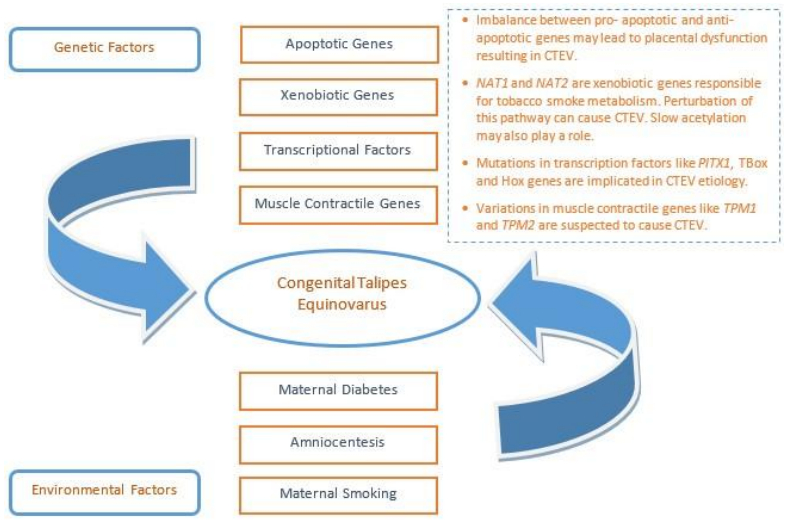
[102] | Jiang B, Zhang Z, Zheng P, et al. (2014) Apoptotic genes expression in placenta of clubfoot-like fetus pregnant rats. Int J Clin Exp Pathol 7: 677-684. |
[103] |
Alderman BW, Takahashi ER, LeMier MK (1991) Risk indicators for talipes equinovarus in Washington State, 1987-1989. Epidemiology 2: 289-292. doi: 10.1097/00001648-199107000-00009
![]() |
[104] | Chung CS, Nemechek RW, Larsen IJ, et al. (1969) Genetic and epidemiological studies of clubfoot in Hawaii. General and medical considerations. Hum Hered 19: 321-342. |
[105] |
Moorthi RN, Hashmi SS, Langois P, et al. (2005) Idiopathic talipes equinovarus (ITEV) (clubfeet) in Texas. Am J Med Genet A 132A: 376-380. doi: 10.1002/ajmg.a.30505
![]() |
[106] |
Miedzybrodzka Z (2003) Congenital talipes equinovarus (clubfoot): a disorder of the foot but not the hand. J Anat 202: 37-42. doi: 10.1046/j.1469-7580.2003.00147.x
![]() |
[107] |
Irani RN, Sherman MS (1972) The pathological anatomy of idiopathic clubfoot. Clin Orthop Relat Res 84: 14-20. doi: 10.1097/00003086-197205000-00004
![]() |
[108] |
Bacino CA, Hecht JT (2014) Etiopathogenesis of equinovarus foot malformations. Eur J Med Genet 57: 473-479. doi: 10.1016/j.ejmg.2014.06.001
![]() |
[109] |
Parker SE, Mai CT, Strickland MJ, et al. (2009) Multistate study of the epidemiology of clubfoot. Birth Defects Res A Clin Mol Teratol 85: 897-904. doi: 10.1002/bdra.20625
![]() |
[110] |
Rogers JM (2009) Tobacco and pregnancy. Reprod Toxicol 28: 152-160. doi: 10.1016/j.reprotox.2009.03.012
![]() |
[111] |
Lambers DS, Clark KE (1996) The maternal and fetal physiologic effects of nicotine. Semin Perinatol 20: 115-126. doi: 10.1016/S0146-0005(96)80079-6
![]() |
[112] |
Hecht JT, Ester A, Scott A, et al. (2007) NAT2 variation and idiopathic talipes equinovarus (clubfoot). Am J Med Genet A 143A: 2285-2291. doi: 10.1002/ajmg.a.31927
![]() |
[113] |
Sommer A, Blanton SH, Weymouth K, et al. (2011) Smoking, the xenobiotic pathway, and clubfoot. Birth Defects Res A Clin Mol Teratol 91: 20-28. doi: 10.1002/bdra.20742
![]() |
[114] | 114. Engell V, Damborg F, Andersen M, et al. (2006) Club foot: a twin study. J Bone Joint Surg Br 88: 374-376. |
[115] |
de Andrade M, Barnholtz JS, Amos CI, et al. (1998) Segregation analysis of idiopathic talipes equinovarus in a Texan population. Am J Med Genet 79: 97-102. doi: 10.1002/(SICI)1096-8628(19980901)79:2<97::AID-AJMG4>3.0.CO;2-K
![]() |
[116] |
Honein MA, Paulozzi LJ, Moore CA (2000) Family history, maternal smoking, and clubfoot: an indication of a gene-environment interaction. Am J Epidemiol 152: 658-665. doi: 10.1093/aje/152.7.658
![]() |
[117] |
Gurnett CA, Alaee F, Kruse LM, et al. (2008) Asymmetric lower-limb malformations in individuals with homeobox PITX1 gene mutation. Am J Hum Genet 83: 616-622. doi: 10.1016/j.ajhg.2008.10.004
![]() |
[118] |
Alvarado DM, McCall K, Aferol H, et al. (2011) Pitx1 haploinsufficiency causes clubfoot in humans and a clubfoot-like phenotype in mice. Hum Mol Genet 20: 3943-3952. doi: 10.1093/hmg/ddr313
![]() |
[119] |
Yong BC, Xun FX, Zhao LJ, et al. (2016) A systematic review of association studies of common variants associated with idiopathic congenital talipes equinovarus (ICTEV) in humans in the past 30 years. Springerplus 5: 896-016-2353-8. eCollection 2016. doi: 10.1186/s40064-016-2353-8
![]() |
[120] |
Rodriguez-Esteban C, Tsukui T, Yonei S, et al. (1999) The T-box genes Tbx4 and Tbx5 regulate limb outgrowth and identity. Nature 398: 814-818. doi: 10.1038/19769
![]() |
[121] | Alvarado DM, Aferol H, McCall K, et al. (2010) Familial isolated clubfoot is associated with recurrent chromosome 17q23.1q23.2 microduplications containing TBX4. Am J Hum Genet 87: 154-160. |
[122] | Lu W, Bacino CA, Richards BS, et al. (2012) Studies of TBX4 and chromosome 17q23.1q23.2: an uncommon cause of nonsyndromic clubfoot. Am J Med Genet A 158A: 1620-1627. |
[123] |
Alnemri ES, Livingston DJ, Nicholson DW, et al. (1996) Human ICE/CED-3 protease nomenclature. Cell 87: 171. doi: 10.1016/S0092-8674(00)81334-3
![]() |
[124] |
Heck AL, Bray MS, Scott A, et al. (2005) Variation in CASP10 gene is associated with idiopathic talipes equinovarus. J Pediatr Orthop 25: 598-602. doi: 10.1097/01.bpo.0000173248.96936.90
![]() |
[125] |
Ester AR, Tyerman G, Wise CA, et al. (2007) Apoptotic gene analysis in idiopathic talipes equinovarus (clubfoot). Clin Orthop Relat Res 462: 32-37. doi: 10.1097/BLO.0b013e318073c2d9
![]() |
[126] |
Daher S, Guimaraes AJ, Mattar R, et al. (2008) Bcl-2 and Bax expressions in pre-term, term and post-term placentas. Am J Reprod Immunol 60: 172-178. doi: 10.1111/j.1600-0897.2008.00609.x
![]() |
[127] |
Peebles DM (2004) Fetal consequences of chronic substrate deprivation. Semin Fetal Neonatal Med 9: 379-386. doi: 10.1016/j.siny.2004.03.008
![]() |
[128] |
Sundberg K, Bang J, Smidt-Jensen S, et al. (1997) Randomised study of risk of fetal loss related to early amniocentesis versus chorionic villus sampling. Lancet 350: 697-703. doi: 10.1016/S0140-6736(97)02449-5
![]() |
[129] |
Cederholm M, Haglund B, Axelsson O (2005) Infant morbidity following amniocentesis and chorionic villus sampling for prenatal karyotyping. BJOG 112: 394-402. doi: 10.1111/j.1471-0528.2005.00413.x
![]() |
[130] |
Mark M, Rijli FM, Chambon P (1997) Homeobox genes in embryogenesis and pathogenesis. Pediatr Res 42: 421-429. doi: 10.1203/00006450-199710000-00001
![]() |
[131] |
McGinnis W, Krumlauf R (1992) Homeobox genes and axial patterning. Cell 68: 283-302. doi: 10.1016/0092-8674(92)90471-N
![]() |
[132] |
Dobbs MB, Gurnett CA, Pierce B, et al. (2006) HOXD10 M319K mutation in a family with isolated congenital vertical talus. J Orthop Res 24: 448-453. doi: 10.1002/jor.20052
![]() |
[133] |
Shrimpton AE, Levinsohn EM, Yozawitz JM, et al. (2004) A HOX gene mutation in a family with isolated congenital vertical talus and Charcot-Marie-Tooth disease. Am J Hum Genet 75: 92-96. doi: 10.1086/422015
![]() |
[134] | Weymouth KS, Blanton SH, Bamshad MJ, et al. (2011) Variants in genes that encode muscle contractile proteins influence risk for isolated clubfoot. Am J Med Genet A 155A: 2170-2179. |
[135] |
McKillop DF, Geeves MA (1993) Regulation of the interaction between actin and myosin subfragment 1: evidence for three states of the thin filament. Biophys J 65: 693-701. doi: 10.1016/S0006-3495(93)81110-X
![]() |
[136] | Gordon AM, Homsher E, Regnier M (2000) Regulation of contraction in striated muscle. Physiol Rev 80: 853-924. |
[137] |
Weymouth KS, Blanton SH, Powell T, et al. (2016) Functional Assessment of Clubfoot Associated HOXA9, TPM1, and TPM2 Variants Suggests a Potential Gene Regulation Mechanism. Clin Orthop Relat Res 474: 1726-1735. doi: 10.1007/s11999-016-4788-1
![]() |
[138] |
Castaneda C, Nalley K, Mannion C, et al. (2015) Clinical decision support systems for improving diagnostic accuracy and achieving precision medicine. J Clin Bioinforma 5: 4-015-0019-3. eCollection 2015. doi: 10.1186/s13336-015-0019-3
![]() |
[139] |
Rehm HL (2013) Disease-targeted sequencing: a cornerstone in the clinic. Nat Rev Genet 14: 295-300. doi: 10.1038/nrg3463
![]() |
[140] |
Richards S, Aziz N, Bale S, et al. (2015) Standards and guidelines for the interpretation of sequence variants: a joint consensus recommendation of the American College of Medical Genetics and Genomics and the Association for Molecular Pathology. Genet Med 17: 405-424. doi: 10.1038/gim.2015.30
![]() |
[141] |
Green RC, Berg JS, Grody WW, et al. (2013) ACMG recommendations for reporting of incidental findings in clinical exome and genome sequencing. Genet Med 15: 565-574. doi: 10.1038/gim.2013.73
![]() |