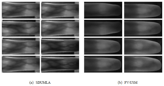
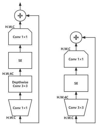
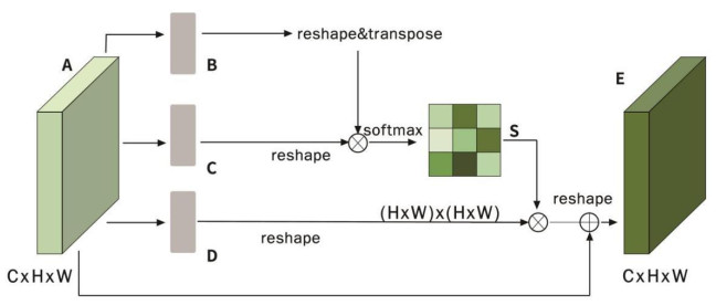
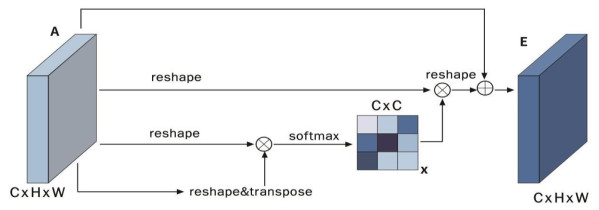
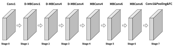
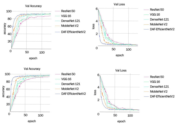
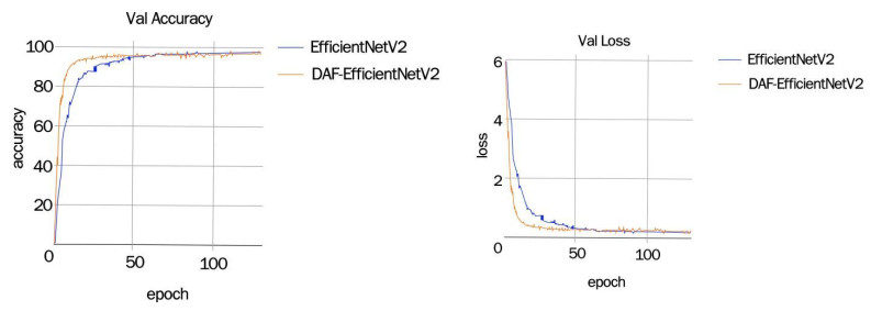
Deep learning is an important technology in the field of image recognition. Finger vein recognition based on deep learning is one of the research hotspots in the field of image recognition and has attracted a lot of attention. Among them, CNN is the most core part, which can be trained to get a model that can extract finger vein image features. In the existing research, some studies have used methods such as combination of multiple CNN models and joint loss function to improve the accuracy and robustness of finger vein recognition. However, in practical applications, finger vein recognition still faces some challenges, such as how to solve the interference and noise in finger vein images, how to improve the robustness of the model, and how to solve the cross-domain problem. In this paper, we propose a finger vein recognition method based on ant colony optimization and improved EfficientNetV2, using ACO to participate in ROI extraction, fusing dual attention fusion network (DANet) with EfficientNetV2, and conducting experiments on two publicly available databases, and the results show that the recognition rate using the proposed method on the FV-USM dataset reaches The results show that the proposed method achieves a recognition rate of 98.96% on the FV-USM dataset, which is better than other algorithmic models, proving that the method has good recognition rate and application prospects for finger vein recognition.
Citation: Xiao Ma, Xuemei Luo. Finger vein recognition method based on ant colony optimization and improved EfficientNetV2[J]. Mathematical Biosciences and Engineering, 2023, 20(6): 11081-11100. doi: 10.3934/mbe.2023490
Deep learning is an important technology in the field of image recognition. Finger vein recognition based on deep learning is one of the research hotspots in the field of image recognition and has attracted a lot of attention. Among them, CNN is the most core part, which can be trained to get a model that can extract finger vein image features. In the existing research, some studies have used methods such as combination of multiple CNN models and joint loss function to improve the accuracy and robustness of finger vein recognition. However, in practical applications, finger vein recognition still faces some challenges, such as how to solve the interference and noise in finger vein images, how to improve the robustness of the model, and how to solve the cross-domain problem. In this paper, we propose a finger vein recognition method based on ant colony optimization and improved EfficientNetV2, using ACO to participate in ROI extraction, fusing dual attention fusion network (DANet) with EfficientNetV2, and conducting experiments on two publicly available databases, and the results show that the recognition rate using the proposed method on the FV-USM dataset reaches The results show that the proposed method achieves a recognition rate of 98.96% on the FV-USM dataset, which is better than other algorithmic models, proving that the method has good recognition rate and application prospects for finger vein recognition.

| [1] |
F. Radzi, M. Khalil-Hani, R. Bakhteri, Finger-vein biometric identification using convolutional neural network, Turkish J. Electr. Eng. Comput. Sci., 24 (2016), 1863–1878. https://doi.org/10.3906/elk-1311-43 doi: 10.3906/elk-1311-43

|
| [2] |
R. Das; E. Piciucco, E. Maiorana, P. Campisi, Convolutional neural network for Finger-Vein-Based biometric identification, IEEE Trans. Inform. Forensics Secur., 14 (2018), 360–373. https://doi.org/10.1109/TIFS.2018.2850320 doi: 10.1109/TIFS.2018.2850320
|
| [3] |
K. J. Noh, J. Choi, J. S. Hong, K. R. Park, Finger-Vein recognition based on densely connected convolutional network using score-level fusion with shape and texture images, IEEE Access, 8 (2020), 96748–96766. doi: 10.1109/ACCESS.2020.2996646
|
| [4] |
D. Zhao, H. Ma, Z. Yang, J. Li, W. Tian, Finger vein recognition based on lightweight CNN combining center loss and dynamic regularization, Infrared Phys. Technol., 105 (2020), 103221. https://doi.org/10.1016/j.infrared.2020.103221 doi: 10.1016/j.infrared.2020.103221
|
| [5] | Hao, Z.; Fang, P.; Yang, H. Finger vein recognition based on multi-task Learning, in Proceedings of the 2020 5th International Conference on Mathematics and Artificial Intelligence, (2020), 133–140. https://doi.org/10.1145/3395260.3395277 |
| [6] |
Y. Lu, S. Xie, S. Wu, Exploring competitive features using deep convolutional neural network for finger vein recognition, IEEE Access, 7 (2019), 35113–35123. https://doi.org/10.1109/ACCESS.2019.2902429 doi: 10.1109/ACCESS.2019.2902429
|
| [7] | R. S. Kuzu, E. Maiorana, P. Campisi, Vein-Based biometric verification using transfer learning, in Proceedings of the 2020 43rd International Conference on Telecommunications and Signal Processing (TSP), (2020), 403–409. https://doi.org/10.1109/TSP49548.2020.9163491 |
| [8] |
Z. Xu, M. M. Kamruzzaman, J. Shi, Method of generating face image based on text description of generating adversarial network, J. Electr. Imaging, 31 (2022), 051411. https://doi.org/10.1117/1.JEI.31.5.051411 doi: 10.1117/1.JEI.31.5.051411
|
| [9] |
Y. Zhang, W. Li, L. Zhang, X. Ning, L. Sun, Y. Lu, Adaptive learning gabor filter for finger-vein recognition, IEEE Access, 7 (2019), 159821–159830. https://doi.org/10.1109/ACCESS.2019.2950698 doi: 10.1109/ACCESS.2019.2950698
|
| [10] |
B. Hou, R. Yan, Convolutional autoencoder model for finger-vein verification, IEEE Trans. Instrum. Meas., 69 (2020), 2067–2074. https://doi.org/10.1109/TIM.2019.2921135 doi: 10.1109/TIM.2019.2921135
|
| [11] |
B. Bharadwaj, J. S. Banu, M. Madiajagan, M. R. Ghalib, O. Castillo, A. Shankar, GPU-Accelerated implementation of a genetically optimized image encryption algorithm, Soft Comput., 25 (2021), 14413–14428. https://doi.org/10.1007/s00500-021-06225-y doi: 10.1007/s00500-021-06225-y
|
| [12] |
G. Wang, C. Sun, A. Sowmya, Multi-weighted co-occurrence descriptor encoding for vein recognition, IEEE Trans. Inform. Forensics Secur., 15 (2019), 375–390. https://doi.org/10.1109/TIFS.2019.2922331 doi: 10.1109/TIFS.2019.2922331
|
| [13] |
P. Jayapriya, K. Umamaheswari, Finger knuckle biometric feature selection based on the FIS_DE optimization algorithm, Neural Comput. Appl., 34 (2022), 5535–5547. https://doi.org/10.1007/s00521-021-06705-0 doi: 10.1007/s00521-021-06705-0
|
| [14] | W. Liu, W. Li, L. Sun, L. Zhang, P. Chen, Finger vein recognition based on deep learning, in 2017 12th IEEE conference on industrial electronics and applications (ICIEA), (2017), 205–210. https://doi.org/10.1109/ICIEA.2017.8282842 |
| [15] |
Y. Liu, J. Ling, Z. Liu, J. Shen, C. Gao, Finger vein secure biometric template generation based on deep learning, Soft Comput., 22 (2018), 2257–2265. https://doi.org/10.1007/s00500-017-2487-9 doi: 10.1007/s00500-017-2487-9
|
| [16] |
W. Yang, S. Wang, J. Hu, G. Zheng, J. Yang; C. Valli, Securing deep learning based edge finger vein biometrics with binary decision diagram, IEEE Trans. Ind. Inform., 15 (2019), 4244–4253. https://doi.org/10.1109/TII.2019.2900665 doi: 10.1109/TII.2019.2900665
|
| [17] |
J. Zhang, Z. Lu, M. Li, H. Wu, GAN-based image augmentation for finger-vein biometric recognition, IEEE Access, 7 (2019), 183118–183132. https://doi.org/10.1109/ACCESS.2019.2960411 doi: 10.1109/ACCESS.2019.2960411
|
| [18] | Y. Zhang, Z. Liu, Research on finger vein recognition based on sub-convolutional neural network, in 2020 International Conference on Computer Network, Electronic and Automation (ICCNEA), (2020), 211–216. https://doi.org/10.1109/ICCNEA50255.2020.00051 |
| [19] |
S. Liu, E. Huang, Y. Xu, K. Wang, D. K. Jain, Computation of facial attractiveness from 3D geometry, Soft Comput., 26 (2022), 10401–10407. https://doi.org/10.1007/s00500-022-07324-0 doi: 10.1007/s00500-022-07324-0
|
| [20] |
G. K. Sidiropoulos, P. Kiratsa, P. Chatzipetrou, G. A. Papakostas, Feature extraction for finger-vein-based identity recognition, J. Imaging, 7 (2021), 89. https://doi.org/10.3390/jimaging7050089 doi: 10.3390/jimaging7050089
|
| [21] |
S. Daas, A. Yahi, T. Bakir, M. Sedhane, M. Boughazi, E. Bourennane, Multimodal biometric recognition systems using deep learning based on the finger vein and finger knuckle print fusion, IET Image Proc., 14 (2020), 3859–3868. https://doi.org/10.1049/iet-ipr.2020.0491 doi: 10.1049/iet-ipr.2020.0491
|
| [22] |
J. Zhang, C. Li, S. Kosov, M. Grzegorzek, K. Shirahama, T. Jiang, et al., LCU-Net: A novel low-cost U-Net for environmental microorganism image segmentation, Pattern Recognit., 115 (2021), 107885. https://doi.org/10.1016/j.patcog.2021.107885 doi: 10.1016/j.patcog.2021.107885
|
| [23] |
J. Zhang, C. Li, Y. Yin, J. Zhang, M. Grzegorzek, Applications of artificial neural networks in microorganism image analysis: a comprehensive review from conventional multilayer perceptron to popular convolutional neural network and potential visual transformer, Artif. Intel. Rev., 56 (2023), 1013–1070. https://doi.org/10.1007/s10462-022-10192-7 doi: 10.1007/s10462-022-10192-7
|

Figures(15) / Tables(3)

Xiao Ma, Xuemei Luo. Finger vein recognition method based on ant colony optimization and improved EfficientNetV2[J]. Mathematical Biosciences and Engineering, 2023, 20(6): 11081-11100. doi: 10.3934/mbe.2023490


 DownLoad: