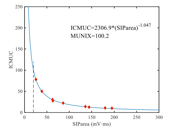
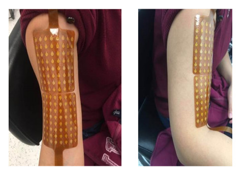
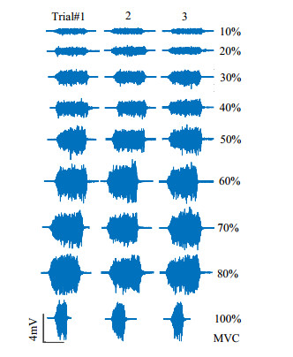
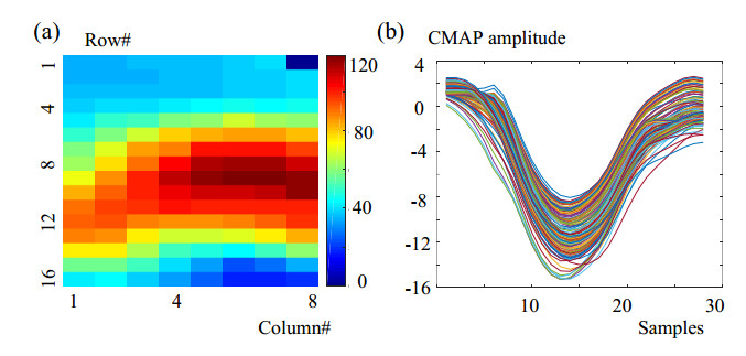
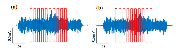
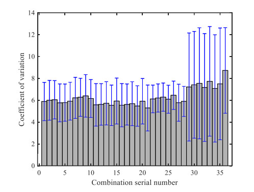
Repeatability is an important attribute of motor unit number index (MUNIX) technology. This paper proposes an optimal contraction force combination for MUNIX calculation in an effort to improve the repeatability of this technology. In this study, the surface electromyography (EMG) signals of the biceps brachii muscle of eight healthy subjects were initially recorded with high-density surface electrodes, and the contraction strength was the maximum voluntary contraction force of nine progressive levels. Then, by traversing and comparing the repeatability of MUNIX under various combinations of contraction force, the optimal combination of muscle strength is determined. Finally, calculate MUNIX using the high-density optimal muscle strength weighted average method. The correlation coefficient and the coefficient of variation are utilized to assess repeatability. The results show that when the muscle strength combination is 10, 20, 50 and 70% of the maximum voluntary contraction force, the repeatability of MUNIX is greatest, and the correlation between MUNIX calculated using this combination of muscle strength and conventional methods is high (PCC > 0.99), the repeatability of the MUNIX method improved by 11.5–23.8%. The results indicate that the repeatability of MUNIX differs for various combinations of muscle strength and that MUNIX, which is measured with a smaller number and lower-level contractility, has greater repeatability.
Citation: Qun Xu, Suqi Xue, Farong Gao, Qiuxuan Wu, Qizhong Zhang. Evaluation method of motor unit number index based on optimal muscle strength combination[J]. Mathematical Biosciences and Engineering, 2023, 20(2): 3854-3872. doi: 10.3934/mbe.2023181
Repeatability is an important attribute of motor unit number index (MUNIX) technology. This paper proposes an optimal contraction force combination for MUNIX calculation in an effort to improve the repeatability of this technology. In this study, the surface electromyography (EMG) signals of the biceps brachii muscle of eight healthy subjects were initially recorded with high-density surface electrodes, and the contraction strength was the maximum voluntary contraction force of nine progressive levels. Then, by traversing and comparing the repeatability of MUNIX under various combinations of contraction force, the optimal combination of muscle strength is determined. Finally, calculate MUNIX using the high-density optimal muscle strength weighted average method. The correlation coefficient and the coefficient of variation are utilized to assess repeatability. The results show that when the muscle strength combination is 10, 20, 50 and 70% of the maximum voluntary contraction force, the repeatability of MUNIX is greatest, and the correlation between MUNIX calculated using this combination of muscle strength and conventional methods is high (PCC > 0.99), the repeatability of the MUNIX method improved by 11.5–23.8%. The results indicate that the repeatability of MUNIX differs for various combinations of muscle strength and that MUNIX, which is measured with a smaller number and lower-level contractility, has greater repeatability.

| [1] |
A. Amin Lari, A. A. Ghavanini, H. R. Bokaee, A review of electrophysiological studies of lower motor neuron involvement in amyotrophic lateral sclerosis, Neurol. Sci. , 40 (2019), 1125–1136. https://doi.org/10.1007/s10072-019-03832-4 doi: 10.1007/s10072-019-03832-4

|
| [2] |
C. E. Candela, L. R. Cecilia, M. R. Samira, C. C. Carlos, A. C. M. Isabel, B. C. Emilia, et al., Venous thromboembolism in amyotrophic lateral sclerosis. A prospective quasi-experimental study, Thromb. Res. , 211 (2022), 81–84. https://doi.org/10.1016/j.thromres.2022.01.002 doi: 10.1016/j.thromres.2022.01.002
|
| [3] |
J. Nijssen, L. H. Comley, E. Hedlund, Motor neuron vulnerability and resistance in amyotrophic lateral sclerosis, Acta Neuropathol. , 133 (2017), 863–885. https://doi.org/10.1007/s00401-017-1708-8 doi: 10.1007/s00401-017-1708-8
|
| [4] | Z. Maria, A. Anna, Lower and upper motor neuron involvement and their impact on disease prognosis in amyotrophic lateral sclerosis, Neural Regen. Res. , 17 (2022), 65–73. https://doi.org/10.4103%2F1673-5374.314289 |
| [5] |
S. D. Nandedkar, D. S. Nandedkar, P. E. Barkhaus, E. V. Stalberg, Motor unit number index (MUNIX), IEEE Trans. Biomed. Eng. , 51 (2004), 2209–2211. https://doi.org/10.1109/TBME.2004.834281 doi: 10.1109/TBME.2004.834281
|
| [6] |
S. D. Nandedkar, P. E. Barkhaus, E. V. Stalberg, Motor unit number index (MUNIX): Principle, method and findings in healthy subjects and in patients with motor neuron disease, Muscle Nerve, 42 (2010), 798–807. https://doi.org/10.1002/mus.21824 doi: 10.1002/mus.21824
|
| [7] |
W. A. Boekestein, H. J. Schelhaas, M. J. A. M. van Putten, D. F. Stegeman, M. J. Zwarts, J. P. van Dijk, Motor unit number index (MUNIX) versus motor unit number estimation (MUNE): A direct comparison in a longitudinal study of ALS patients, Clin. Neurophysiol. , 123 (2012), 1644–1649. https://doi.org/10.1016/j.clinph.2012.01.004 doi: 10.1016/j.clinph.2012.01.004
|
| [8] |
C. Neuwirth, P. E. Barkhaus, C. Burkhardt, J. Castro, D. Czell, M. de Carvalho, et al., Tracking motor neuron loss in a set of six muscles in amyotrophic lateral sclerosis using the motor unit number index (MUNIX): A 15-month longitudinal multicentre trial, J. Neurol. Neurosurg. Psychiatry, 86 (2015), 1172–1179. https://doi.org/10.1136/jnnp-2015-310509 doi: 10.1136/jnnp-2015-310509
|
| [9] |
J. Furtula, B. Johnsen, P. B. Christensen, K. Pugdahl, C. Bisgaard, M. K. Christensen, et al., MUNIX and incremental stimulation MUNE in ALS patients and control subjects, Clin. Neurophysiol. , 124 (2013), 610–618. https://doi.org/10.1016/j.clinph.2012.08.023 doi: 10.1016/j.clinph.2012.08.023
|
| [10] |
C. Neuwirth, S. Nandedkar, E. Stalberg, P. E. Barkhaus, M. de Carvalho, J. Furtula, et al., Motor unit number index (MUNIX): A novel neurophysiological marker for neuromuscular disorders; test-retest reliability in healthy volunteers, Clin. Neurophysiol. , 122 (2011), 1867–1872. https://doi.org/10.1016/j.clinph.2011.02.017 doi: 10.1016/j.clinph.2011.02.017
|
| [11] |
N. Dias, X. H. Li, C. Zhang, Y. C. Zhang, Innervation asymmetry of the external anal sphincter in aging characterized from high-density intra-rectal surface EMG recordings, Neurourol. Urodyn. , 37 (2018), 2544–2550. https://doi.org/10.1002/nau.23809 doi: 10.1002/nau.23809
|
| [12] |
R. Gunther, C. Neuwirth, J. C. Koch, P. Lingor, N. Braun, R. Untucht, et al., Motor unit number index (MUNIX) of hand muscles is a disease biomarker for adult spinal muscular atrophy, Clin. Neurophysiol. , 130 (2019), 315–319. https://doi.org/10.1016/j.clinph.2018.11.009 doi: 10.1016/j.clinph.2018.11.009
|
| [13] |
S. Verma, J. Forte, M. Ritchey, D. Shah, Motor unit number index in children with later-onset spinal muscular atrophy, Muscle Nerve, 62 (2020), 633–637. https://doi.org/10.1002/mus.26909 doi: 10.1002/mus.26909
|
| [14] |
C. Neuwirth, C. Burkhardt, J. Alix, J. Castro, M. de Carvalho, M. Gawel, et al., Quality control of motor unit number index (MUNIX) measurements in 6 muscles in a single-subject "round-robin" setup, Plos One, 11 (2016), 1–11. https://doi.org/10.1371/journal.pone.0153948 doi: 10.1371/journal.pone.0153948
|
| [15] |
S. W. Ahn, S. H. Kim, J. E. Kim, S. M. Kim, S. H. Kim, K. S. Park, et al., Reproducibility of the motor unit number index (MUNIX) in normal controls and amyotrophic lateral sclerosis patients, Muscle Nerve, 42 (2010), 808–813. https://doi.org/10.1002/mus.21765 doi: 10.1002/mus.21765
|
| [16] |
C. Neuwirth, N. Braun, K. G. Claeys, R. Bucelli, M. Weber, Implementing motor unit number index (MUNIX) in a large clinical trial: Real world experience from 27 centres, Clin. Neurophysiol. , 129 (2018), 1756–1762. https://doi.org/10.1016/j.clinph.2018.04.614 doi: 10.1016/j.clinph.2018.04.614
|
| [17] |
C. Neuwirth, S. Nandedkar, E. Stalberg, M. Weber, Motor unit number index (MUNIX): A novel neurophysiological technique to follow disease progression in amyotrophic lateral sclerosis, Muscle Nerve, 42 (2010), 379–384. https://doi.org/10.1002/mus.21707 doi: 10.1002/mus.21707
|
| [18] |
M. L. Escorcio-Bezerra, A. Abrahao, I. de Castro, M. A. T. Chieia, L. A. de Azevedo, D. S. Pinheiro, et al., MUNIX: Reproducibility and clinical correlations in amyotrophic lateral sclerosis, Clin. Neurophysiol. , 127 (2016), 2979–2984. https://doi.org/10.1016/j.clinph.2016.06.011 doi: 10.1016/j.clinph.2016.06.011
|
| [19] |
C. Boulay, D. Emilien, F. Audic, B. Chabrol, A. Shahram, Motor unit number index: A potential electrophysiological biomarker for pediatric spinal muscular atrophy, Muscle Nerve, 64 (2021), 445–453. https://doi.org/10.1016/j.clinph.2016.06.011 doi: 10.1016/j.clinph.2016.06.011
|
| [20] |
S. D. Nandedkar, P. E. Barkhaus, E. V. Stalberg, Reproducibility of MUNIX in patients with amyotrophic lateral sclerosis, Muscle Nerve, 44 (2011), 919–922. https://doi.org/10.1002/mus.22204 doi: 10.1002/mus.22204
|
| [21] |
D. Fathi, B. Mohammadi, R. Dengler, S. Boselt, S. Petri, K. Kollewe, Lower motor neuron involvement in ALS assessed by motor unit number index (MUNIX): Long-term changes and reproducibility, Clin. Neurophysiol. , 127 (2016), 1984–1988. https://doi.org/10.1016/j.clinph.2015.12.023 doi: 10.1016/j.clinph.2015.12.023
|
| [22] |
G. Alessio, G. S. Jayne, J. M. Wakeling, Identification of regional activation by factorization of high-density surface EMG signals: A comparison of principal component analysis and non-negative matrix factorization, J. Electromyogr. Kinesiol. , 41 (2018), 116–123. https://doi.org/10.1016/j.jelekin.2018.05.002 doi: 10.1016/j.jelekin.2018.05.002
|
| [23] |
A. Konstantin, T. Yu, R. L. Carpentier, Y. Aoustin, D. Farina, Simulation of motor unit action potential recordings from intramuscular multichannel scanning electrodes, IEEE Trans. Biomed. Eng. , 67 (2020), 2005–2014. https://doi.org/10.1109/TBME.2019.2953680 doi: 10.1109/TBME.2019.2953680
|
| [24] |
A. Matran-Fernandez, I. J. R. Martínez, R. Poli, C. Cipriani, L. Citi, SEEDS, simultaneous recordings of high-density EMG and finger joint angles during multiple hand movements, Sci. Data, 6 (2019), 1–10. https://doi.org/10.1038/s41597-019-0200-9 doi: 10.1038/s41597-019-0200-9
|
| [25] |
Y. Peng, Y. C. Zhang, Improving the repeatability of motor unit number index (MUNIX) by introducing additional epochs at low contraction levels, Clin. Neurophysiol. , 128 (2017), 1158–1165. https://doi.org/10.1016/j.clinph.2017.03.044 doi: 10.1016/j.clinph.2017.03.044
|
| [26] |
F. Miralles, MUNIX value dependence on surface electromyogram properties, Clin. Neurophysiol. , 130 (2019), 2287–2289. https://doi.org/10.1016/j.clinph.2019.08.030 doi: 10.1016/j.clinph.2019.08.030
|
| [27] |
S. G. Boe, D. W. Stashuk, W. F. Brown, T. J. Doherty, Decomposition-based quantitative electromyography: Effect of force on motor unit potentials and motor unit number estimates, Muscle Nerve, 31 (2005), 365–373. https://doi.org/10.1002/mus.20266 doi: 10.1002/mus.20266
|
| [28] | C. Neuwirth, M. Weber, The motor unit number index (MUNIX)-A new electrophysiological marker to estimate the number of motor neurons: A literature review, Klinische Neurophysiologie, 44 (2013), 132–139. |
| [29] |
G. Malgorzata, K. K. Magdalena, Does the MUNIX method reflect clinical dysfunction in amyotrophic lateral sclerosis: A practical experience, Medicine, 95 (2016), 1–5. https://doi.org/10.1097/MD.0000000000003647 doi: 10.1097/MD.0000000000003647
|
| [30] |
F. Fatehi, A. M. Grapperon, D. Fathi, E. Delmont, S. Attarian, The utility of motor unit number index: A systematic review, Neurophysiol. Clin., 48 (2018), 251–259. https://doi.org/10.1016/j.neucli.2018.09.001 doi: 10.1016/j.neucli.2018.09.001
|
| [31] |
M. Drey, C. Grösch, C. Neuwirth, J. M. Bauer, C. C. Sieber, The motor unit number index (MUNIX) in sarcopenic patients, Experimental Gerontology, 48 (2013), 381–384. https://doi.org/10.1016/j.exger.2013.01.011 doi: 10.1016/j.exger.2013.01.011
|
| [32] |
M. L. Escorcio-Bezerra, A. S. B. Oliveira, N. I. D. Braga, G. M. Manzano, Improving the reproducibility of motor unit number index, Muscle Nerve, 55 (2017), 635–638. https://doi.org/10.1002/mus.25260 doi: 10.1002/mus.25260
|
| [33] |
S. W. Ahn, Applicability of the digital instrument to improve the reproducibility of motor unit number index, Ann. Clin. Neurophysiol., 20 (2018), 26–30. https://doi.org/10.14253/acn.2018.20.1.26 doi: 10.14253/acn.2018.20.1.26
|
| [34] |
Y. Peng, J. B. He, B. Yao, S. Li, P. Zhou, Y. C. Zhang, Motor unit number estimation based on high-density surface electromyography decomposition, Clin. Neurophysiol., 127 (2016), 3059–3065. https://doi.org/10.1016/j.clinph.2016.06.014 doi: 10.1016/j.clinph.2016.06.014
|
| [35] |
J. P. van Dijk, J. H. Blok, B. G. Lapatki, I. N. van Schaik, M. J. Zwarts, D. F. Stegeman, Motor unit number estimation using high-density surface electromyography, Clin. Neurophysiol., 119 (2008), 33–42. https://doi.org/10.1016/j.clinph.2007.09.133 doi: 10.1016/j.clinph.2007.09.133
|
| [36] |
S. H. Nawab, S. S. Chang, C. Luca, High-yield decomposition of surface EMG signals, Clin. Nurophysiol., 121 (2010), 1602–1615. https://doi.org/10.1016/j.clinph.2009.11.092 doi: 10.1016/j.clinph.2009.11.092
|
| [37] |
K. A. Mazurek, R. David, A. Nicholas, J. J. Foxe, E. G. Freedman, Utilizing high-density electroencephalography and motion capture technology to characterize sensorimotor integration while performing complex actions, IEEE Trans. Neural Syst. Rehabil. Eng., 28 (2020), 287–296. https://doi.org/10.1109/TNSRE.2019.2941574 doi: 10.1109/TNSRE.2019.2941574
|
| [38] |
A. Holobar, D. Zazula, Multichannel blind source separation using convolution kernel compensation, IEEE Trans. Signal Proces., 55 (2007), 4487–4496. https://doi.org/10.1109/TSP.2007.896108 doi: 10.1109/TSP.2007.896108
|
| [39] |
A. Holobar, D. Farina, Blind source identification from the multichannel surface electromyogram, Physiol. Meas., 35 (2014). https://doi.org/10.1088/0967-3334/35/7/R143 doi: 10.1088/0967-3334/35/7/R143
|
| [40] |
W. Qi, H. Su, A cybertwin based multimodal network for ECG patterns monitoring using deep learning, IEEE Trans. Ind. Inf., 18 (2022), 6663–6670. https://doi.org/10.1109/TⅡ.2022.3159583 doi: 10.1109/TⅡ.2022.3159583
|
| [41] |
Y. Ning, X. Zhu, S. Zhu, Y. Zhang, Surface EMG decomposition based on K-means clustering and convolution kernel compensation, IEEE J. Biomed. Health Inf., 19 (2015), 471–477. https://doi.org/10.1109/JBHI.2014.2328497 doi: 10.1109/JBHI.2014.2328497
|
| [42] |
H. Su, W. Qi, Y. Hu, H. R. Karimi, G. Ferrigno, E. D. Momi, An incremental learning framework for human-like redundancy optimization of anthropomorphic manipulators, IEEE Trans. Ind. Inf., 18 (2022), 1864–1872. https://doi.org/10.1109/TⅡ.2020.3036693 doi: 10.1109/TⅡ.2020.3036693
|
| [43] |
F. R. Gao, Y. Y. Cao, C. Zhang, Y. C. Zhang, A preliminary study of effects of channel number and location on the repeatability of Motor Unit Number Index (MUNIX), Front. Neurol., 11 (2020), 191. https://doi.org/10.3389/fneur.2020.00191 doi: 10.3389/fneur.2020.00191
|
| [44] |
M. Gawel, M. Kuzma-Kozakiewicz, Does the MUNIX method reflect clinical dysfunction in Amyotrophic Lateral Sclerosis: A practical experience, Medicine, 95 (2016), 1–5. https://doi.org/10.1097/MD.0000000000003647 doi: 10.1097/MD.0000000000003647
|
| [45] |
S. Li, J. Liu, M. Bhadane, P. Zhou, W. Z. Rymer, Activation deficit correlates with weakness in chronic stroke: Evidence from evoked and voluntary EMG recordings, Clin. Neurophysiol., 125 (2014), 2413–2417. https://doi.org/10.1016/j.clinph.2014.03.019 doi: 10.1016/j.clinph.2014.03.019
|
| [46] |
S. D. Nandedkar, P. E. Barkhaus, E. V. Stalberg, C. Neuwirth, M. Weber, Motor unit number index: Guidelines for recording signals and their analysis, Muscle Nerve, 58 (2018), 374–380. https://doi.org/10.1002/mus.26099 doi: 10.1002/mus.26099
|
| [47] |
M. Atzori, A. Gijsberts, C. Castellini, B. Caputo, H. Müller, Electromyography data for non-invasive naturally-controlled robotic hand prostheses, Sci. Data, 1 (2014), 140053. https://doi.org/10.1038/sdata.2014.53 doi: 10.1038/sdata.2014.53
|
| [48] |
R. D. Kaya, R. L. Hoffman, B. C. Clark, Reliability of a modified motor unit number index (MUNIX) technique, J. Electromyogr. Kinesiol., 24 (2014), 18–24. https://doi.org/10.1016/j.jelekin.2013.10.005 doi: 10.1016/j.jelekin.2013.10.005
|
| [49] |
M. L. Escorcio-Bezerra, A. Abrahao, D. Santos-Neto, N. I. D. Braga, A. S. B. Oliveira, G. M. Manzano, Why averaging multiple MUNIX measures in the longitudinal assessment of patients with ALS?, Clin. Neurophysiol., 128 (2017), 2392–2396. https://doi.org/10.1016/j.clinph.2017.09.104 doi: 10.1016/j.clinph.2017.09.104
|
| [50] |
C. Neuwirth, S. Nandedkar, E. Stalberg, P. E. Barkhaus, M. de Carvalho, J. Furtula, et al., Motor unit number index (MUNIX): Reference values of five different muscles in healthy subjects from a multi-centre study, Clin. Neurophysiol., 122 (2011), 1895–1898. https://doi.org/10.1016/j.clinph.2011.05.014 doi: 10.1016/j.clinph.2011.05.014
|
| [51] |
E. Delmont, F. Wang, J. P. Lefaucheur, A. Puma, C. Breniere, G. Beaudonnet, et al., Motor unit number index as an individual biomarker: Reference limits of intra-individual variability over time in healthy subjects, Clin. Neurophysiol., 131 (2020), 2209–2215. https://doi.org/10.1016/j.clinph.2020.06.019 doi: 10.1016/j.clinph.2020.06.019
|
| [52] |
S. D. Nandedkar, P. E. Barkhaus, E. V. Stalberg, Motor unit number index (MUNIX) and compound muscle action potential amplitude: A reappraisal, Clin. Neurophysiol., 130 (2019), 2010–2011. https://doi.org/10.1016/j.clinph.2019.07.021 doi: 10.1016/j.clinph.2019.07.021
|
| [53] |
A. A. Okhovat, S. Advani, B. Ziaadini, A. Panahi, S. Salehizadeh, S. Nafissi, et al., The value of MUNIX as an objective electrophysiological biomarker of disease progression in chronic inflammatory demyelinating polyneuropathy, Muscle Nerve, 65 (2022), 433–439. https://doi.org/10.1002/mus.27498 doi: 10.1002/mus.27498
|
| [54] |
H. Su, W. Qi, J. Chen, D. Zhang, Fuzzy approximation-based task-space control of robot manipulators with remote center of motion constraint, IEEE Trans. Fuzzy Syst., 30 (2022), 1564–1573. https://doi.org/10.1109/TFUZZ.2022.3157075 doi: 10.1109/TFUZZ.2022.3157075
|
| [55] |
S. W. Lee, K. M. Wilson, B. A. Lock, D. G. Kamper, Subject-specific myoelectric pattern classification of functional hand movements for stroke survivors, IEEE Trans. Neural Syst. Rehabil. Eng., 19 (2011), 558–566. https://doi.org/10.1109/TNSRE.2010.2079334 doi: 10.1109/TNSRE.2010.2079334
|
| [56] |
A. Manfredo, C. Matteo, M. Henning, Deep learning with convolutional neural networks applied to electromyography data: A resource for the classification of movements for prosthetic hands, Front. Neurorobot., 10 (2016), 1–10. https://doi.org/10.3389/fnbot.2016.00009 doi: 10.3389/fnbot.2016.00009
|
| [57] |
H. Su, W. Qi, Z. Li, Z. Chen, G. Ferrigno, E. D. Momi, Deep neural network approach in EMG-based force estimation for human-robot interaction, IEEE Trans. Artif. Intell., 2 (2021), 404–412. https://doi.org/10.1109/TAI.2021.3066565 doi: 10.1109/TAI.2021.3066565
|
Figures(6) / Tables(5)

Qun Xu, Suqi Xue, Farong Gao, Qiuxuan Wu, Qizhong Zhang. Evaluation method of motor unit number index based on optimal muscle strength combination[J]. Mathematical Biosciences and Engineering, 2023, 20(2): 3854-3872. doi: 10.3934/mbe.2023181


 DownLoad: