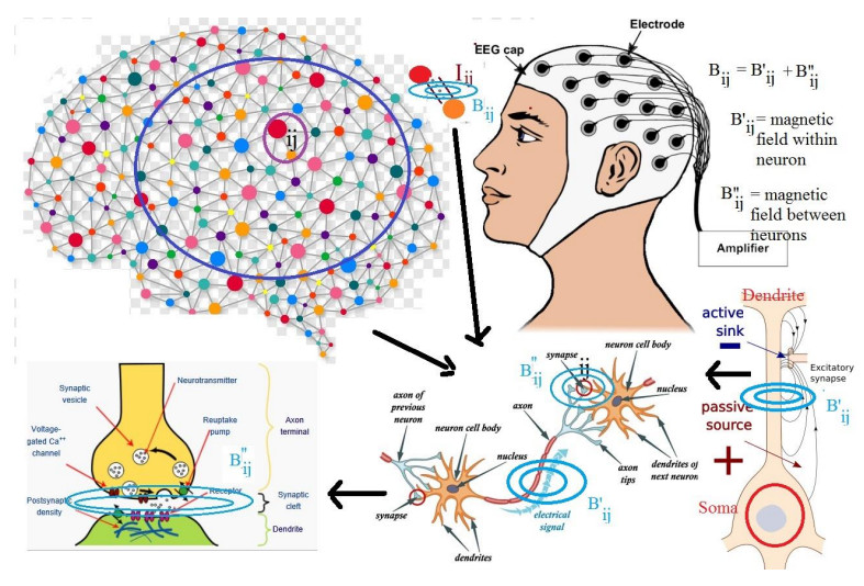
It is known that differences between potentials of soma, dendrites and different parts of neural structures may be the origin of electroencephalogram (EEG) waves. These potentials may be produced by some excitatory synapses and currents of charges between neurons and then thereafter may themselves cause the emergence of new synapses and electrical currents. These currents within and between neurons emit some electromagnetic waves which could be absorbed by electrodes on the scalp, and form topographic images. In this research, a model is proposed which formulates EEG topographic parameters in terms of the charge and mass of exchanged particles within neurons, those which move between neurons, the number of neurons and the length of neurons and synapses. In this model, by knowing the densities of the frequencies in different regions of the brain, one can predict the type, charge and velocity of particles which are moving along neurons or are exchanged between neurons.
Citation: Massimo Fioranelli, O. Eze Aru, Maria Grazia Roccia, Aroonkumar Beesham, Dana Flavin. A model for analyzing evolutions of neurons by using EEG waves[J]. Mathematical Biosciences and Engineering, 2022, 19(12): 12936-12949. doi: 10.3934/mbe.2022604
It is known that differences between potentials of soma, dendrites and different parts of neural structures may be the origin of electroencephalogram (EEG) waves. These potentials may be produced by some excitatory synapses and currents of charges between neurons and then thereafter may themselves cause the emergence of new synapses and electrical currents. These currents within and between neurons emit some electromagnetic waves which could be absorbed by electrodes on the scalp, and form topographic images. In this research, a model is proposed which formulates EEG topographic parameters in terms of the charge and mass of exchanged particles within neurons, those which move between neurons, the number of neurons and the length of neurons and synapses. In this model, by knowing the densities of the frequencies in different regions of the brain, one can predict the type, charge and velocity of particles which are moving along neurons or are exchanged between neurons.

| [1] |
A. Mucci, A. Üçok, M. Ø. Nielsen, Electrophysiological and neuroimaging research on negative symptoms: Future challenges, Clin. EEG Neurosci., 49 (2018), 3–5. https://doi.org/10.1177/1550059417748074 doi: 10.1177/1550059417748074

|
| [2] |
F. C. Morabito, D. Labate, F. L. Foresta, A. Bramanti, G. Morabito, I. Palamara, Multivariate multi-scale permutation entropy for complexity analysis of Alzheimer's disease EEG, Entropy, 14 (2012), 1186–1202. https://doi.org/10.3390/e14071186 doi: 10.3390/e14071186
|
| [3] | D. F. Salisbury, Stimulus processing awake and asleep: Similarities and differences in electrical CNS responses, in Sleep onset: Normal and abnormal processes (eds. R. D. Ogilvie and J. R. Harsh, American Psychological Association, (1994), 289–308. https://doi.org/10.1037/10166-017 |
| [4] |
F. C. Morabito, D. Labate, A. Bramanti, F. La Foresta, G. Morabito, I. Palamara, et al., Enhanced compressibility of EEG signal in Alzheimer's disease patients, IEEE Sensors J., 13 (2013), 3255–3262. https://doi: 10.1109/JSEN.2013.2263794 doi: 10.1109/JSEN.2013.2263794
|
| [5] | F. C. Morabito, D. Labate, G. Morabito, I. Palamara, H. Szu, Monitoring and diagnosis of Alzheimer's disease using noninvasive compressive sensing EEG, in Proc. SPIE 8750, Independent Component Analyses, Compressive Sampling, Wavelets, Neural Net, Biosystems, and Nanoengineering XI, 87500Y, (2013). https://doi.org/10.1117/12.2020886 |
| [6] |
A. Mucci, U. Volpe, E. Merlotti, P. Bucci, S. Galderisi, Pharmaco-EEG in Psychiatry, Clin. EEG Neurosci., 37 (2006), 81–98. https://doi: 10.1177/155005940603700206 doi: 10.1177/155005940603700206
|
| [7] |
M. Christoph, T. K Michel, EEG microstates as a tool for studying the temporal dynamics of whole-brain neuronal networks: A review, NeuroImage, 180 (2018), 577–593. https://doi.org/10.1016/j.neuroimage.2017.11.062. doi: 10.1016/j.neuroimage.2017.11.062
|
| [8] |
A. Jabès, G. Klencklen, P. Ruggeri, C. M. Michel, P. B. Lavenex, P. Lavenex, Resting‐state EEG microstates parallel age-related differences in allocentric spatial working memory performance, Brain Topogr., 34 (2021), 442–460. https://doi.org/10.1007/s10548-021-00835-3 doi: 10.1007/s10548-021-00835-3
|
| [9] |
L. Bréchet, D. Brunet, L. Perogamvros, G. Tonini, C. M. Michel, EEG microstates of dreams, Sci. Rep., 10 (2020), 17069. https://doi.org/10.1038/s41598-020-74075-z doi: 10.1038/s41598-020-74075-z
|
| [10] |
W. J. Bosl, H. Tager-Flusberg, C. A. Nelson, EEG analytics for early detection of autism spectrum disorder: A data-driven approach, Sci. Rep., 8 (2018), 6828. https://doi.org/10.1038/s41598-018-24318-x doi: 10.1038/s41598-018-24318-x
|
| [11] |
T. H. Pham, J. Vicnesh, J. K. E. Wei, S. L. Oh, N. Arunkumar, E. W. Abdulhay, et al., Autism spectrum disorder diagnostic system using HOS Bispectrum with EEG signals., Int. J. Environ. Res. Public Health, 17 (2020), 971. https://doi.org/10.3390/ijerph17030971 doi: 10.3390/ijerph17030971
|
| [12] |
V. Bairagi, EEG signal analysis for early diagnosis of Alzheimer disease using spectral and wavelet based features, Int. J. Inf. Tecnol., 10 (2018), 403–412. https://doi.org/10.1007/s41870-018-0165-5 doi: 10.1007/s41870-018-0165-5
|
| [13] |
A. Markovic, P. Achermann, T. Rusterholz, L. Tarokh, Heritability of sleep EEG topography in adolescence: Results from a longitudinal twin Study, Sci. Rep., 8 (2018), 7334. https://doi.org/10.1038/s41598-018-25590-7 doi: 10.1038/s41598-018-25590-7
|
| [14] |
A. Bersagliere, R. D. Pascual-Marqui, L. Tarokh, P. Achermann, Mapping slow waves by EEG topography and source localization: Effects of sleep deprivation, Brain Topogr., 31 (2018), 257–269. https://doi.org/10.1007/s10548-017-0595-6 doi: 10.1007/s10548-017-0595-6
|
| [15] |
B. Kim, E. Hwang, R. E. Strecker, J. H. Choi, Y. Kim, Differential modulation of NREM sleep regulation and EEG topography by chronic sleep restriction in mice, Sci. Rep., 10 (2020). https://doi.org/10.1038/s41598-019-54790-y doi: 10.1038/s41598-019-54790-y
|
| [16] |
M. A. Rahman, A. Anjum, M. M. H. Milu, F. Khanam, M. S. Uddin, M. N. Mollah, Emotion recognition from EEG-based relative power spectral topography using convolutional neural network, Array, 11 (2021), 100072. https://doi.org/10.1016/j.array.2021.100072 doi: 10.1016/j.array.2021.100072
|
| [17] |
M. Xu, J. Yao, Z. Zhang, R. Li, B. Yang, C. Y. Li, et al., Learning EEG topographical representation for classification via convolutional neural network, Pattern Recogn., 105 (2020), 107390. https://doi.org/10.1016/j.patcog.2020.107390 doi: 10.1016/j.patcog.2020.107390
|
| [18] |
S. Scarpelli, C. Marzano, A. D'Atri, M. Gorgoni, M. Ferrara, L. De Gennaro, State- or trait-like individual differences in dream recall: Preliminary findings from a within-subjects study of multiple nap REM sleep awakenings, Front. Psychol., 6 (2015), 928. https://doi.org/10.3389/fpsyg.2015.00928 doi: 10.3389/fpsyg.2015.00928
|


Figures(3) / Tables(3)

Massimo Fioranelli, O. Eze Aru, Maria Grazia Roccia, Aroonkumar Beesham, Dana Flavin. A model for analyzing evolutions of neurons by using EEG waves[J]. Mathematical Biosciences and Engineering, 2022, 19(12): 12936-12949. doi: 10.3934/mbe.2022604


 DownLoad: