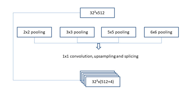
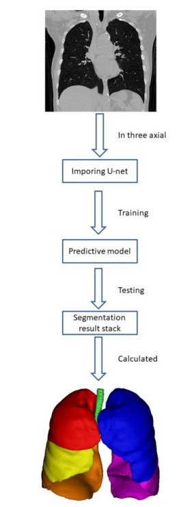
Pulmonary segmentectomy is one of the advanced techniques in thoracic surgery, but it is difficult to understand and master because of its complex anatomical structure. The purpose of this study is to explore the application effect of three-dimensional (3D) image reconstruction based on an improved U-net network in the anatomy of thoracic surgery. In this study, a total of 40 standardization training residents of thoracic surgery in our hospital were randomly divided into two groups. The control group was taught by conventional thin-slice CT images, while the observation group was taught by 3D image reconstruction based on the improved U-net network. After the training process was completed, the teaching effect was compared between these two groups. Using the improved U-net network model, 3D reconstruction of pulmonary segments can be realized quickly. Compared with the control group, the individual and total objective scores in the observation group were higher. The satisfaction of learning interest, content understanding, clinical thinking mode, and understanding of operation process in the observation group was higher than that of the control group. From the results, we concluded that the 3D image reconstruction technology based on the improved U-net network could help students master the anatomical structure of pulmonary segments and improve their learning interest and clinical thinking ability.
Citation: Xuefei Deng, Yu Liu, Hao Chen. Three-dimensional image reconstruction based on improved U-net network for anatomy of pulmonary segmentectomy[J]. Mathematical Biosciences and Engineering, 2021, 18(4): 3313-3322. doi: 10.3934/mbe.2021165
Pulmonary segmentectomy is one of the advanced techniques in thoracic surgery, but it is difficult to understand and master because of its complex anatomical structure. The purpose of this study is to explore the application effect of three-dimensional (3D) image reconstruction based on an improved U-net network in the anatomy of thoracic surgery. In this study, a total of 40 standardization training residents of thoracic surgery in our hospital were randomly divided into two groups. The control group was taught by conventional thin-slice CT images, while the observation group was taught by 3D image reconstruction based on the improved U-net network. After the training process was completed, the teaching effect was compared between these two groups. Using the improved U-net network model, 3D reconstruction of pulmonary segments can be realized quickly. Compared with the control group, the individual and total objective scores in the observation group were higher. The satisfaction of learning interest, content understanding, clinical thinking mode, and understanding of operation process in the observation group was higher than that of the control group. From the results, we concluded that the 3D image reconstruction technology based on the improved U-net network could help students master the anatomical structure of pulmonary segments and improve their learning interest and clinical thinking ability.

| [1] |
Y. Mao, D. Yang, J. He, M. J. Krasna, Epidemiology of lung cancer, Surg. Oncol. Clin., 25 (2016), 439–445. doi: 10.1016/j.soc.2016.02.001

|
| [2] | F. Nasim, B. F. Sabath, G. A. Eapen, Lung cancer, Med. Clin. North Am., 103 (2019), 463–473. |
| [3] |
L. Kutob, F. Schneider, Lung cancer staging, Surg. Pathol. Clin., 13 (2020), 57–71. doi: 10.1016/j.path.2019.10.003
|
| [4] | A. E. Abbas, Surgical management of lung cancer: history, evolution, and modern advances, Curr. Oncol. Rep., 20 (2018), 1–7. |
| [5] |
Y. Chen, J. Zhang, Q. Chen, T. Li, K. Chen, Q. Yu, et al., Three-dimensional printing technology for localised thoracoscopic segmental resection for lung cancer: a quasi-randomised clinical trial, World J. Surg. Oncol., 18 (2020), 1–9. doi: 10.1186/s12957-019-1767-5
|
| [6] | F. Guo, G. Zhu, J. Shen, Y. Ma, Health risk stratification based on computed tomography pulmonary artery obstruction index for acute pulmonary embolism, Sci. Rep., 8 (2018). |
| [7] |
K. Suzuki, H. Saji, K. Aokage, S. Watanabe, M. Okada, J. Mizusawa, et al., Comparison of pulmonary segmentectomy and lobectomy: Safety results of a randomized trial, J. Thorac. Cardiovasc. Surg., 158 (2019), 895–907. doi: 10.1016/j.jtcvs.2019.03.090
|
| [8] |
J. Wu, X. Wu, W. Zeng, D. Guo, Z. Fang, L. Chen, et al., Chest CT findings in patients with coronavirus disease 2019 and its relationship with clinical features, Invest. Radiol., 55 (2020), 257–261. doi: 10.1097/RLI.0000000000000670
|
| [9] |
X. Huang, S. Yue, C. Wang, H. Wang, Optimal three-dimensional reconstruction for lung cancer tissues, Technol. Health Care, 25 (2017), 423–434. doi: 10.3233/THC-171345
|
| [10] | Y. Yagi, R. G. Aly, K. Tabata, A. Barlas, N. Rekhtman, T. Eguchi, et al., Three-dimensional histologic, immunohistochemical, and multiplex immunofluorescence analyses of dynamic vessel co-option of spread through air spaces in lung adenocarcinoma, J. Thorac. Oncol., 15 (2020), 589–600. |
| [11] |
S. Hu, E. A. Hoffman, J. M. Reinhardt, Automatic lung segmentation for accurate quantitation of volumetric X-ray CT images, IEEE Trans. Med. Imaging, 20 (2001), 490–498. doi: 10.1109/42.929615
|
| [12] | J. Long, E. Shelhamer, T. Darrell, Fully convolutional networks for semantic segmentation, in Proceedings of the IEEE conference on computer vision and pattern recognition, (2015), 3431–3440. |
| [13] | G. Cathelain, B. Rivet, S. Achard, J. Bergounioux, F. Jouen, U-net neural network for heartbeat detection in ballistocardiography, in 2020 42nd Annual International Conference of the IEEE Engineering in Medicine & Biology Society (EMBC), (2020), 465–468. |
| [14] | T. Falk, D. Mai, R. Bensch, Ö. Çiçek, A. Abdulkadir, Y. Marrakchi, et al., U-Net: deep learning for cell counting, detection, and morphometry, Nat. Methods, 16 (2019), 67–70. |
| [15] | C. Kou, W. Li, W. Liang, Z. Yu, J. Hao, Microaneurysms segmentation with a U-Net based on recurrent residual convolutional neural network, J. Med. Imaging (Bellingham), 6 (2019), 025008. |
| [16] |
S. Liu, Y. Li, J. Zhou, J. Hu, N. Chen, Y. Shang, et al., Segmenting nailfold capillaries using an improved U-net network, Microvasc. Res., 130 (2020), 104011. doi: 10.1016/j.mvr.2020.104011
|
| [17] |
F. R. Hirsch, G. V. Scagliotti, J. L. Mulshine, R. Kwon, W. J. Curran, Y. Wu, et al., Lung cancer: current therapies and new targeted treatments, Lancet, 389 (2017), 299–311. doi: 10.1016/S0140-6736(16)30958-8
|
| [18] |
H. Hoy, T. Lynch, M. Beck, Surgical treatment of lung cancer, Crit. Care Nurs. Clin. North Am., 31 (2019), 303–313. doi: 10.1016/j.cnc.2019.05.002
|
| [19] |
G. S. Jones, D. R. Baldwin, Recent advances in the management of lung cancer, Clin. Med., 18 (2018), s41–s46. doi: 10.7861/clinmedicine.18-2-s41
|
| [20] |
S. H. Hyun, M. S. Ahn, Y. W. Koh, S. J. Lee, A machine-learning approach using PET-based radiomics to predict the histological subtypes of lung cancer, Clin. Nucl. Med., 44 (2019), 956–960. doi: 10.1097/RLU.0000000000002810
|
| [21] | S. Nakazawa, K. Shimizu, A. Mogi, H. Kuwano, VATS segmentectomy: past, present, and future, Gen. Thorac. Cardiovasc. Surg., 66 (2018), 81–90. |
| [22] |
G. S. Skloot, The effects of aging on lung structure and function, Clin. Geriatr. Med., 33 (2017), 447–457. doi: 10.1016/j.cger.2017.06.001
|


Figures(4) / Tables(2)

Xuefei Deng, Yu Liu, Hao Chen. Three-dimensional image reconstruction based on improved U-net network for anatomy of pulmonary segmentectomy[J]. Mathematical Biosciences and Engineering, 2021, 18(4): 3313-3322. doi: 10.3934/mbe.2021165


 DownLoad: