Review

Development of expression systems for the production of recombinant human Fas ligand extracellular domain derivatives using Pichia pastoris and preparation of the conjugates by site-specific chemical modifications: A review

  • Received: 20 November 2017 Accepted: 17 January 2018 Published: 22 January 2018
  • Human Fas ligand extracellular domain (hFasLECD) is a biomedically important glycoprotein with three potential N-linked carbohydrate-chain attachment sites. hFasLECD can induce an apoptotic cell-death in many malignant cells. Hence, the creation of novel molecular tools exhibiting useful biological activities, based on the exploitation of this protein domain as their components, opens up a great possibility of the advancements in future medical applications. This review mainly focuses on the development of expression systems for obtaining various derivatives of recombinant hFasLECD using Pichia pastoris and the preparation of the conjugates by site-specific chemical modifications of the expressed products. Firstly, a brief introduction of human Fas ligand protein and an overview of the previous works, on the heterologous expression systems for recombinant hFasLECD as well as the associated derivatives aimed at medical applications, were described. Then, the experimental results, obtained during our investigations into the development of the expression systems for the recombinant hFasLECD derivatives using chemically synthesized artificial genes in Pichia pastoris, were summarized. After that, the current state of the methodology for preparation of the hFasLECD conjugates by site-specific chemical modifications, and the functional characterizations of the prepared conjugates, were presented. Finally, conclusions, including a relevant discussion and future perspectives, are provided.

    Citation: Michiro Muraki. Development of expression systems for the production of recombinant human Fas ligand extracellular domain derivatives using Pichia pastoris and preparation of the conjugates by site-specific chemical modifications: A review[J]. AIMS Bioengineering, 2018, 5(1): 39-62. doi: 10.3934/bioeng.2018.1.39

    Related Papers:

    [1] Murtaza Shabbir Hussain, Gabriel M Rodriguez, Difeng Gao, Michael Spagnuolo, Lauren Gambill, Mark Blenner . Recent advances in bioengineering of the oleaginous yeast Yarrowia lipolytica. AIMS Bioengineering, 2016, 3(4): 493-514. doi: 10.3934/bioeng.2016.4.493
    [2] Sezer Okay, Mehmet Sezgin . Transgenic plants for the production of immunogenic proteins. AIMS Bioengineering, 2018, 5(3): 151-161. doi: 10.3934/bioeng.2018.3.151
    [3] Bashir Sajo Mienda, Faezah Mohd Salleh . Bio-succinic acid production: Escherichia coli strains design from genome-scale perspectives. AIMS Bioengineering, 2017, 4(4): 418-430. doi: 10.3934/bioeng.2017.4.418
    [4] Mark Warburton, Hossam Omar Ali, Wai Choon Liong, Arona Martin Othusitse, Amir Zaki Abdullah Zubir, Steve Maddock, Tuck Seng Wong . OneClick: A Program for Designing Focused Mutagenesis Experiments. AIMS Bioengineering, 2015, 2(3): 126-143. doi: 10.3934/bioeng.2015.3.126
    [5] Lindsay Arnold, Tian-Bo Yang, Rachel Chen . A thermal responsive affinity ligand for precipitation of sialylated proteins. AIMS Bioengineering, 2016, 3(1): 92-102. doi: 10.3934/bioeng.2016.1.92
    [6] Liwei Chen, Jaslyn Lee, Wei Ning Chen . The use of metabolic engineering to produce fatty acid-derived biofuel and chemicals in Saccharomyces cerevisiae: a review. AIMS Bioengineering, 2016, 3(4): 468-492. doi: 10.3934/bioeng.2016.4.468
    [7] Torsten Schüling, Alina Eilers, Thomas Scheper, Johanna Walter . Aptamer-based lateral flow assays. AIMS Bioengineering, 2018, 5(2): 78-102. doi: 10.3934/bioeng.2018.2.78
    [8] Azhar Alhasawi, Vasu D. Appanna . Enhanced extracellular chitinase production in Pseudomonas fluorescens: biotechnological implications. AIMS Bioengineering, 2017, 4(3): 366-375. doi: 10.3934/bioeng.2017.3.366
    [9] Pawel Jajesniak, Tuck Seng Wong . From genetic circuits to industrial-scale biomanufacturing: bacterial promoters as a cornerstone of biotechnology. AIMS Bioengineering, 2015, 2(3): 277-296. doi: 10.3934/bioeng.2015.3.277
    [10] Wiebke Wesseling . Beneficial biofilms in marine aquaculture? Linking points of biofilm formation mechanisms in Pseudomonas aeruginosa and Pseudoalteromonas species. AIMS Bioengineering, 2015, 2(3): 104-125. doi: 10.3934/bioeng.2015.3.104
  • Human Fas ligand extracellular domain (hFasLECD) is a biomedically important glycoprotein with three potential N-linked carbohydrate-chain attachment sites. hFasLECD can induce an apoptotic cell-death in many malignant cells. Hence, the creation of novel molecular tools exhibiting useful biological activities, based on the exploitation of this protein domain as their components, opens up a great possibility of the advancements in future medical applications. This review mainly focuses on the development of expression systems for obtaining various derivatives of recombinant hFasLECD using Pichia pastoris and the preparation of the conjugates by site-specific chemical modifications of the expressed products. Firstly, a brief introduction of human Fas ligand protein and an overview of the previous works, on the heterologous expression systems for recombinant hFasLECD as well as the associated derivatives aimed at medical applications, were described. Then, the experimental results, obtained during our investigations into the development of the expression systems for the recombinant hFasLECD derivatives using chemically synthesized artificial genes in Pichia pastoris, were summarized. After that, the current state of the methodology for preparation of the hFasLECD conjugates by site-specific chemical modifications, and the functional characterizations of the prepared conjugates, were presented. Finally, conclusions, including a relevant discussion and future perspectives, are provided.


    1. Introduction


    1.1. Human Fas ligand

    Human Fas ligand (hFasL) is a glycosylated, Type 2 single-spanning membrane protein, which locates its carboxyl-terminal side domain to the outside of the cells, and is encoded on human chromosome number 1 (1q23) [1,2]. A schematic structural composition of hFasL relevant to the contents of this paper is presented in Figure 1. hFasL can induce an apoptosis in numerous malignant human cells expressing Fas receptor (FasR/CD95/APO-1) [3,4], and belongs to a protein group designated as tumor necrosis factor (TNF) superfamily [5]. The proteins inducing an apoptotic cell-death by binding to death receptors, such as FasR, are generally called death ligands. Fas ligand (FasL/CD95L/CD178/APO-1L) is a key member of death ligands, which also includes TNFα, lymphotoxin α (TNFβ), TRAIL (CD253/APO-2L) and TWEAK (CD255/APO-3L). Due to its medically remarkable biological activity, FasL has been extensively investigated as a candidate for therapeutic proteins with the aim of the contribution for the treatment of many serious human diseases, such as cancers, rheumatoid arthritis and other various auto-immune diseases represented by systemic lupus erythematosus [6,7,8,9], since its discovery and identification as a cell-death inducing protein via an extrinsic cellular pathway [10,11]. A number of reviews and books, which cover the basic characteristics of FasL-FasR signaling in the immune system and its potentials for medical applications have been published [12,13,14]. The history of discovery and cloning of the encoding gene, the basic signal transduction pathway in the cells as well as main physiological roles and pathological functions in the human body are mostly covered in these reference literatures.

    Figure 1. Schematic structural composition of wild-type hFasL. MEM, membrane-spanning region; TRI, essential region for the trimerization of subunits; TNFHD, TNF homology domain. The positions of the Asn residues consisting of N-glycosylation sites were indicated by arrows. NH2 and COOH indicate the amino-terminal end and the carboxyl-terminal end, respectively.

    In this review, the current state of the development of expression systems for the production of recombinant hFasL extracellular domain (hFasLECD) derivatives using Pichia pastoris and the preparation of the conjugates by site-specific chemical modifications will be presented.

    In Figure 2, the amino acid sequence of the extracellular domain part of human FasL protein is presented. hFasLECD has long been predicted to exist as a homo-trimer of identical subunits under the physiological conditions, and to possess a 3D structure showing a bell-shaped top-truncated pyramid with the height of approximately 6 nm [5]. The first detailed 3D structure of hFasLECD was experimentally revealed as a complex with human decoy receptor 3 (hDcR3) [15,16] using X-ray crystallography, and the 3D structural model was deposited to Protein Data Bank (PDB) with an identification number (ID) of 4msv in 2013. However, to date, neither 3D structure of sole wild-type hFasLECD molecule nor the complex of hFasLECD with human FasR extracellular domain (hFasRECD) has been published. Alternatively, the non-complexed and the complexed structures with hDcR3 of a mutant hFasLECD, which contains the substitution mutation of the region from the 164th to the 169th amino acid sequence (AspThrTyrGlyIleVal) with the corresponding sequence in human TLIA (HisGluLeuGlyLeuAla), belonging also to the TNF ligand superfamily, have been clarified by X-ray crystallography, and deposited to PDB with the IDs of 5l19 and 5l36, respectively [16]. Therefore, the structural change of wild-type hFasLECD during the formation of the complex with hDcR3 as well as the details of differences in the ligand-receptor interaction modes between the complex with hFasRECD and that with hDcR3 still remains to be clarified.

    Figure 2. Amino acid sequence of wild type hFasLECD. The sequence is expressed by single letter codes of the amino acid residues, and the β-sheet type secondary structure regions are shown in blue underbars. Each residue number of the amino acids mentioned in the text is described.

    In Figure 3, the 3D structure of the hFasLECD in complex with hDcR3 is shown. In this Figure, the amino acid residues mentioned in the following sections of this paper, namely the amino-terminal residue (Leu143 in this model), the Asn residues (Asn184, Asn250 and Asn260) consisting of the three potential N-linked carbohydrate-chain attachment sites and the two Cys residues (Cys202 and Cys233) forming a single intra-molecular disulfide bond, were colored differently from other parts of the molecule. According to the 3D structure revealed by X-ray crystallography, many well-extended β-sheet type secondary structures were identified in the region between the 139th and the 281st amino acid residues (Figure 2). This suggested the structural rigidness of the above-mentioned region, supported by many hydrogen-bonding interactions. Also, the results of the identified β-sheet type secondary structure regions obtained from the crystallographic analysis exhibited a fair agreement with the previous results predicted by a sequence homology search analysis of hFasLECD in comparison with other TNF superfamily ligand proteins [5]. Among the structural parts of hFasL, the extracellular domain plays a role in triggering the signal transduction for apoptosis induction by binding to hFasR, expressed on the surface of target cells, specifically.

    Figure 3. 3D structure of the complex between hFasLECD and hDcR3. The structures were drawn using the atomic coordinate data (ID: 4msv) deposited in PDB. Colors: cyan, hFasLECD part (from Leu143 to Leu281); orange, the N-terminal residue (Leu143) in this model; red, the Asn residues located at the three potential N-linked carbohydrate-chain attachment sites; yellow, the Cys residues forming an intra-molecular disulfide bond (Cys202 and Cys233); white, hDcR3 part.

    1.2. Heterologous expression systems developed for hFasLECD

    In 2012, the author published a review paper, which described the developmental state of heterologous expression systems for death ligands and death receptors including hFasL and hFasR [17]. In the present review, an overview limited to hFasLECD including the later advancement is described. The analysis of the encoding gene revealed that hFasL protein is composed of the amino-terminal side intracellular domain (80 amino acid residues), the intermediate membrane spanning region mainly comprised of hydrophobic amino acid residues (LeuCysLeuLeuValMetPhePheMetValLeuValAlaLeuValGlyLeuGlyLeuGlyMetPhe, 22 residues) and the carboxyl-terminal side extracellular domain (179 amino acid residues) (Figure 1) [1,2]. No secretion signal sequence exists at the amino-terminal site, thus the membrane spanning region sequence was considered to play a role of the signaling sequence, which determines the location of the carboxyl-terminal domain to the outside of the cells. The biologically active whole hFasL protein exists as a homo-trimer of identical subunits, and the normally-folded expressed products of the extracellular domain part alone also form trimers. For the purpose of a straight production of hFasLECD part alone in a biologically active form, it is advantageous to use a secretory expression system of eukaryotes. To date, a methylotrophic yeast, P. pastoris [18,19], and a cellular slime mold, Dictyostelium discoideum [20,21], have been exploited for the expression as the heterologous hosts other than mammalian cells. In these systems, it is expected that the glycosylation at the potential three N-linked carbohydrate-chain attachment sites occurs, as the result of a post-translational modification of the expressed products.

    On the other hand, in the cases of an intracellular expression system using prokaryotic bacterial hosts such as Escherichia coli, lacking a glycosylation system, no glycosylation occurs, and the expressed proteins fused with thioredoxin existed either as incorrectly folded insoluble inclusion bodies [22] or as a soluble product [21]. The attempt of extracellular expression of a protein carrying hFasLECD in E. coli, using the signal sequence of alkaline phosphatase, resulted in the secretion of a soluble but inactive product into the medium [18]. In the X-ray crystallographic analysis of hFasLECD in complex with hDcR3, soluble samples of wild-type and mutant hFasLECDs, prepared by the refolding of the insoluble inclusion bodies produced in the E. coli cells, were used for the complex formation with hDcR3, which was produced by an insect secretory expression system using S2 cells derived from Drosophila melanogaster [16]. According to the description in the paper, sole hFasLECD was difficult to crystallize because of its aggregation propensity. This suggested a significant contribution of the glycosylation, observed in the products expressed using eukaryotic secretory systems, to the solubility of hFasLECD in aqueous medium.


    1.3. hFasLECD derivatives aimed at medical applications

    In 2014, the author published a review paper, which partly covered the state of development concerning the derivatives of hFasLECD and hFasRECD, aimed at medical applications [23]. In the present review, a brief overview limited to hFasLECD including the later progress is described. It is suggested that a single trimer of hFasLECD subunits cannot strongly induce an apoptosis of target cells, even after binding to hFasR, and that it is important to bring together at least two neighboring FasR on the cellular membrane, for the efficient assembly of an apoptosis-executing signaling complex [24]. Therefore, a variety of derivatives to promote the formation of higher-ordered assemblies have been devised thus far. Among them, MegaFasL, which was made by a fusion with the collagen-like domain of human adiponectin, formed a hexamer of hFasLECD subunits by the dimerization of the trimers. This derivative showed an apoptosis inducing activity against the glioma cells [25] and the glioblastoma stem-like cells [26], which are the origin of clinical, malignant human brain tumors. MegaFasL has been industrially manufactured using Chinese hamster ovary cells (CHO cells) [27], and a Phase Ⅰ and Ⅱ clinical trial studies on the safety and the pharmacokinetics of rising doses in relapsed/refractory multiple myeloma patients (Clinical Trial.gov, ID: NCT03196947) is now ongoing [28]. Another trial for derivation of hFasLECD by fusing the D1, Ig and D2 domain of a human leukemia inhibitory factor receptor, gp190, was also investigated. The fusion proteins with the Ig domain formed a hexamer and a dodecamer, and exhibited the therapeutic effect of prolonged life toward immune-deficient mice transplanted with an epidemoid carcinoma cells [29].

    In the process of developing hFasLECD derivatives as therapeutic agents, one of the most important points to be considered will be the reduction of harmful side effects on the sensitive healthy organs such as the liver. In order to overcome this problem, numerous endeavors have been made to install an ability of specific delivery to the target cells. Among them, a representative strategy is to produce the fusion proteins between hFasLECD and scFv-type antibody domain, which specifically recognize the abundant antigens expressed on the target cancer cells, using gene-fusion technologies. For examples, CD20 on B-cells derived lymphoma cells and CD7 on T-cells derived leukemia cells were employed as the target antigens for the fusion proteins. The recombinant fusion proteins, produced by mammalian cells including HEK293 cells and CHO cells, were examined for the treatment of diseases, and certain therapeutic efficacies have been confirmed by the experiments using in vitro cultured cells and also in vivo animal bodies. The hFasLECD conjugates with functional proteins including those other than the fusion proteins with the scFv-type antibodies, prepared by gene fusion methods aiming at medical applications, were summarized in Table 1 [30,31,32,33,34,35,36,37,38,39,40,41,42,43].

    Table 1. hFasLECD gene-fusion type conjugates with functional proteins for targeting cell-surface proteins, aimed at medical applications.
    Fused proteins Targeted cell-surface molecules Examined cells Reference
    scFv-FAP FAP (a fibroblast activation protein) FAP expressing tumor cells (HT1080 and HeLa) [30]
    scFv-Rit CD20 (B-cells associated antigen) B-cell derived tumor cells (BJAB, Ramos, JY, CA5-1, Jiyoyo, DEV, SJO, NALM-6, Raji, PR-1, Z-138), T-cells derived tumor cells (MOLT16) [31]
    scFv-CD7 CD7 (T-cells associated antigen) T-cells derived tumor cells (Jurkat, CEM, CD7-positive transfectant of Ramos, MOLT16, HuT78), B-cells derived tumor cells (Ramos, Raji) [32]
    scFv-TAG72 TAG72 (tumor cells associated mucin-like glycoprotein) Oral squamous tumor cells (Cal-27, Detroit-562, RPMI-2650, HSC-4, SAS), Human Lang epithelial cells (WT-38), Human oral keratinocytes cells (HOK), TAG72 expressing tumor cells (A20, HeLa, Jurkat) [33,34]
    scFv-TAL6 TAL6 (epithelial cells associated membrane protein) TAL6 expressing tumor cells (HeLa, 231, DKBK3,468, MCF7, A20, Jurkat) [34]
    scFv-F8 EDA domain of fiblonectin Murine F9 teratocarcinoma cells [35]
    CD40 CD40 (T-cells associated antigen) B-cells derived tumor cells (Dauji, Raji, JY), T-cells derived tumor cells (Jurkat-CD40L+, Jurkat-CD40L) [36]
    CTLA4 B7-1 and B7-2 B-cells derived tumor cells (Dauji, Raji, JY), T-cells derived tumor cells (Jurkat-CD40L+, Jurkat-CD40L), lymphoma cells (HL60, RPMI8226), renal tumor cells (A498), liver tumor cells (SK-Hep1), fibroblast-like synoviocytes, murine allografts (corneal, cardiac) [36,37,38,39,40]
    E3C1 domain of Del1 Extracellular matrix of cancer cells Human oral squamous cancer cells (SCCKN) planted in mouse [41]
    Extracellular segments of HLA-A*02:01 allele fused to β2-microglobulin Cellular endothelial protein C receptor CD32+ L-cells transfectant, T-cells derived tumor cells (Jurkat) [42]
    Vγ4 chain and Vδ5 chain of human γδTCR Cellular endothelial protein C receptor T-cells derived tumor cells (Jurkat) [42]
    Streptavidin Receptors on diabetogenic T cells splenocytes Diabetogenic splenocytes from insulin dependent type 1 diabetes model mouse [43]
     | Show Table
    DownLoad: CSV

    The yeast P. pastoris is easy to use in recombinant expression experiments, since it belongs to single-cell microorganisms as is the same with E. coli. As a eukaryote, it also possesses the advanced secretion machinery with an excellent quality control system, which is similar to that of mammalian cells, at the same time. Therefore, this organism has been frequently used for the production of biologically active heterologous recombinant proteins, including human origin proteins, by secretory expressions [44,45,46]. In fact, the recombinant hFasLECD secreted from P. pastoris showed an apoptosis inducing activity against the target cells, expressing FasR on their surfaces [18,47].


    2. Development of expression systems for the preparation of recombinant hFasLECD derivatives using P. pastoris


    2.1. Construction of secretory expression systems

    P. pastoris is a methylotrophic yeast, hence it is possible to cultivate this microorganism using methanol as a sole carbon source for nutrition. The first recombinant expression of biologically active hFasLECD in P. pastoris, which employed P. pastoris GS115 strain for the expression, was published in 1997 [18]. For the induction of secretion in P. pastoris, the promoter region gene derived from P. pastoris alcohol oxidase 1 (AOX1) and the following prepro signal sequence gene from S. cerevisiae α-mating factor peptide (αMF) were fused in front of a cDNA derived gene of the amino-acid residues from the 103rd to the 281st of wild-type hFasLECD. The expression using a very high cell density (OD600 = 100) of the starting culture was induced in buffered methanol-complex medium (BMMY medium), and the resulting secretion amount was reported to be 100 mg l–1 [44].

    We used chemically synthesized artificial genes for the construction of a secretory expression system of hFasLECD in P. pastoris [19]. The same strain of P. pastoris GS115 was used. The artificial hFasLECD gene, designed by the optimal codons used in abundantly expressed genes of yeast proteins, was fused after the same AOX1 promoter region gene derived from P. pastoris and the same prepro signal sequence gene of the αMF peptide derived from S. cerevisiae. The assembled genes were inserted between the restriction endonuclease recognition-sites, Xho I and Not I, in a plasmid vector for the secretory expression, phttps://www.aimspress.com/aimspress-data/aimsboa/2018/1/PICZαA, which contains an antibiotic, Zeocin, resistant marker gene [19]. An example of the artificial gene sequences inserted into the expression vector was presented in Figure 4. The induction of the expression was also conducted in the same BMMY medium (500 ml) under the condition of 302.5 K, 96 h cultivation using a triple-baffled, 3 l volume glass flask. Although the cell density of the starting culture before expression induction was less than one tenth of the above mentioned preceding study [18], the maximum secretion level reached was considerably low, and amounted to only 1–2 mg l–1 in this study. In the following experiments described in sections 2.2 and 2.3, we always used essentially the same cultivation conditions for the expression induction.

    Figure 4. Artificial synthetic gene for the expression of the amino-terminal FLAG-(Lys)3GlyCys(Gly)4tagged [Δ(103-138), Asn184Gln + Asn250Gln] hFasLECD mutant in P. pastoris. The secretion-signal cleavage point was indicated by a red arrow. The FLAG sequence region in the amino-terminal tag sequence was shown in green. The reactive Cys residue in the tag sequence and the Gln residues used for the deletion of the potential N-glycosylation sites were shown in red. The other part of the tag sequence was shown in blue. The numbers above the amino acid residues indicate the positions in the primary sequence of wild-type hFasL. The Cys residues composing the intramolecular disulfide bridge and the Asn residue at the remaining N-glycosylation site were underlined. The restriction endonucleases used for the insertion into the expression plasmid were described. Ter: termination codon.

    2.2. Consequences of the addition of tag sequences and the deletion of carbohydrate-chain attachment sites

    For the purpose of increasing the secretion amount, we have examined several modifications of the expression unit. The first trial was the addition of tag sequences. After consideration about the utilization possibility of affinity chromatography in the purification step of the expressed products, either AspTyrLysAspAspAspAspLys (FLAG) tag sequence or (His)6 tag sequence was fused to the amino-terminal site or the carboxyl-terminal site of the hFasLECD encoding gene, always concomitant with an intervening (Gly)5 sequence as the flexible spacer [19]. Consequently, as compared to the case of non-tagged hFasLECD, the secretion amount increased only in the case of the modified hFasLECD that was fused with the amino-terminal FLAG-(Gly)5 tag sequence.

    Another modification target was concerning the Asn residues at three potential N-linked carbohydrate chain attachment sites (Asn184, Asn250 and Asn260), existing in the hFasLECD gene (Figure 1). Either a single, double or triple substitution mutagenesis of the Asn residues with Gln residues, starting from the amino-terminal side, was introduced to the amino-terminally FLAG-(Gly)5 tagged hFasLECD [19]. As a result, judged by the density of the bands in SDS-PAGE analysis, the Asn184Gln mutant was most efficiently secreted from P. pastoris among the wild-type and the three mutants of hFasLECD, and the production yield after purification reached approximately 5 mg l–1. On the other hand, the secretion amount of the amino-terminally (His)6-(Gly)5 tagged derivative significantly decreased. Further, almost no secretion was observed in the cases of both of the carboxyl-terminally tagged derivatives and the derivative lacking all N-glycosylation sites, which suggested seriously bad effects of these modifications on the normal folding in the secretion process of P. pastoris.

    Among the deletion mutations concerning the N-glycosylation sites, the effect of the Asn260Gln mutation was especially substantial. The derivative retaining the N-glycosylation site at Asn260 alone secreted almost the same amount of the product as that of the wild-type hFasLECD. In contrast, the derivative retaining the Asn250 site alone did not seem to secrete a stable product, even though it is the same with respect to the residual of the single N-linked carbohydrate-chain attachment site. As long as the five independent single colonies of the latter mutant were analyzed by SDS-PAGE, all showed either a faint band of the product or the disappearance of the product band during cultivation, which indicated the decomposition or the insolubility of the secreted product. These results suggested that the N-glycosylation at the Asn260 is extremely important for the normal folding and the stability of hFasLECD molecule. Also, the comparison of the band density at the molecular-weight positions of the secreted products, regarding the wild-type and the mutant hFasLECD genes using an SDS-PAGE analysis, revealed that the effect of the deletion of the Asn184 attachment site was insignificant and the ratio of the product containing the glycosylation at this site was small in the case of the expression of the wild-type hFasLECD, as well as that the effect of deletion of the Asn250 site was substantial and the secreted products were mostly glycosylated at this site in the cases of the expression of wild-type and Asn184Gln mutant hFasLECDs. Overall, the above results presented that the functional characteristics and the performance of the three potential N-glycosylation sites in hFasLECD largely depended on their locations in the 3D structure (Figure 3).

    For the purpose of seeking the importance of the structural integrity concerning the attached carbohydrate chain at the Asn260 site, the expression experiment with respect to one of the most efficiently secreted mutants in P. pastoris GS115 strain, [Δ(103-138), Asn184Gln + Asn250Gln] (see details in the section 2.3), was also conducted using P. pastoris SuperMan5 strain (BioGrammatics Co.), which lacks the OCH1 gene responsible for the elongation of outer carbohydrate chain in yeast and attaches only a partial, core chain called Man5 (Man5GlcNAc2) [48]. As far as judged by the SDS-PAGE analysis of the cultivation medium, no clear band of the secreted product was detected. This revealed that the structural integrity of the carbohydrate chain attached at the Asn260 site is essential for the normal folding process in the secretory pathway of P. pastoris, and also strongly suggested that the recognition of the parts except for the core carbohydrate-chain part by N-linked carbohydrate-chains trimming enzymes, such as glucosidases Ⅰ and Ⅱ, and some lectin-like molecular chaperons, such as calnexin and calreticulin, plays essential roles for the secretion process [49]. The abnormally folded hFasLECD proteins were thought to be processed by the endoplasmic reticulum associated degradation mechanism, which constitutes the quality control system for secretory proteins in P. pastoris [50].

    At the early stage of the expression study, the author intended to crystallize the product to solve the 3D structure of the sole hFasLECD molecule by X-ray analysis. Hence, the digestion of the attached carbohydrate chains, existing in the Asn184Gln and the (Asn184Gln + Asn250Gln) mutant hFasLECDs, using Endo Hf glycosidase under the non-denaturing conditions was examined for the possibility of producing carbohydrate-chain trimmed hFasLECDs with uniform chemical structures [51]. The experiments revealed that the carbohydrate-chain attached at the Asn260 site was fairly more resistant to the enzymatic digestion as compared to the carbohydrate-chain attached at the Asn250 site, requiring a much more amount of the enzyme and the reaction time. This result was consistent with the substantial contribution of the carbohydrate chain attached at the Asn260 site to the molecular stability and the efficient secretion of hFasLECD, mentioned above. Further, the resulted derivative of [Δ(103-138), Asn184Gln + Asn250Gln] mutant possessing a single N-acetylglucosamine (GlcNAc) residue alone, produced by the digestion with Endo Hf, was prone to cause an aggregation, which produced an insoluble material during the process to make a concentrated sample for the crystallization trial experiments. A MALDI-TOF mass-spectrometry analysis of this carbohydrate-chain trimmed derivative showed that the measured molecular weight was identical to the chemical structure of the hFasLECD mutant containing a single GlcNAc residue [51], suggesting the absence of other post-translational modifications such as the attachment of O-linked carbohydrate chains.


    2.3. Effects of the amino-terminal region deletion on the secretory production yield

    The structural part of the amino acid residues from the 103rd to the 138th of hFasLECD was known to be unnecessary for the formation of the trimetric structure of the subunits, which is essential for its binding activity toward hFasR, from the analysis of a series of amino-terminal part deletion mutants [2,52]. Therefore, in order to clarify the deletion effect of this region on the production of hFasLECD in P. pastoris, the secretory expression of the hFasLECD genes containing the deletion mutation of the 103rd—the 138th residues in combination with the substitution mutation of either Asn184Gln or the (Asn184Gln + Asn250Gln) were conducted [51]. Interestingly, a large increase in the secretion amount of the product was observed with either case. The purification yield amounted to approximately 24 mg l–1 in the case of [Δ(103-138), Asn184Gln + Asn250Gln] mutant. A unique Pro residue rich sequence, ProSerProProPro, located at the carboxyl-terminal part of the (103–138) region (Figure 2) can be a rate-determining factor in the normal folding process of hFasLECD. It is possible that the large increase in the secretion amount of the products was achieved due to the removal of this limiting factor, in the case of the amino-terminal region deletion mutants.


    2.4. Purification using cation-exchange chromatography

    In the preceding study using P. pastoris as the expression host mentioned in the section 2.1, a Con A-agarose affinity chromatography was used for the initial step of purification, since the secreted product was a glycoprotein possessing high mannose-type carbohydrate-chains [18]. Whereas, the author employed carbohydrate-chain independent purification methods using cation-exchange chromatography, since we planned to make the products of uniform chemical structures by the trimming of the carbohydrate chains, as described above. In the amino acid sequence of wild-type hFasLECD, basic amino-acid residues (Lys and Arg) are more abundant as compared to acidic amino acid residues (Asp and Glu). Twenty basic residues and sixteen acidic residues, and seventeen basic residues and twelve acidic residues are present in the regions from the 103rd to the 281st amino acid residues and the 139th to the 281st amino acid residues, respectively (Figure 2). The theoretical isoelectric points of the former and the latter regions, calculated using ExPASy Proteomics Server [53], were 8.96 and 9.15, respectively.

    Cation-exchange chromatography columns, such as Hi-Trap S, Resource S and Mono S (GE healthcare), worked effectively in the purification steps, since all the derivatives of hFasLECD were considered stable under the condition of pH 5.5. More specifically, the samples in the cultivation medium were first coarsely fractionated by a Hi-Trap S column after a thorough buffer-exchange using 50 mM sodium acetate (pH 5.3–pH 5.6), and then further purified by the chromatography columns with a higher resolution performance, such as Resource S or Mono S. The amino-terminally FLAG-(Gly)5 tagged [Δ(103-138), Asn184Gln + Asn250Gln] mutant, which simultaneously contains the deletions of the amino-terminal region and a couple of N-glycosylation sites of hFasLECD, exhibited a peak profile much sharper than that of wild-type hFasLECD in the cation-exchange chromatography, in addition to its high secretion amount [19]. Therefore, this mutant was adopted as the basic molecule for the preparation of the derivatives holding further modifications. In relation to the purification step using cation-exchange chromatography, a modified amino-terminal tag sequence, FLAG-(Lys)3GlyCys(Gly)4, which contains an insertion of three Lys residues between FLAG and GlyCys(Gly)4 (Figure 4), was also devised to make the resolution more efficient in the initial fractionation step using the Hi-Trap S column chromatography, concerning the production of the Cys residue-containing starting derivatives for site-specific chemical modifications [54], described later in the section 3.


    2.5. Confirmation of the specific binding activity toward hFasRECD

    The biological activities of hFasL represented by the apoptosis induction are usually demonstrated via the specific binding toward hFasR, which is known as the sole execution-type receptor for the signal transduction by hFasL [12,13,14]. Therefore, it is basically possible to judge the potential for showing the biological activities against malignant human cells, such as cancer cells, in the derivatives of hFasLECD produced by P. pastoris, from the existence of the specific binding activity toward hFasRECD. Hence, it was necessary for us to obtain an efficient probe molecule containing hFasRECD for the detection of this activity. The fusion protein of hFasRECD with human IgG1-Fc domain (hFasRECD-Fc) is a representative molecule suitable for this purpose. Purified samples of recombinant hFasRECD-Fc for research purposes were available as the commercial reagents including the one expressed in mouse NS0 cells (R & D), however, all commercially available products were too expensive to use in our experiments at ease. Thus, we decided to develop a new preparation method by ourselves. An initial trial for the secretory production of the hFasRECD part alone in P. pastoris using a chemically synthesized artificial gene, composed of the yeast optimal codons, as a substitute for the hFasLECD gene failed, and resulted in essentially no secretion of the product into the medium. Then, we changed our strategy to produce the recombinant hFasRECD-Fc molecule using insect expression systems, by following a preceding study, which conducted a successful production of the fusion protein between mouse FasRECD and human IgG1 Fc domain [55]. After obtaining a promising result in the secretory expression of hFasRECD-Fc with a fair purification yield (6.7 mg l–1) using Spodoptera frugipera 9 (sf9) cells-cultivation system, a further experiment was performed using the secretory expression into the hemolymph of a silkworm (Bombyx mori) larvae, which can be expected to produce a higher amount of the product than the sf9-cells system in a fairly good probability [56]. This silkworm expression system worked well and 22.5 mg of the purified sample was obtained from 26 ml of the hemolymph of the recombinant silkworm.

    The hemolymph of silkworm contained large amounts of endogenous proteins, which can be an obstacle for the efficient purification. However, in the case of hFasRECD-Fc, the purification was rather easily performed, since a high-performance Protein G conjugated prepacked affinity chromatography column (Hi-Trap Protein G HP, GE healthcare), possessing the specific binding activity toward the Fc domain, was commercially available. Another versatile point of hFasRECD-Fc is that it is applicable to the detection of the specific binding activity toward hFasLECD by the co-immunoprecipitation experiments using a Protein A/Protein G conjugated agarose or magnetic beads. In the co-immunoprecipitation experiments, all the hFasLECD derivatives prepared using P. pastoris so far exhibited a strong binding activity toward hFasLECD. With respect to the site-specific mutations in hFasRECD-Fc, it was possible to prepare an N-linked carbohydrate-chain deficient mutant by replacing a couple of the Asn residues (Asn102 and Asn120) composing the N-glycosylation sites of hFasRECD with Gln residues, using the silkworm larvae secretory expression system [47]. The secretion amount of this mutant was lower than the case of wild type hFasRECD-Fc, however the same purification procedures were successfully applied. The glycosylation deficient hFasRECD-Fc mutant was examined for the binding activity toward the derivative of the amino-terminal tag free (Asn184Gln + Asn250Gln) mutant hFasLECD, which concomitantly contained the further trimming modification of the remaining single N-linked carbohydrate-chain to a GlcNAc residue, using Endo Hf, as well. The co-immunoprecipitation experiment presented a strong binding activity between them, which excluded the possibility of direct participation of the carbohydrate chains in the specific interactions between hFasLECD and hFasRECD [47].

    In connection with the development of the hFasRECD-Fc derivatives, another derivative containing a thrombin protease recognition site (AlaAlaAlaProArgGlySerAla) in the hinge region between the hFasRECD part and the Fc domain part, named hFasRECD-T-Fc, was also examined for the secretory expression in silkworm larvae [57]. A fairly high expression was observed with hFasRECD-T-Fc again, and the purification yield amounted to 13.5 mg from 25 ml of the recombinant silkworm's hemolymph. The hFasRECD part was isolated from the expressed hFasRECD-T-Fc by the digestion with thrombin, and was further purified to homogeneity by the fractionation using a cation-exchange chromatography column. The specific interactions between hFasLECD and the isolated hFasRECD was confirmed by the size-exclusion chromatography analysis of the sample mixtures using the absorbance measurements detected at two wavelengths (215 nm and 280 nm), since the isolated hFasRECD domain does not contain any tryptophan residue.


    2.6. Development of a disposable culture-bag system

    As adopted in the secretory production experiments of hFasLECD mutants using P. pastoris described above, one of the most representative strategies in constructing an efficient expression unit is to use the inducible strong promoter sequence of the AOX-1 gene derived from P. pastoris, as a device for the induction expression using methanol, and also to employ the prepro signal sequence of the αMF gene derived from S. cerevisiae, as a tool for secretion [58]. In the cultivation of P. pastoris using methanol, it is important to secure an enough supply of either air or pure oxygen gas for the efficient conversion of methanol to formaldehyde in the initial step of the metabolism. The affinity of oxygen gas, a substrate of the alcohol oxidase in the oxidation reaction, toward the enzyme is not always strong [59]. Based on this fact, the author developed a cultivation system using a commercial disposable culture-bag made of polypropylene with a total volume of 5 l or 10 l, which exploits a forced aeration using a small diaphragm-type pump [47]. In Figure 5, the conceptual scheme and a picture of the device used for the cultivation are presented. In this system, up to 2.5 l or 5 l volume of the cultivation at a time is possible, since practically less than a half of the total volume of the disposable culture-bags can be used as the cultivation media. As an example, with regard to the amino-terminal tag free [Δ(103-138), Asn184Gln + Asn250Gln] derivative of hFasLECD, the final purification yield of the secreted product, using the disposable culture-bag system, increased approximately threefold as compared with that using a conventional baffled glass-flask of 3 l volume (cultivation volume: 500 ml) [19,47]. The cultivation system using the disposable culture-bag was easily transferable from the baffled glass-flask system, since the same rotary shaker apparatus for the cultivation and the same medium for the induction expression were usable [60]. The author employed this system for obtaining the starting samples used in the site-specific chemical conjugation experiments.

    Figure 5. Disposable culture-bag system. Left panel, conceptual scheme; right panel, picture of the device. Created by modifying the Figure 1b in [47] (© Michiro Muraki, licensed under CC BY 2.0).

    3. Preparation of hFasLECD conjugates by site-specific chemical modifications


    3.1. Development of the methodology for site-specific chemical conjugations

    In considering the functional improvements of hFasLECD by site-specific chemical conjugations, it is essential to maintain the apoptosis inducing activity caused via the specific binding to hFasRECD after the conjugations. To realize this aimed design, it is inevitable to understand the interaction modes in the complex between hFasLECD and hFasRECD, from the 3D structural viewpoint. A trimeric hFasLECD, consisting of three identical subunits, is considered to form a complex with a monomeric hFasRECD in a ratio of 1:3, by analogy with the results in the 3D structural analysis of the complexes of the homologous death ligands–death receptors, such as TRAIL –DR5 and TNFα–TNFR2 complexes [5,61].

    According to the results of the 3D structural analysis of the hFasLECD–hDcR3 complex, the amino-terminal part of hFasLECD locates fairly apart from the binding interface with the receptor (Figure 3). Therefore, if we connect a hydrophilic peptide tag to the amino-terminal end of hFasLECD, it was reasonable to predict in advance that the peptide tag would locate exposed into the medium without disturbing the ligand binding to the receptor. Based on this prediction, a Cys residue, which can play a role of the foothold for the site-specific chemical modifications, was introduced into the region of the continued Gly residues in the FLAG-(Gly)5 tag sequence of the hFasLECD derivative to make the FLAG-GlyCys(Gly)4 tag. By conducting the insertion of the Cys residue in this way, the author also wished that the following chemical modifications would not affect the functional integrity of the FLAG tag sequence, and moreover that the added Cys residue would locate at the position distant from the body of hFasLECD as possible, which should facilitate the chemical modification reactions by enhancing the exposure of the Cys residue into the medium.

    The secretory expression experiments of the mutant hFasLECDs containing the Cys residue in the tag sequence using P. pastoris revealed that a significant part of the subunits, constituting the trimer, was secreted into the culture medium as a SS-bonded form, however the resulting modified trimer was possible to be purified using the same procedures as performed for the derivatives containing the tag sequences devoid of the reactive Cys residue [47,54]. The wild-type hFasLECD possesses an intra-molecularly buried SS-bond, connecting Cys202 and Cys233 residues (Figure 3). Hence, in order to perform the site-specific modifications of the Cys residue in the amino-terminal tag sequence alone, it is necessary to maintain the side-chain of the Cys residue in the tag sequence as the state of a reactive free sulfhydryl (SH) group, without reducing the intra-molecularly buried SS-bond. Tris-(2-carboxyethyl)phosphine (TCEP) was effectively used for this purpose. As differed from the cases using SH group(s) containing reducing agents such as 2-mercaptoethanol (2ME) and dithiothreitol (DTT), TCEP can selectively reduce a SS-bond exposed to the medium into free SH groups, which are reactive with various maleimide group containing compounds, by choosing an adequate reagent concentration and the reaction temperature [62]. Also, TCEP does not cause an exchange of SS-bonds as 2ME and DTT, therefore the reduced product can be directly used in the next step reaction without removing the excess reductant, in principle.

    A couple of conjugation reaction strategies were examined for the chemical modification of the reactive Cys residue to obtain the aimed products, thus far. One was the single-step direct ligation reaction with a large excess amount of the maleimide derivative of the aimed molecule for the conjugation, and the other was the two-step ligation reaction employing the inverse-electron deficient Diels-Alder (iEDDA) reaction between trans-cyclooctene (TCO) group and 6-methyl-1, 2, 4, 5-tetrazine (MTZ) group, which was known as one of the most efficient click chemistry reactions [63,64]. The structures of the commercial chemical reagents, which was used for the derivation reactions using the ethyleneglycol oligomer reagents containing either the TCO group or the MTZ group, and for the quenching reactions toward the residual unreacted TCO and MTZ groups after the conjugation reactions, are shown in Figure 6. The iEDDA reactions were performed after conducting the preceding modification reaction of the Cys residue containing hFasLECD mutants with a large excess amount of TCO-PEG3-Maleimide or MTZ-PEG4-Maleimide (Figure 6).

    Figure 6. Structures of the commercial chemical reagents containing either TCO group or MTZ group. The reagents used for the derivation of protein molecules and the quenching reactions after the conjugation reactions are shown.

    3.2. Preparation and characterization of the conjugates

    Both strategies described above have been examined on the preparations of the site-specific chemical conjugates of the hFasLECD mutants (approximately 60 kDa). The single-step strategy was applied for the preparation of conjugates with N-ethylmaleimide [47], polyethylene glycol (PEG) compounds possessing the maleimide group(s) either at single terminal end or at both terminal ends [47], and fluoresceine-5-maleimide [65]. The two-steps strategy was used for the preparation of fluorescent derivatives by the conjugation of sulfo-Cy3 moiety and the creation of protein–protein conjugates with chicken egg-white avidin (approximately 66 kDa) and rabbit IgG Fab' domain (approximately 55 kDa) [54].

    All fluorescent derivatives showed a characteristic emission spectrum depending on the chemical structures of the attached fluorescent groups. While the average number of the conjugated fluorescent group per a trimer of hFasLECD subunits was 2.5 in the case of the fluorescein moiety introduced by the single-step conjugation reaction, the corresponding number was decreased to 1.5 and 1.6 in the cases of the sulfo-Cy3 moiety introduced by the two-step conjugation reactions using the MTZ group containing sulfo-Cy3 derivatives (sulfo-Cy3-MTZ) and the TCO group containing sulfo-Cy3 derivatives (sulfo-Cy3-TCO), respectively. All the fluorescent conjugates exhibited a strong binding activity toward hFasRECD in the co-immunoprecipitation experiments, which indicated that the site-specific chemical conjugation strategy based on the 3D structural analysis of the ligand–receptor complex was essentially effective for the retention of the hFasRECD binding activity. However, a slightly larger tendency toward dissociation during the elution in the size-exclusion chromatography column was observed for the complex of the sulfo-Cy3 conjugate of hFasLECD, produced using sulfo-Cy3-TCO, with hFasRECD-Fc, as compared with the complexes produced using the fluorescein conjugate [65] and the sulfo-Cy3 conjugate using sulfo-Cy3-MTZ [54]. This suggested the possibility of a small increase in the steric hindrance in the former case. Consequently, in the following conjugation experiments with protein molecules, the TCO group containing derivative of hFasLECD, hFasLECD-PEG3-TCO, was chosen for the reactions.

    In Figure 7, the 3D structures of the proteins (only a single Fab part with regard to the rabbit IgG derived Fab' domain) used for the protein-protein conjugation experiments are shown. The MTZ group containing derivative of avidin (Avidin-PEG4-MTZ) was synthesized by reacting the nine Lys residues, existing per each of the four identical subunits, with eightfold molar excess amount of the tetraethyleneglycol compound possessing a MTZ group and a sulfo-N-hydroxysuccinimide ester group at either end (MTZ-PEG4-sulfoNHS ester, Figure 6). In theory, it was expected that multiple MTZ groups were introduced on the molecular surface of avidin at random positions. As expected, the SDS-PAGE analysis of the conjugation product obtained by the reaction with hFasLECD-PEG3-TCO suggested the presence of multiple MTZ groups possessing Lys residues per a single subunit [54]. On the other hand, the MTZ group containing derivative of rabbit IgG derived Fab' domain (rFab'-PEG4-MTZ) was synthesized by reacting the freshly prepared, unpaired Cys residue at the carboxyl-terminal end of the Fab' domain, which was obtained by the treatment of the (Fab')2 fragment derived from a whole rabbit IgG molecule, with a large excess molar amount of MTZ-PEG4-Maleimide (Figure 6) [54]. Therefore, the number of the introduced MTZ group was expected to be one per domain in this case.

    Figure 7. 3D structures of hFasLECD, chicken avidin and rabbit IgG derived Fab. All 3D structures were drawn using the atomic coordinate data deposited in PDB [PDB ID: hFasLECD, 4msv (partially extracted); chicken avidin, 1ave; rabbit IgG derived Fab, 4hbc]. The molecules are colored differently depending on the kind of the composing atoms (grey, carbon; red, oxygen; blue, nitrogen; yellow, sulfur).

    The size-exclusion column chromatography analysis of the four different kinds of the reaction mixtures, which were obtained by incubating hFasLECD-PEG3-TCO (2.5 mg ml–1) with 1.0, 1.2, 1.5 or 3.0 molar excess amount of Avidin-PEG4-MTZ (4.3 mg ml–1) for an hour at 301 K, showed the appearance of several clearly resolved peaks from that of the original sample in the region of the higher molecular-weight. No essential difference in the chromatography profile of the peaks regarding the conjugated products was observed among the samples, irrespective of the mixing ratio of the components. The highest peak was considered to be the 1:1 adduct. After the conjugation reaction, the unreacted MTZ groups remaining on the surface of avidin molecule were quenched with a large molar excess amount of a TCO group containing primary amine (TCO-NH2·HCl, Figure 6), and further fractionated to isolate the peak component corresponding to the quenched 1:1 adduct. This isolated conjugate sample retained the strong binding activity toward hFasRECD-Fc. The sample was also confirmed to possess a strong biotin-binding activity in the co-immunoprecipitation experiment using a biotin conjugated goat-derived anti rabbit IgG (H & L) antibody, as well as by the complex formation experiment with a biotin conjugated fluorescent dye [54].

    On the other hand, the reaction mixtures, which were obtained by incubating hFasLECD-PEG3-TCO (2.5 mg ml–1) with either 1.0, 2.0, 3.0 or 5.0 molar excess amount of rFab'-PEG4-MTZ (2.3 mg ml–1) for an hour at 298 K, showed a remarkable difference in the chromatography profiles. The peak of the 1:1 adduct was the highest among the peaks of the conjugation products in the case that equimolar amount of rFab'-PEG4-MTZ was used, and the peaks of the 1:2 and the 1:3 adducts prevailed in the case that 5.0 molar excess amount of rFab'-PEG4-MTZ was used [54]. After the quenching reactions using a large molar excess amount of an MTZ group containing primary amine (MTZ-PEG4-NH2·HCl, Figure 6), the 1:1 adduct alone, and the mixture of the 1:2 and the 1:3 adducts were confirmed to retain a strong binding activity toward hFasRECD-Fc. Both samples were also confirmed to possess the Fab' domain(s) with the structural integrity [54].

    From the results of the size-exclusion column chromatography analysis, it was revealed that significantly large amounts of unconjugated molecules, 45% and 62% of the original samples of the MTZ group containing derivatives, still remained in the reaction mixtures comprising equimolar amounts of hFasLECD-PEG3-TCO and the MTZ components after an hour of the incubation, with respect to the cases using Avidin-PEG4-MTZ and rFab'-PEG4-MTZ, respectively. In the latter case, no essential change in the amount of the remaining unconjugated sample was also observed, even after the doubled reaction time. These results indicated that there was a certain limit on the increase in the efficiency of protein–protein conjugation reactions, which suggested that further improvements in the linker part design, such as the employment of longer spacers, will be required for an increase in the conjugation efficiency.

    Finally, the biological activity, concerning some of the hFasLECD derivatives with the site-specific chemical conjugations described above, has been examined at the cellular level. The amino-terminally FLAG-tagged hFasLECD conjugated with N-ethylmaleimide was examined for the cytotoxicity against a human colorectal cancer cell line, HT-29 cells, and a strong cytotoxic activity by the crosslinking with anti-FLAG M2 antibody was observed (Figure 8) [47]. Further investigations into the cell-death inducing activity with regard to the fluorescent derivatives and the conjugates with protein molecules are also now in progress.

    Figure 8. Cytotoxic activity of the amino-terminal FLAG-GlyCys(Gly)4 tagged hFasLECD, conjugated with N-ethylmaleimide, against HT-29 cells. Cell morphology after 72 h treatment in the presence of anti-FLAG M2 antibody (2 μg/ml) was presented. Note the appearance of the dead cells as dense shrank particles in the hFasLECD sample. Created by modifying the Figure 10a in [47] (© Michiro Muraki, licensed under CC BY 2.0).

    4. Conclusions

    The artificial evolution of the expression systems for obtaining useful hFasLECD derivatives in P. pastoris and the molecular manipulation of the expressed product using site-specific chemical modifications, aimed at medical applications, are still in their infancy, and we are at the stage of developing efficient methodologies for them. hFasLECD has the potential to exhibit unique medically important biological functions, represented by apoptosis induction, which makes this glycoprotein highly attractive as an effective therapeutic agent, targeting on the serious diseases unmet with good enough interventions, still at the present time. In fact, even though the same cell-death induction via the binding to hFasR, the relative activity of the recombinant hFasLECD produced by P. pastoris was at least 20-fold higher than an agonistic antibody specific to FasR, named Jo2, and exhibited a much less toxicity to the liver [6,18]. Nevertheless, as speculated from the death incidents due to acute hepatic failures, caused by the systemic administration of either an agonistic antibody against mouse FasR [66] or hFasLECD to mice [18], the treatment using a sole hFasLECD alone seems to be rather impractical. Isn't it possible to prevent the unwanted side-effects by attaching an advanced target specificity to death ligands? As a promising clue for answering to this proposition, a number of fusion proteins of death ligands including hFasLECD with functional proteins such as scFv-type antibodies, raised against the cell-surface molecules, have already been developed for the targeting to the malignant cells, to date (Table 1) [67,68].

    Recently, many biorthogonal reactions, which are also suitable for the conjugations with proteins by site-specific chemical modifications, have been developed. Among them, the iEDDA reaction between MTZ and TCO groups are highly effective for the protein–protein conjugations, because of its extremely fast kinetics [54,69]. The strategy for obtaining the conjugates with other proteins by site-specific chemical modifications may be applicable not only to Fab' fragments but also to other antibody formats including whole immunoglobulin molecules. The conjugates with avidin are expected to bind many biotin moiety-containing molecules strongly, which will enable us to prepare a variety of the stable complexes with many kinds of functional molecules [70]. Although it is necessary to take the possible immunogenicity into consideration, this provides the significant potentials for a number of medical applications using a cell-surface molecule targeting [71,72,73,74]. Furthermore, it is known that the sensitivity of cancer cells against the hFasL-induced cytotoxicity was significantly enhanced by the action of clinical anti-cancer drugs [75,76], cytokines [77,78] and radiation [79], through the increase of the number of cell-surface hFasR. The level of soluble hFasR in serum has been suggested to be applicable to detect people possessing a high risk for cancers at the very early stage, as a valid biomarker [80]. Soluble hFasR was also associated with the patients at a risk for development of a group of acute kidney injury [81]. These results provide the fluorescent hFasLECD derivatives with the possibility of applications as the molecular tools for developing novel diagnostic systems of the diseases.

    On the other hand, in the chemical modification of protein molecules, especially in the cases of preparing the conjugates with other functional proteins such as antibody fragments, the existence of a certain limitation on the increase in the conjugation efficiency is expected, as judged by the experimental results obtained so far. Therefore, in general, a large amount of the purified starting samples will be required for the chemical conjugation experiments. There is a high probability to produce the recombinant glycoproteins at more reasonable prices using the secretion systems of P. pastoris, as compared to the systems using mammalian cells. This makes the harnessing of P. pastoris attractive to the development of novel therapeutic proteins including those exploiting post-chemical modifications, in spite of the possible problems arising from the difference in the carbohydrate-chain structures compared to human glycoproteins [82,83].


    Acknowledgements

    This work was supported by a grant for operating expenses from the Ministry of Economy, Trade and Industry, Japan. The 3D structures of protein molecules were depicted using the atomic coordinate data and the molecular graphics program, jV, provided by PDB Japan. The structures of chemical compounds were drawn by ACD/Chemsketch (Freeware) 2016.1.1. The author thanks these providers for their great help. Also, all of the members of National Institute of Advanced Industrial Science and Technology, Japan, especially Dr. Kiyonori Hirota, are gratefully appreciated for their assistance in publishing this review paper.


    Conflict of interest

    The author declares that he has no competing interests in this paper.


    [1] Takahashi T, Tanaka M, Inazawa J, et al. (1994) Human Fas ligand: gene structure, chromosomal location and species specificity. Int Immunol 6: 1567–1574. doi: 10.1093/intimm/6.10.1567
    [2] Linkermann A, Qian J, Lettau M, et al. (2005) Considering Fas ligand as a target for therapy. Expert Opin Ther Tar 9: 119–134. doi: 10.1517/14728222.9.1.119
    [3] Yonehara S, Ishii A, Yonehara M (1989) A cell-killing monoclonal antibody (anti-Fas) to a cell surface antigen co-downregulated with the receptor of tumor necrosis factor. J Exp Med 169: 1747–1756. doi: 10.1084/jem.169.5.1747
    [4] Nagata S (1999) Fas ligand-induced apoptosis. Annu Rev Genet 33: 29–55. doi: 10.1146/annurev.genet.33.1.29
    [5] Bodmer JL, Schneider P, Tschopp J (2002) The molecular architecture of the TNF superfamily. Trends Biochem Sci 27: 19–26. doi: 10.1016/S0968-0004(01)01995-8
    [6] Timmer T, de Vries EG, de Jong S (2002) Fas receptor-mediated apoptosis: a clinical application? J Pathol 196: 125–134. doi: 10.1002/path.1028
    [7] Peter ME, Hadji A, Murmann AE (2015) The role of CD95 and CD95 ligand in cancer. Cell Death Differ 22: 549–559. doi: 10.1038/cdd.2015.3
    [8] Calmon-Hamaty F, Audo R, Combe B, et al. (2015) Targeting the Fas/FasL system in rheumatoid arthritis therapy: promising or risky? Cytokine 75: 228–233. doi: 10.1016/j.cyto.2014.10.004
    [9] Wu J, Wilson J, He J, et al. (1996) Fas ligand mutation in a patient with systemic lupus erythematosus and lymphoproliferative disease. J Clin Invest 98: 1107–1113. doi: 10.1172/JCI118892
    [10] Suda T, Takahashi T, Golstein P, et al. (1993) Molecular cloning and expression of the Fas ligand, a novel member of the tumor necrosis factor family. Cell 75: 1169–1178. doi: 10.1016/0092-8674(93)90326-L
    [11] Nagata S (1997) Apoptosis by death factor. Cell 88: 355–365. doi: 10.1016/S0092-8674(00)81874-7
    [12] Wajant H (2006) Fas signaling. New York: Springer Science+Business Media, 1–160.
    [13] Strasser A, Jost PJ, Nagata S (2009) The many roles of Fas receptor signaling in the immune system. Immunity 30: 180–192. doi: 10.1016/j.immuni.2009.01.001
    [14] Bremer E (2013) Targeting of the tumor necrosis factor receptor superfamily for cancer immunotherapy. ISRN Oncol 2013: 371854.
    [15] Hsieh SL, Lin WW (2017) Decoy receptor 3: an endogenous immunomodulator in cancer growth and inflammatory reactions. J Biomed Sci 24: 39. doi: 10.1186/s12929-017-0347-7
    [16] Liu W, Ramagopal U, Cheng H, et al. (2016) Crystal structure of the complex of human FasL and its decoy receptor DcR3. Structure 24: 2016–2023. doi: 10.1016/j.str.2016.09.009
    [17] Muraki M (2012) Heterologous production of death ligands' and death receptors' extracellular domains: structural features and efficient systems. Protein Peptide Lett 19: 867–879. doi: 10.2174/092986612801619606
    [18] Tanaka M, Suda T, Yatomi T, et al. (1997) Lethal effect of recombinant human Fas ligand in mice pretreated with Propionibacterium acnes. J Immunol 158: 2303–2309.
    [19] Muraki M (2006) Secretory expression of synthetic human Fas ligand extracellular domain gene in Pichia pastoris: influences of tag addition and N-glycosylation site deletion, and development of a purification method. Protein Expres Purif 50: 137–146. doi: 10.1016/j.pep.2006.08.006
    [20] Lu Y, Knol JC, Linskens MHK, et al. (2004) Production of the soluble human Fas ligand by Dictyostelium discoideum cultivated on a synthetic medium. J Biotechnol 108: 243–251. doi: 10.1016/j.jbiotec.2003.12.006
    [21] Luo Z, Xu Z, Zhuo S, et al. (2012) Production, purification and cytotoxity of soluble human Fas ligand expressed by Escherichia coli and Dictyostelium discoideum. Biochem Eng J 62: 86–91. doi: 10.1016/j.bej.2012.01.002
    [22] Sun KH, Sun GH, Tsai CY, et al. (2005) Expression, purification, refolding, and characterization of recombinant human soluble-Fas ligand from Escherichia coli. Enzyme Microb Tech 36: 527–534. doi: 10.1016/j.enzmictec.2004.11.013
    [23] Muraki M (2014) Disulfide-bridged proteins with potential for medical applications: therapeutic relevance, sample preparation and structure-function relationships. Integr Mol Med 1: 38–56.
    [24] Holler N, Tardivel A, Kovacsovicsbankowski M, et al. (2003) Two adjacent trimeric Fas ligands are required for Fas signaling and formation of a death-inducing signaling complex. Mol Cell Biol 23: 1428–1440. doi: 10.1128/MCB.23.4.1428-1440.2003
    [25] Eisele G, Roth P, Hasenbach K, et al. (2011) APO010, a synthetic hexameric CD95 ligand, induces human glioma cell death in vitro and in vivo. Neuro-Oncology 13: 155–164. doi: 10.1093/neuonc/noq176
    [26] Eisele G, Wolpert F, Decrey G, et al. (2013) APO010, a synthetic hexameric CD95 ligand, induces death of human glioblastoma stem-like cells. Anticancer Res 33: 3563–3571.
    [27] Oncology venture: APO010, 2012. Available from: http://www.oncologyventure.com/pipeline/apo010/.
    [28] ClinicalTrials.gov: ID NCT03196947, Safety and pharmacokinetics of rising doses of APO010 in relapsed/refractory multiple myeloma patients selected by DRP (SMR-3184). Available from: https://clinicaltrials.gov/ct2/show/NCT03196947?recrs=ab&cond=APO010&rank=1.
    [29] Daburon S, Devaud C, Costet P, et al. (2013) Functional characterization of a chimeric soluble Fas ligand polymer with in vivo anti-tumor activity. Plos One 8: e54000. doi: 10.1371/journal.pone.0054000
    [30] Samel D, Müller D, Gerspach J, et al. (2003) Generation of a FasL-based proapoptotic fusion protein devoid of systemic toxicity due to cell-surface antigen-resticted activation. J Biol Chem 278: 32077–32082. doi: 10.1074/jbc.M304866200
    [31] Bremer E, Ten CB, Samplonius DF, et al. (2008) Superior activity of fusion protein scFvRit:sFasL over cotreatment with rituximab and Fas agonists. Cancer Res 68: 597–604. doi: 10.1158/0008-5472.CAN-07-5171
    [32] Bremer E, Ten CB, Samplonius DF, et al. (2006) CD7-restricted activation of Fas-mediated apoptosis: a novel therapeutic approach for acute T-cell leukemia. Blood 107: 2863–2870. doi: 10.1182/blood-2005-07-2929
    [33] Chien MH, Chang WM, Lee WJ (2017) A Fas ligand (FasL)-fused humanized antibody against tumor-associated glycoprotein 72 selectively exhibits the cytotoxic effect against oral cancer cells with a low FasL/Fas ratio. Mol Cancer Ther 16: 1102–1113. doi: 10.1158/1535-7163.MCT-16-0314
    [34] Chan DV, Sharma R, Ju CYA, et al. (2013) A recombinant scFv-FasLext as a targeting cytotoxic agent against human Jurkat-Ras cancer. J Biomed Sci 20: 16. doi: 10.1186/1423-0127-20-16
    [35] Hemmerle T, Hess C, Venetz D, et al. (2014) Tumor targeting properties of antibody fusion proteins based on different members of the murine tumor necrosis superfamily. J Biotechnol 172: 73–76. doi: 10.1016/j.jbiotec.2013.12.010
    [36] Orbach A, Rachmilewitz J, Shani N, et al. (2010) CD40.FasL and CTLA-4.FasL fusion proteins induce apoptosis in malignant cell lines by dual signaling. Am J Pathol 177: 3159–3168.
    [37] Aronin A, Amsili S, Progozhina T, et al. (2014) Highly efficient in-vivo Fas-mediated apoptosis of B-cell lymphoma by hexameric CTLA4-FasL. J Hematol Oncol 7: 64. doi: 10.1186/s13045-014-0064-6
    [38] Zhang W, Wang B, Wang F, et al. (2012) CTLA4-FasL fusion product suppresses proliferation of fibroblast-like synoviocytes and progression of adjuvant-induced arthritis in rats. Mol Immunol 50: 150–159. doi: 10.1016/j.molimm.2012.01.007
    [39] Shi W, Chen M, Xie L (2007) Prolongation of corneal allograft survival by CTLA4-FasL in a murine model. Graef Arch Clin Exp 245: 1691–1697. doi: 10.1007/s00417-007-0606-5
    [40] Feng YG, Jin YZ, Zhang QY (2005) CTLA4-Fas ligand gene transfer mediated by adenovirus induce long-time survival of murine cardiac allografts. Transpl P 37: 2379–2381. doi: 10.1016/j.transproceed.2005.03.022
    [41] Kitano H, Mamiya A, Kokubun S, et al. (2012) Efficient nonviral gene therapy with FasL and Del1 fragments in mice. J Gene Med 14: 642–650. doi: 10.1002/jgm.2682
    [42] Morello A, Daburon S, Castroviejo M, et al. (2013) Enhancing production and cytotoxic activity of polymeric soluble FasL-based chimeric proteins by concomitant expression of soluble FasL. Plos One 8: e73375. doi: 10.1371/journal.pone.0073375
    [43] Franke DDH, Yolcu ES, Alard P, et al. (2007) A novel multimeric form of FasL modulates the ability of diabetogenic T cells to mediate type 1 diabetes in an adoptive transfer model. Mol Immunol 44: 2884–2892. doi: 10.1016/j.molimm.2007.01.014
    [44] Cereghino JL, Cregg JM (2000) Heterologous protein expression in the methylotrophic yeast Pichia pastoris. FEMS Microbiol Rev 24: 45–66. doi: 10.1111/j.1574-6976.2000.tb00532.x
    [45] Daly R, Hearn MTW (2005) Expression of heterologous proteins in Pichia pastoris: a useful experimental tool in protein engineering and production. J Mol Recogit 18: 119–138. doi: 10.1002/jmr.687
    [46] Ahmad M, Hirtz M, Pichler H, et al. (2014) Protein expression in Pichia pastoris: recent achievements and perspectives for heterologous protein production. Appl Microbiol Biot 98: 5301–5317. doi: 10.1007/s00253-014-5732-5
    [47] 
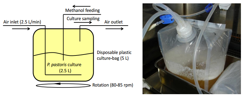
Muraki M (2014) Improved production of recombinant human Fas ligand extracellular domain in Pichia pastoris: yield enhancement using disposable culture-bag and its application to site-specific chemical modifications. BMC Biotechnol 14: 19. doi: 10.1186/1472-6750-14-19
    [48] Jacobs PP, Geysens S, Vervecken W, et al. (2009) Engineering complex-type N-glycosylation in Pichia pastoris using glycoswitch technology. Nat Protoc 4: 58–70. doi: 10.1038/nprot.2008.213
    [49] Helenius A, Aebi M (2004) Roles of N-linked glycans in endoplasmic reticulum. Annu Rev Biochem 73: 1019–1049. doi: 10.1146/annurev.biochem.73.011303.073752
    [50] Pfeffer M, Maurer M, Stadlmann J, et al. (2012) Intracellular interactome of secreted antibody Fab fragment in Pichia pastoris reveals its routes of secretion and degradation. Appl Microbiol Biot 93: 2503–2512. doi: 10.1007/s00253-012-3933-3
    [51] Muraki M (2008) Improved secretion of human Fas ligand extracellular domain by N-terminal part truncation in Pichia pastoris and preparation of the N-linked carbohydrate chain trimmed derivative. Protein Expres Purif 60: 205–213.
    [52] Orlinick JR, Elkon KB, Chao MV (1997) Separate domains of the human Fas ligand dictate self-association and receptor binding. J Biol Chem 51: 32221–32229.
    [53] Gasteiger E, Hoogland C, Gattiker A, et al. (2005) Protein identification and analysis tools on the ExPASy server, In: Walker JM, The Proteomics Protocols Handbook, Totowa: Humana Press, 571–607.
    [54] 
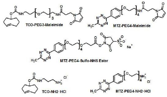
Muraki M, Hirota K (2017) Site-specific chemical conjugation of human Fas ligand extracellular domain using trans-cyclooctene-methyltetrazine reactions. BMC Biotechnol 17: 56. doi: 10.1186/s12896-017-0381-2
    [55] Mahiou J, Abastado JP, Cabanie L, et al. (1998) Soluble FasR ligand-binding domain: high-yield production of active fusion and non-fusion recombinant proteins using the baculovirus/insect cell system. Biochem J 330: 1051–1058. doi: 10.1042/bj3301051
    [56] Muraki M, Honda S (2010) Efficient production of human Fas receptor extracellular domain-human IgG1 heavy chain Fc domain fusion protein using baculovirus/silkworm expression system. Protein Expres Purif 73: 209–216. doi: 10.1016/j.pep.2010.05.007
    [57] Muraki M, Honda S (2011) Improved isolation and purification of functional human Fas receptor extracellular domain using baculovirus-silkworm expression system. Protein Expres Purif 80: 102–109. doi: 10.1016/j.pep.2011.07.002
    [58] Vogl T, Glieder A (2013) Regulation of Pichia pastoris promoters and its consequences for protein production. New Biotechnol 30: 385–404. doi: 10.1016/j.nbt.2012.11.010
    [59] Allais JJ, Louktibi A, Baratti J (1980) Oxidation of methanol by the yeast, Pichia pastoris. Purification and properties of the alcohol oxidase. Agric Biol Chem 44: 2279–2289.
    [60] Muraki M (2014) Secretory production of recombinant proteins in methylotrophic yeast Pichia pastoris using a disposable culture-bag. PSSJ Arch 7: e078.
    [61] Vanamee ÉS, Faustmann DL (2018) Structural principles of tumor necrosis factor superfamily signaling. Sci Signal 11: eaao4910. doi: 10.1126/scisignal.aao4910
    [62] Hermanson GT (2013) Chapter 10: Fluorescent Probes, In: Bioconjugate techniques (Third Ed.), London: Academic Press, 395–463.
    [63] Wu H, Devaraj NK (2016) Inverse electron-demand Diels-Alder biorthogonal reactions. Top Curr Chem 374: 3. doi: 10.1007/s41061-015-0005-z
    [64] Mayer S, Lang K (2017) Tetrazines in inverse-electron-demand Diels-Alder cycloadditions and their use in biology. Synthesis-Stuttgart 49: 830–848.
    [65] Muraki M (2016) Preparation of a functional fluorescent human Fas ligand extracellular domain derivative using a three-dimensional structure guided site-specific fluorochrome conjugation. SpringerPlus 5: 997. doi: 10.1186/s40064-016-2673-8
    [66] Ogasawara J, Watanabe-Fukunaga R, Adachi M, et al. (1993) Lethal effect of the anti-Fas antibody in mice. Nature 364: 806–809. doi: 10.1038/364806a0
    [67] Wajant H, Gerspach J, Pfizenmaier K (2013) Engineering death receptor ligands for cancer therapy. Cancer Lett 332: 163–174. doi: 10.1016/j.canlet.2010.12.019
    [68] Villa-Morales M, Fernández-Piqueras J (2012) Targeting the Fas/FasL signaling pathway in cancer therapy. Expert Opin Ther Tar 16: 85–101. doi: 10.1517/14728222.2011.628937
    [69] Oliveira BL, Guo Z, Bernardes GJL (2017) Inverse electron demand Diels-Alder reactions in chemical biology. Chem Soc Rev 46: 4895–4950. doi: 10.1039/C7CS00184C
    [70] Green NM (1963) Avidin. 4. Stability at extremes of pH and dissociation into sub-units by guanidine hydrochloride. Biochem J 89: 609–620.
    [71] Paganelli G, Magnani P, Zito F, et al. (1991) Three-step monoclonal antibody tumor targeting in carcinoembryonic antigen positive patients. Cancer Res 51: 5960–5966.
    [72] Penichet ML, Kang YS, Pardridge WM, et al. (1999) An antibody-avidin fusion protein specific for the transferrin receptor serves as a delivery vehicle for effective brain targeting: initial applications in anti-HIV antisense drug delivery to brain. J Immunol 163: 4421–4426.
    [73] Schultz J, Lin Y, Sanderson J, et al. (2000) A tetravalent single-chain antibody-streptavidin fusion protein for pretargeted lymphoma therapy. Cancer Res 60: 6663–6669.
    [74] Mohsin H, Jia F, Bryan JN, et al. (2011) Comparison of pretargeted and conventional CC49 radioimmunotherapy using 149Pm, 166Ho, and 177Lu. Bioconjugate Chem 22: 2444–2452. doi: 10.1021/bc200258x
    [75] Micheau O, Solary E, Hammann A, et al. (1997) Sensitization of cancer cells treated with cytotoxic drugs to Fas-mediated cytotoxity. J Natl CancerI 89: 783–789. doi: 10.1093/jnci/89.11.783
    [76] Yang D, Torres CM, Bardhan K, et al. (2012) Decitabine and vorinostat cooperate to sensitize colon carcinoma cells to Fas ligand-induced apoptosis in vitro and tumor suppression in vivo. J Immunol 188: 4441–4449. doi: 10.4049/jimmunol.1103035
    [77] Galenkamp KMO, Carriba P, Urresti J, et al. (2015) TNFα sensitizes neuroblastoma cells to FasL-, cisplatin- and etoposide-induced cell death by NF-ΚB-mediated expression of Fas. Mol Cancer 14: 62. doi: 10.1186/s12943-015-0329-x
    [78] Xu X, Fu XY, Plate J (1998) IFN-γ induces cell growth inhibition by Fas-mediated apoptosis: requirement of STAT1 protein for up-regulation of Fas and FasL expression. Cancer Res 58: 2832–2837.
    [79] Horton JK, Siamakpour-Reihani S, Lee CT, et al. (2015) Fas death receptor: a breast cancer subtype-specific radiation response biomarker and potential therapeutic target. Radiat Res 184: 456–469. doi: 10.1667/RR14089.1
    [80] Tamakoshi A, Nakachi K, Ito Y, et al. (2008) Soluble Fas level and cancer mortality: findings from a nested case-control study within a large-scale prospective study. Int J Cancer 123: 1913–1916. doi: 10.1002/ijc.23731
    [81] Bhatraju PK, Robinson-Cohen C, Mikacenic C, et al. (2017) Circulating levels of soluble Fas (sCD95) are associated with risk for development of a nonresolving acute kidney injury subphenotype. Crit Care 21: 217. doi: 10.1186/s13054-017-1807-x
    [82] Hamilton SR, Gerngross TU (2007) Glycosylation engineering in yeast: the advent of fully humanized yeast. Curr Opin Biotech 18: 387–392. doi: 10.1016/j.copbio.2007.09.001
    [83] Liu L, Stadheim A, Hamuro L, et al. (2011) Pharmacokinetics of IgG1 monoclonal antibodies produced in humanized Pichia pastoris with specific glycoforms: a comparative study with CHO produced materials. Biologicals 39: 205–210. doi: 10.1016/j.biologicals.2011.06.002
  • This article has been cited by:

    1. Michiro Muraki, Kiyonori Hirota, Confirmation of covalently-linked structure and cell-death inducing activity in site-specific chemical conjugates of human Fas ligand extracellular domain, 2018, 11, 1756-0500, 10.1186/s13104-018-3501-8
    2. Michiro Muraki, Sensitization to cell death induced by soluble Fas ligand and agonistic antibodies with exogenous agents: A review, 2020, 7, 2375-1576, 122, 10.3934/medsci.2020011
  • Reader Comments
  • © 2018 the Author(s), licensee AIMS Press. This is an open access article distributed under the terms of the Creative Commons Attribution License (http://creativecommons.org/licenses/by/4.0)
通讯作者: 陈斌, bchen63@163.com
  • 1. 

    沈阳化工大学材料科学与工程学院 沈阳 110142

  1. 本站搜索
  2. 百度学术搜索
  3. 万方数据库搜索
  4. CNKI搜索

Metrics

Article views(6795) PDF downloads(1168) Cited by(2)

Article outline

Figures and Tables

Figures(8)  /  Tables(1)

Other Articles By Authors

/

DownLoad:  Full-Size Img  PowerPoint
Return
Return

Catalog