Review Topical Sections

Smart systems related to polypeptide sequences

  • Received: 29 December 2015 Accepted: 03 March 2016 Published: 09 March 2016
  • Increasing interest for the application of polypeptide-based smart systems in the biomedical field has developed due to the advantages given by the peptidic sequence. This is due to characteristics of these systems, which include: biocompatibility, potential control of degradation, capability to provide a rich repertoire of biologically specific interactions, feasibility to self-assemble, possibility to combine different functionalities, and capability to give an environmentally responsive behavior. Recently, applications concerning the development of these systems are receiving greater attention since a targeted and programmable release of drugs (e.g. anti-cancer agents) can be achieved. Block copolymers are discussed due to their capability to render differently assembled architectures. Hybrid systems based on silica nanoparticles are also discussed. In both cases, the selected systems must be able to undergo fast changes in properties like solubility, shape, and dissociation or swelling capabilities. This review is structured in different chapters which explain the most recent advances on smart systems depending on the stimuli to which they are sensitive. Amphiphilic block copolymers based on polyanionic or polycationic peptides are, for example, typically employed for obtaining pH-responsive systems. Elastin-like polypeptides are usually used as thermoresponsive polymers, but performance can be increased by using techniques which utilize layer-by-layer electrostatic self-assembly. This approach offers a great potential to create multilayered systems, including nanocapsules, with different functionality. Recent strategies developed to get redox-, magnetic-, ultrasound-, enzyme-, light- and electric-responsive systems are extensively discussed. Finally, some indications concerning the possibilities of multi-responsive systems are discussed.

    Citation: Lourdes Franco, Luís J. del Valle, Jordi Puiggalí. Smart systems related to polypeptide sequences[J]. AIMS Materials Science, 2016, 3(1): 289-323. doi: 10.3934/matersci.2016.1.289

    Related Papers:

    [1] Jean-Bernard Baillon, Guillaume Carlier . From discrete to continuous Wardrop equilibria. Networks and Heterogeneous Media, 2012, 7(2): 219-241. doi: 10.3934/nhm.2012.7.219
    [2] Edward S. Canepa, Alexandre M. Bayen, Christian G. Claudel . Spoofing cyber attack detection in probe-based traffic monitoring systems using mixed integer linear programming. Networks and Heterogeneous Media, 2013, 8(3): 783-802. doi: 10.3934/nhm.2013.8.783
    [3] Matthieu Canaud, Lyudmila Mihaylova, Jacques Sau, Nour-Eddin El Faouzi . Probability hypothesis density filtering for real-time traffic state estimation and prediction. Networks and Heterogeneous Media, 2013, 8(3): 825-842. doi: 10.3934/nhm.2013.8.825
    [4] Raúl M. Falcón, Venkitachalam Aparna, Nagaraj Mohanapriya . Optimal secret share distribution in degree splitting communication networks. Networks and Heterogeneous Media, 2023, 18(4): 1713-1746. doi: 10.3934/nhm.2023075
    [5] Samitha Samaranayake, Axel Parmentier, Ethan Xuan, Alexandre Bayen . A mathematical framework for delay analysis in single source networks. Networks and Heterogeneous Media, 2017, 12(1): 113-145. doi: 10.3934/nhm.2017005
    [6] Carlos F. Daganzo . On the variational theory of traffic flow: well-posedness, duality and applications. Networks and Heterogeneous Media, 2006, 1(4): 601-619. doi: 10.3934/nhm.2006.1.601
    [7] Leah Anderson, Thomas Pumir, Dimitrios Triantafyllos, Alexandre M. Bayen . Stability and implementation of a cycle-based max pressure controller for signalized traffic networks. Networks and Heterogeneous Media, 2018, 13(2): 241-260. doi: 10.3934/nhm.2018011
    [8] Félicien BOURDIN . Splitting scheme for a macroscopic crowd motion model with congestion for a two-typed population. Networks and Heterogeneous Media, 2022, 17(5): 783-801. doi: 10.3934/nhm.2022026
    [9] Anya Désilles . Viability approach to Hamilton-Jacobi-Moskowitz problem involving variable regulation parameters. Networks and Heterogeneous Media, 2013, 8(3): 707-726. doi: 10.3934/nhm.2013.8.707
    [10] Fethallah Benmansour, Guillaume Carlier, Gabriel Peyré, Filippo Santambrogio . Numerical approximation of continuous traffic congestion equilibria. Networks and Heterogeneous Media, 2009, 4(3): 605-623. doi: 10.3934/nhm.2009.4.605
  • Increasing interest for the application of polypeptide-based smart systems in the biomedical field has developed due to the advantages given by the peptidic sequence. This is due to characteristics of these systems, which include: biocompatibility, potential control of degradation, capability to provide a rich repertoire of biologically specific interactions, feasibility to self-assemble, possibility to combine different functionalities, and capability to give an environmentally responsive behavior. Recently, applications concerning the development of these systems are receiving greater attention since a targeted and programmable release of drugs (e.g. anti-cancer agents) can be achieved. Block copolymers are discussed due to their capability to render differently assembled architectures. Hybrid systems based on silica nanoparticles are also discussed. In both cases, the selected systems must be able to undergo fast changes in properties like solubility, shape, and dissociation or swelling capabilities. This review is structured in different chapters which explain the most recent advances on smart systems depending on the stimuli to which they are sensitive. Amphiphilic block copolymers based on polyanionic or polycationic peptides are, for example, typically employed for obtaining pH-responsive systems. Elastin-like polypeptides are usually used as thermoresponsive polymers, but performance can be increased by using techniques which utilize layer-by-layer electrostatic self-assembly. This approach offers a great potential to create multilayered systems, including nanocapsules, with different functionality. Recent strategies developed to get redox-, magnetic-, ultrasound-, enzyme-, light- and electric-responsive systems are extensively discussed. Finally, some indications concerning the possibilities of multi-responsive systems are discussed.


    Multidrug-resistant bacteria (MDRB) are microorganisms that are resistant to one or more antimicrobial agents. They are usually resistant to all but one or two commercially available antimicrobial agents. This definition includes microbes that have acquired resistance to at least one agent in three or more antimicrobial categories. The MDRB of clinical interest include: Methicillin-resistant Staphylococcus aureus (MRSA), Staphylococcus aureus with resistance to vancomycin [these are Vancomycin-intermediate Staphylococcus aureus (VISA) and Vancomycin-resistant Staphylococcus aureus (VRSA)],Vancomycin-resistant enterococci (VRE), Extended spectrum beta-lactamases (ESBLs) producing gram-negative bacilli, Multidrug-resistant Streptococcus pneumoniae (MDRSP), Carbapenem-resistant Enterobacteriaceae (CRE) and Multidrug-resistant Acinetobacter baumannii [1][3] .

    Infectious diseases caused by MDRB are an important burden globally. They have for centuries been among the leading causes of death, disability, growing challenges to health security and human progress, especially in developing countries [4].

    Although, many new antibacterial drugs have been produced, bacteria exhibiting resistance to them have increased and is becoming a global concern as we are fast running out of therapeutic options [5],[6]. The challenges of antimicrobial resistance are faced in both the health care and community settings, necessitating a broad approach with multiple partners across the continuum of care. For example, 18–33% of MRSA colonized patients subsequently developed MRSA infections. Community-acquired methicillin-resistant Staphylococcus aureus (CA-MRSA) strains also constitute an increasing proportion of hospital-onset MRSA infections. The Centre for Disease Control and Prevention (CDC) estimated that over 2 million illnesses and 23,000 deaths per year are attributable to antibiotic resistance in the United States [3].

    Vancomycin is widely prescribed for the treatment of infections caused by MRSA; but the emergence of VISA and VRSA has been reported by many authors. Really, teicoplanin, daptomycin, linezolid, etc are expensive drugs which are currently prescribed when faced with MRSA with low sensitivity to vancomycin. However, development of resistance to these drugs has been identified worldwide [7][11].

    Usage of plants in fighting against illnesses and diseases has deep roots in man's history. Researchers are interested in plant extracts as medicines because there are several reports regarding the antimicrobial activity of their crude extracts which might be better substitutes for conventional antibiotics. Recent published reports opined that medicinal plants with anti-MRSA activity can be considered for treatment of MRSA infections [8],[12]. This present work is a brief review on MRSA, VISA, VRSA and some medicinal plants with anti-MRSA activities.

    Staphylococcus aureus is a Gram-positive coccoid bacterium. The cells are arranged in irregular grape-like appearance and they are usually found as normal flora in humans and animals. It is ubiquitous in the human population and 30–40% of adults are asymptomatic carriers. It is also a major pathogen of human and can cause a range of infections from mild skin infections and food poisoning, to life threatening infections [13][17].

    Resistance to methicillin by S. aureus was initially observed in 1961 shortly after the antibacterial agent was introduced clinically and since then, there has been a global epidemic of Methicillin-resistant Staphylococcus aureus (MRSA) in both healthcare and community settings [18][20]. MRSA isolates from the UK and Denmark in the early 1960s constituted the very first epidemic MRSA clone soon after methicillin was introduced and it has since emerged as an important pathogen in human medicine [21][23]. Although, methicillin is no longer prescribed for patients and has been replaced by isoxazolyl penicillins, particularly flucloxacillin in the UK, the acronym MRSA has stayed [24]. It is characterized by antibiotic resistance to penicillins, cephalosporins, carbapenems and has tendency of developing resistance to quinolones, aminoglycosides, and macrolides [10],[25],[26].

    The origination of MRSA was as a result of Staphylococcal Cassette Chromosome mec (SCCmec) genes acquired by methicillin-susceptible S. aureus (MSSA). The SCCmec harbours the mecA gene which encodes the penicillin-binding protein (PBP2a) that confers resistance to all β-lactam antibiotics [10],[27][29]. SCCmec also contains the cassette chromosome recombinases (ccr) gene complex. The ccr genes (composed of ccrC or a pair of ccrA and ccrB) encode recombinases mediating integration and excision of SCCmec into or from the chromosome. The ccr genes and surrounding genes form the ccr gene complex. In addition to ccr and mec gene complexes, SCCmec contains a few other genes and various other mobile genetic elements such as: insertion sequences, transposons and plasmids [30],[31].

    Eleven different types of SCCmec (I-XI) and five allotypes of the ccr gene complexes (ccrAB1, ccrAB2, ccrAB3, ccrAB4 and ccrC) have been reported. Generally, SCCmec types I, II, III, VI and VIII are called hospital-acquired MRSA or (HA-MRSA). Types IV, V and VII as community-acquired (CA-MRSA) while types IX, X and XI as livestock-associated MRSA (LA-MRSA) [31],[32]. Expression of methicillin resistance in S. aureus is commonly under regulatory control by mecI or blaI gene. The mecI and blaI repressors are controlled by the mecRI and blaRI transducers [20].

    MRSA remains a major public health concern worldwide and a therapeutic challenge as the antibacterial drugs effective for treatment are scanty and costly. The changing epidemiology of MRSA infections, varying resistance to commonly used antibiotics and involvement in hospital and community infections are influencing the use and clinical outcomes of currently available anti-infective agents [33].

    Vancomycin is an antibacterial agent that inhibits cell wall production by binding with the D-alanyl-D-alanine C terminus of the bacterial cell wall precursors, and subsequently preventing cross-linking by transpeptidation. Vancomycin acts extracellularly and inhibits late-stage peptidoglycan biosynthesis which results in the intracellular accumulation of UDP-linked MurNAc-pentapeptide precursors. The vancomycin complex involves a number of hydrogen bonds between the peptide component of vancomycin and the D-Ala-D-Ala residue. Any process that interferes with vancomycin binding to D-Ala-D-Ala residues in the cell wall will decrease the potency of the drug [13],[36].

    Vancomycin was widely utilized for the treatment of MRSA infections and has led to the emergence of vancomycin-intermediate and vancomycin-resistant S. aureus (VISA and VRSA) [37]. This also triggered off alarms in the medical community as S. aureus causes life-threatening infections in hospitalized and non-hospitalized patients [38]. Vancomycin-intermediate S. aureus (VISA), heterogeneous vancomycin-intermediate S. aureus (hVISA) and vancomycin-resistant S. aureus (VRSA) are the three classes of S. aureus that are resistant to vancomycin which have emerged in different locations of the world [39].

    Vancomycin-intermediate S. aureus (VISA) was first reported from Japan in 1996 with reduced susceptibility to vancomycin (having a Minimum Inhibitory Concentration (MIC) of 8 mg/L). It has now spread to other hospitals in Asia, France, Brazil, USA, United Kingdom, etc [40]. S. aureus vancomycin breakpoints were redefined by the Clinical and Laboratory Standards Institute (CLSI) in 2006 as follows: resistant at MIC ≥ 16 µg/ml, intermediate at 4–8 µg/ml and susceptible at ≤ 2 µg/ml [34][36].

    VISA isolates emerged as a result of mutations (not their acquisition of foreign genetic elements) in MRSA isolates during treatment of patients with vancomycin. The comparison of vancomycin-susceptible and -resistant isolates to the VISA isolates showed that the mutations often occurred in the walkR, vraSR, rpoB (ribosomal) genes and the yvqF/vraSR system. Usually, the relevant mutated genes seemed to be directly or indirectly involved with the biosynthesis/metabolism of the staphylococcal cell wall [41].

    Often, there were treatment failures when VISA infections were treated with vancomycin [41]. It was observed that under vancomycin selective pressure usually during treatment, the VISA strains with a vancomycin MIC of 8 µg/ml have emerged and led to therapy failure. However, the nature of this resistance phenotype (VISA) was unstable especially when vancomycin selective pressure is removed as some strains reverted back to vancomycin-susceptible strains with MIC at 2 µg/ml [36].

    In 1997, the first case of hVISA was reported in Japan. The cultures of hVISA strains contain both low-frequency subpopulations of bacteria with increased vancomycin MIC value and high frequency of bacteria with low vancomycin MIC values (close to those of susceptible strains) [41]. The MIC for hVISA strains was defined by the presence of subpopulations of VISA at a rate of one organism per 105 to 106 organisms [42],[43]. The hVISA strains were detected using vancomycin population analysis profile (PAP) which was proposed as the most accurate method for hVISA detection; however, it is relatively time-consuming and requires the use of a spiral plater. The hVISA strain has generally required formal population analysis using the serial passage of screened isolates of S. aureus on selective agar containing increasing concentrations of vancomycin for its detection [13]. Results are generally not ready until at least 3 to 5 days [36].

    VISA and hVISA strains have thickened cell wall with reduced glycopeptide cross-linking as a result of the complex reorganization of cell wall metabolism. It has been proposed that the thickened cell wall may trap and sequester vancomycin and consequently, interferes with its mode of action [13]. This could be due to alteration in peptidoglycan production leading to increased residues of D alanyl-D-alanine, which bind vancomycin molecules and prevent them from reaching the target sites [18][20].

    In 2002, the first hospital strain of Vancomycin-resistant S. aureus (VRSA) was reported in the United States [44]. The acquisition of vanA gene from vancomycin-resistant enterococci resulted in the emergence of vancomycin-resistant strains of S. aureus (VRSA) with vancomycin MIC value greater than 16 µg/ml [36],[41],[45].

    MRSA has spread worldwide, and its prevalence has increased in both health-care and community environments. The proportion of MRSA varied among countries such as for instance: 0.4% in Sweden [24]; 25% in western part to 50% in southern India [10]; 33%–43% in Nigeria [46]; 37–56% in Greece, Portugal and Romania in 2014 [47]. High prevalence of MRSA with rates greater than 50% has also been reported in hospitals worldwide including in Asia, Malta, North and South America [29],[48]. Variation in the prevalence rates of MRSA was due to different epidemiological factors such as geographical and health system capability in running infection control program [49].

    Akanbi and Mbe [50] reported a prevalence range of 0% to 6% VRSA in southern parts of Nigeria among clinical isolates and also 57.7% in Zaria, northern Nigeria. Goud, et al., [51] reported a vancomycin resistance in 1.4% of S. aureus isolates in southern India. Other countries such as: Australia, Korea, Hong Kong, Scotland, Israel, Thailand, South Africa, etc have also reported S. aureus with vancomycin sensitivity reduction with prevalence ranges from 0–74% [20],[36],[52].

    Currently, there are seven common antibiotics used against MRSA, which are: vancomycin, daptomycin, linezolid, Sulfamethoxazole and trimethoprim (TMP-SMZ), quinupristin-dalfopristin, clindamycin and tigecycline. These antibiotics are gradually losing their efficiency as MRSA strains are developing resistance against them [8],[20],[53]. Presently, the therapeutic alternatives available for treatment of infections caused by MRSA and S. aureus with reduced vancomycin susceptibility are limited. Therefore, there is a global urgency for the development of novel drugs that will be effective in the treatment of S. aureus exhibiting multidrug resistance so as to combat the scourge caused by the microorganism in the globe [52].

    Natural products including medicinal plants have contributed immensely to human health, well-being and development of novel drugs. They are useful natural blueprints for the development of new drugs (especially in western countries) or/and phytomedicines purified to be used for the treatment of disease (commonly in developing countries and Europe) [54]. Medicinal plants can be valuable therapeutic resources. In numerous developing countries, including Nigeria, 80% of patients use home-made phytomedicines to treat infectious diseases. Despite the availability of modern medicine in some communities, the use of medicinal plants has remained high due to their efficacy, popularity and low cost. They also represent sources of potentially important new pharmaceutical substances since all the plants parts are utilized in traditional treatment and can therefore, act as lead compounds (Table 1).

    The applications of phytomedicines for human well being and as blueprints for developing novel useful drugs have drastically increased worldwide in recent years [77].

    The emergence of multidrug-resistant infectious agents associated with over- and inappropriate use of antibiotics has necessitated the World Health Organization (WHO) to acknowledge and pronounce the urgent need to develop novel antimicrobials and/or new approaches to tackle the menace caused by them in the globe; these have subsequently led to the resuscitation of the interest in medicinal plants [78]. The most common bacteria that have been used in susceptibility tests with numerous medicinal plants include: Staphylococcus aureus, methicillin-resistant Staphylococcus aureus (MRSA), vancomycin-resistant Enterococcus (VRE), Pseudomonas aeruginosa Helicobacter pylori, etc [54]. Presently, numerous studies have reported the antibacterial activity of many plant extracts against MRSA. In this study, only fifty-one (51) plants with anti-MRSA activities from thirty-five (35) families were mentioned (Table 1). The minimum inhibitory concentrations (MIC) values of the plants on the tested MRSA strains were between 1.25 µg/ml to 6.30 mg/ml. Twenty-nine of the plants had MIC values < 1.0 mg/ml while the remaining twenty-two MIC values were > 1.0 mg/ml but < 8.0 mg/ml. Extracts exhibiting activities with MIC values below 8 mg/ml are widely accepted to possess some antimicrobial activity while those with values below 1 mg/ml are considered noteworthy [77],[79]. However, most of the plants in this review were not tested on S. aureus strains with reduced vancomycin susceptibility.

    The solvents used for the medicinal plants extraction in this review were ethanol and methanol (Table 1). This is probably because alcoholic extracts have higher antimicrobial activity than aqueous extracts. It has been reported that ethanolic extracts have higher antimicrobial activity than aqueous extracts because of the presence of higher amounts of polyphenols. They are more efficient in cell walls and seeds degradation causing polyphenols to be released from cells. Also, the enzyme polyphenol oxidase, degrades polyphenols in water extracts but is inactive in methanol and ethanol. Moreover, water is a better medium for the growth of microorganisms than ethanol [80].

    Although, methanol is more polar than ethanol but it is not frequently used for plant extraction due to its cytotoxic nature that may give incorrect results [81].

    Extracts of medicinal plants are rich in phytochemicals. Phytochemicals or secondary metabolites are natural protective agents biosynthesized by plants against external stress and pathogenic attack. They are crucial for plant defences and survival. They have been divided into several categories: phenolics, alkaloids, steroids, terpenes, saponins, etc. They exhibit other bioactivities such as antimutagenic, anticarcinogenic, antioxidant, antimicrobial, and anti-inflammatory properties and are therefore responsible for the medicinal potential of plants (Table 2). Hence, from this review, anti-MRSA plants have antibacterial effect on MRSA strains and other medicinal/therapeutic uses as depicted in Table 2.

    Table 1.  Medicinal plants with activities on methicillin-resistant S. aureus (MRSA).
    Botanical name Family Local/Common name Place of collection Plant part used Extracting Solvent MIC /MBC (mg/ml) MRSA MIC /MBC (mg/ml) VRSA References
    Acacia catechu (L. f.) Willd Fabaceae Cutch tree, black catechu Thailand Wood Ethanol 1.6–3.2/25 55,56
    Garcinia mangostana L. Clusiaceae Mangosteen Thailand Fruit shell Ethanol 0.05–0.4/0.1–0.4 55,57
    Impatiens balsamina Balsaminaceae Garden balsam Thailand Leaf Ethanol 6.3/25 55,58
    Peltophorum ptercarpum (DC.) Fabaceae Yellow flame tree Thailand Bark Ethanol 0.1–0.8/6.3 55,59
    Psidium guajava L. Myrtaceae Guava Thailand Leaf Ethanol 0.2–1.6/6.3 55,60
    Punica granatum L. Punicaceae Pomegranate Thailand Fruit shell Ethanol 0.2–0.4/1.6–3.2 55,61
    Uncaria gambir (Hunter) Roxb. Rubiaceae Gambier, White cutch Thailand Leaf, stem Ethanol 0.4–0.8/3.2 55,62
    Walsura robusta Meliaceae Bonlichu Thailand Wood Ethanol 1.6–3.2/25 55,63
    Swietenia mahagoni Meliaceae Mahagoni Malaysia Seed Ethanol 0.2–0.78/0.78–1.56 64
    Tinospora crispa Menispermaceae Patawali Malaysia Stem Ethanol 0.4–0.78/0.78–1.56 64
    Butea monosperma Lam. Fabaceae Flame-of-the-forest India Leaf Ethanol 5.91/13.30 1.16/2.62 65
    Callistemon rigidus R.Br. Myrtaceae Stiff bottlebrush India Leaf Methanol 0.00125–0.08 66
    Acacia albida Del. Fabaceae Gawo Nigeria Stem bark Methanol 3.0/4.0 67
    Anchomanes difformis Engl. Araceae Chakara Nigeria Roots Methanol 4.0/5.0 67
    Boscia senegalensis Del. Capparidaceae Anza Nigeria Roots Methanol 5.0/6.0 67
    Moringa oleifera Lam. Moringacceae Zogale Nigeria Leaf Ethanol 4.0/5.0 67
    Mormodica basalmina Linn. Cucurbitaceae Garahuni Nigeria Whole plant Methanol 4.0/5.0 67
    Nymphaea lotus Linn. Nymphaeaceae White lotus Nigeria Leaf Ethanol 5.0–10.0/10.0–30.0 5.0–10.0/10.0–30.0 68
    Pavetta crassipes K. Schum. Rubiaceae Gadau Nigeria Leaf Methanol 4.0/5.0 67
    Phyllanthus amarus Schum. Thonn. Euphorbiaceae Geron tsuntsaye Nigeria Whole plant Methanol 4.0/5.0 67
    Vernonia blumeoides Hook. f. Asteraceae Bagashi Nigeria Aerial part Ethanol 4.0/5.0 67
    Curcuma xanthorrhiza Zingiberaceae Java ginger Indonesia Rhizome Ethanol 0.5/ND 69
    Kaempferia pandurata Roxb. Zingiberaceae Temu kunci, fingerroot Indonesia Rhizome Ethanol 0.3/ND 69
    Senna alata Fabaceae Candle bush Indonesia Leaf Ethanol 0.5/ND 69
    Mallotus yunnanensis Pax et. Hoffm. Euphorbiaceae - China Tender Branches & leaves (TBL) Ethanol 0.008–0.032/0.064–0.26 70
    Skimmia arborescens Anders. Rutaceae Japanese skimmia China TBL Ethanol 0.016–0.064/0.13–0.26 70
    Cyclobalanopsis austroglauca Y.T. Chang Fagaceae Oak China TBL Ethanol 0.016–0.064/0.13–0.51 70
    Manglietia hongheensis Y.m Shui et. W.H. Chen. Magnoliaceae Magnolia China TBL Ethanol 0.008–0.13/0.032–0.51 70
    Brandisia hancei Hook.f. Scrophulariaceae - China Whole plant Ethanol 0.032–0.064/0.13–0.26 70
    Evodia daneillii (Benn) Hemsl. Rutaceae Bebe tree China TBL Ethanol 0.032–0.064/0.064–0.26 70
    Schima sinensis (Hemsl. et. Wils) Airy-shaw. Theaceae Schima China TBL Ethanol 0.016–0.064/0.064–0.26 70
    Garcinia morella Desr. Clusiaceae Gamboge China Whole plant Ethanol 0.016–0.064/0.064–0.26 70
    Meliosma squamulata Hance. Lauraceae - China TBL Ethanol 0.032–0.064/0.13–0.26 70
    Curculigo orchioides Gaertn. Hypoxidaceae Golden eye-grass China Whole plant Ethanol 0.26–0.51/0.51–>2.05 70
    Euonymus fortunei (Turcz.); Hand. Mazz. Celastraceae Spindle, Winter creeper China Vane Ethanol 0.51/1.02–>2.05 70
    Alnus nepalensis D. Don. Betulaceae Nepalese alder China TBL Ethanol 0.26–1.02/1.02–>2.05 70
    Illicium simonsii Maxim. Illiciaceae - China TBL Ethanol 0.51–1.02/1.02–>2.05 70
    Blumea balsamifer (Linn.) D.C. Asteraceae Sambong China Whole plant Ethanol 0.064–0.26/0.26–1.02 70
    Machilus salicina Hance. Lauraceae Liu ye run nan China TBL Ethanol 0.51–1.02/1.02–>2.05 70
    Schisandra viridis A.c.Smith. Schisandraceae Magnolia vine China Vane Ethanol 0.064–0.26/0.26–1.02 70
    Selaginella tamariscina (Seauv.) Spring. Selaginellaceae Little club moss China Whole plant Ethanol 0.51–1.02/1.02–>2.05 70
    Celastrus orbiculatus Thunb. Celastraceae Chinese bittersweet China Vane Ethanol 0.51–1.02/1.02–>2.05 70
    Polygonum molleD. Don. Polygonaceae Knotweed China Whole plant Ethanol 0.26–0.51/1.02–>2.05 70
    Carex prainii C.B. Clarke Cyperaceae Sedges China Whole plant Ethanol 1.02–2.05/2.05–>2.05 70
    Embelia burmf. Myrsinaceae Baberung, Vidanga China Leaves Ethanol 0.51–1.02/1.02–>2.05 70
    Melianthus major L. Melianthaceae Giant honey flower South Africa Leaves Ethanol 0.78/3.12 71
    Melianthus comosusVahl Melianthaceae Honey flower South Africa Leaves Ethanol 0.39/1.56 71
    Dodonaea angustifolia (L.f.) Benth Sapindaceae Sticky hopbush, sand olive South Africa Leaves Ethanol 0.59/1.17 71
    Withania somnifera L. Solanaceae Ashwagandha, Winter cherry South Africa Roots & leaves Ethanol 1.56/>6.25 71,72,73
    Quercus infectoria Olivier Fagaceae “Machika or Oak galls South Africa Nutgalls Ethanol 0.4–3.2/3.2–6.3 74
    Thymus vulgaris L. Lamiaceae Thyme Peru Leaves Essential oil 0.057/ND 75,76

    Key: ND- Not done; MIC- Minimum inhibitory concentration; MBC- Minimum bactericidal concentration; VRSA- Vancomycin-resistant S. aureus

     | Show Table
    DownLoad: CSV
    Table 2.  Anti-MRSA plants with their phytochemical contents and medicinal uses.
    Medicinal Plant Phytochemical content Medicinal uses References
    Acacia catechu tannins, flavonoids, amino acids , saponins, triterpenoids Cold, cough, diarrhea, piles,fever. ulcers, boils,etc 82,83
    Garcinia mangostana Xanthones and phenolics (tannins) skin infections, wounds, dysentery, urinary disorders, cystitis and gonorrhoea 84,85
    Impatiens balsamina flavanoids, triterpenoids, glycosides, fatty acids & alkaloids diuretic, emetic, laxative, demulcent and tonic 86
    Peltophorum ptercarpum fatty acids, amino acids, terpenoids, phenolics, flavonoids, alkaloids, steroids etc. stomatitis, insomnia, skin troubles, constipation, ringworm, insomnia, dysentery, muscular pains, sores, and skin disorders 87
    Psidium guajava Tannins, Steroids, Alkaloids, glycosides, vitamins, carbohydrates diarrhea, sore throat, vomiting, stomach upset, vertigo etc. 88
    Punica granatum Tannins, Alkaloids, glycosides, vitamins, carbohydrates, flavanoids, saponins, triterpenoids sore throats, coughs, urinary infections, digestive disorders, skin disorders, arthritis, expel worms 89
    Uncaria gambir tannins, catechin, gambiriins wounds and ulcers, fevers, headaches, gastrointestinal illnesses, bacterial/fungal infections, diarrhoea, sore throat 90,91
    Walsura robusta Sesquiterpenoid 10-nitro-isodauc-3-en-15-al, 10-oxo-isodauc-3-en-15-al Antibacterial, antimicrobial, astringent, diarrhea 92,93
    Swietenia mahagoni Alkaloids, terpenoids, anthraquinone, cardiac glycosides, saponins, phenols, flavonoids, etc Hypertension, diabetes, malaria, amoebiasis, cough, chest pain, tuberculosis, antibacterial 64,94
    Tinospora crispa Triterpenes, flavones o-glycosides (apigenine), picroretoside, berberine, palmatine, picroretine & resin Fever, jaundice, hyperglycemia, wounds, intestinal worms, skin infections, antibacterial activity 64
    Butea monosperma Tannins, Saponins, Alkaloids, Glycosides, Carbohydrates hepatoprotective, antidiabetic, antihelmintic, antimicrobial, antitumour, antiulcer, inflammatory diseases, wound healing, etc 65
    Callistemon rigidus Tannins & phenolic compounds, Lipids & fats, Steroids, Alkaloids, Saponins, Terpenoids Treatment of cough, bronchitis and respiratory tract infections 66,95
    Acacia albida Alkaloids, tannins, saponins, phenols, flavonoids respiratory infections, skin infections, digestive disorders, malaria and other fevers, toothache in humans and eye infections in livestock. 96
    Anchomanes difformis Alkaloids, tannins, saponins cough, respiratory diseases, dysentery 97,98
    Boscia senegalensis Alkaloids, anthraquinone, cardiac glycosides, saponins, phenols, tannins, etc Anticancer and ulcer swellings 98
    Moringa oleifera anthraquinone, cardiac glycosides, saponins, phenols, tannins, flavonoids Asthma, eye infections, migraine, headache, febrifuge, abortifacient 98
    Mormodica basalmina resins, alkaloids, flavonoids, glycosides, steroids, terpenes, cardiac glycoside, saponins anti-HIV, anti-plasmodial,anti-diarrheal, anti-septic, anti-bacterial, anti-viral, anti-inflammatory, anti-microbial, etc 99
    Nymphaea lotus phenols, tannins, saponins, alkaloids and steroids aphrodisiac, anodyne, astringent, cardiotonic, sedative, analgesic and as anti-inflammatory agent. 68,100
    Pavetta crassipes flavonoids, sugars, tannins, saponins, glycosides, alkaloids and polyphenols respiratory infections and abdominal disorders,gonorrhoeae,cough remedy 101,102
    Phyllanthus amarus lignans, flavonoids, hydrolysable tannins (ellagitannins), polyphenols, triterpenes, sterols and alkaloids. used in the problems of stomach, genitourinary system, liver, kidney and spleen. It is bitter, astringent, stomachic, diuretic, febrifuge and antiseptic 103
    Vernonia blumeoides glycosides, saponins, alkaloids, tannins, flavonoids, steroids/terpenes treatment of various human ailments including parasitic (malaria) and infectious diseases 104
    Curcuma xanthorrhiza Alkaloids, terpenoids, cardiac glycosides, saponins, phenols, flavonoids, coumarin Treatment of liver damage, hypertension, diabetes, and cancer. 105,106
    Kaempferia pandurata Flavonoids, such as pinostrobin, pinocembrin, alpinetin, cardamonin, etc Treatment of cough, stomach distended, diuretic, anti-anthelminthic, uterus inflammation, vaginal infection 107,108
    Senna alata flavonoids, tannic acid, anthocyanin, alkaloids, quercetin and coumarins Antimicrobial, antifungal, ringworm, asthma, aphthous ulcers 109
    Mallotus yunnanensis Polyphenols, tannins, flavonoids, coumarins, various terpenoids hepatitis, sore, otitis media, stomach and duodenal ulcer, enlarged spleen and boils swelling, hematuria leucorrhea and traumatic bleeding 70
    Skimmia arborescens alkaloids, coumarins, triterpenoids, phenols HBV (skimmianine), rheumatoid, paralysisa, beriberi, and containing toxic substances 70
    Cyclobalanopsis austroglauca None astringing sores, carbuncles, dysentery, hemostasis and vaginal discharge 70
    Manglietia hongheensis Alkaloids vomiting, diarrhea, dysentery, constipation and geriatric hacking cough 70
    Brandisia hancei hydroxytyrosol derivatives and glycosides jaundice, boils, swelling, tuberculosis injury, hematemesis, osteomyelitis, periostitis, rheumatism and pain 70
    Evodia daneillii alkaloids, flavonoid glycosides, flavaprin, limonoids diarrhea, abdominal pain and vomiting 70
    Schima sinensis benzoquinone, tannins, phenols, lignans, flavonoids, triterpenoids furuncle and swelling 70
    Garcinia Morella phenols (gambogic acid), flavonoids (xanthones),triterpenoids wound rot, carbuncle, tinea, ulcer and sore, anthelminthic and containing toxic substances 70
    Meliosma squamulata Triterpenoids scabies, carbuncle boils swollen poison, hemorrhoids, enterobiasis, beriberi, rheumatoid, and snake bite 70
    Curculigo orchioides triterpenoids, lignans, flavonoids, alkaloids, stereoids diarrhea, ulcer, pus and muscles atrophy 70
    Euonymus fortune alkaloids, triterpenoids, flavonoids chronic diarrhea, dysentery, dispersing blood stasis and traumatic bleeding 70
    Alnus nepalensis tannins, triterpenoids, flavonoids, phenols bleeding of the nose, enteritis and dysentery 70
    Illicium simonsii terpenoids, lignans, flavonoids, phenols scabies, bladder hernias, mixed cropping of edible spices and containing toxic substances 70
    Blumea balsamifera flavonoids, simple terpenoids anti-rheumatism, ringworm and sores, dysentery, detoxification and snake bite 70
    Machilus salicina alkaloids, lignans carbuncle, furunculosis and sore pain 70
    Schisandra viridis lignans, triterpenoids, organic acids urticaria, herpes zoster, rheumatism and analgesia 70
    Selaginella tamariscina flavonoids, phenol glycosides, trehalose inflammation, pharyngolaryngitis and bacteriostasis 70
    Celastrus orbiculatus sesquiterpene, flavonoids dysentery, multiple abscess, Herpes zoster, detoxification, inflammatory, cellulites and snake bite 70
    Polygonum molle tannins, flavonoids, alkaloids carbuncle, swollen abscess, fistula and scrofula 70
    Carex prainii alkaloids, polyphenols, flavonoids antipyresis, diuretic and chyluria 70
    Embelia burm quinones, triterpenoids, flavonoids heat clearing and detoxicating, pharyngitis, dysentery, diarrhea, furuncle ulcer, skin itching, swelling and pain of hemorrhoids, etc 70
    Melianthus major quercetin 3-O-β-galactoside-6-gallate, kaempferol 3-O-α-arabinopyranoside wound healing and sores 71,110
    Melianthus comosus Triterpenoids wound healing, sores, skin inflammation, snakebite 110,111,112
    Dodonaea angustifolia diterpenoids, flavonoids skin infections and irritations, inflammation, tuberculosis and pneumonia 110,113
    Withania somnifera withanolides, alkaloids, chlorogenic acid, glycosides, glucose, tannins, and flavonoids anti-inflammatory, antimicrobial, antitumour, anti-convulsant, sedative 72,73
    Quercus infectoria tannin, saponin, gallic acid and ellagic acid hemorrhages, chronic diarrhea, dysentery, Skin disease, sore throat 114,115,116
    Thymus vulgaris alkaloids, carbohydrates and glycosides, flavonoid, resins, saponins, tannins, sterols and triterpenes headache, fevers, ulcers, arthritis, microbial infections even cancers 117

     | Show Table
    DownLoad: CSV

    The therapeutic properties of these medicinal plants obtained from their phytochemicals could be employed for drug development [118]. The antibacterial (anti-MRSA) activity of these plants is attributed to their phytochemical contents. For instance, flavonoids complex with bacterial cell wall, extracellular and soluble protein while tannins inactivate microbial adhesions, enzymes and cell envelop proteins [55],[67][69],[119].

    Although, these anti-MRSA plants are likely promising candidates for drug development for MRSA infections, it has been reported that most plants contain potentially toxic, mutagenic, and/or carcinogenic substances. Therefore, it is highly recommended that medicinal plants undergo a critical sequential antimicrobial, pharmacological, and toxicology screening to ascertain their safety and selection as good candidates for novel drug development [77],[79],[120].

    S. aureus is a common microorganism that is widely spread in the human population with many being asymptomatic carriers. It can also cause life-threatening infections and its strains have evolved into MRSA and strains with reduced vancomycin susceptibility (VISA, hVISA and VRSA). These strains cause infections and diseases that are either difficult to treat or resistant to the empiric antibiotics usually prescribed for treatment. The globe is running short of drugs/antibiotics available for therapy as a result of infections associated with this organism.

    Many research studies have reported that some medicinal plants in different countries have anti-MRSA activities due to their phytochemical contents. These plants can be employed as alternative candidates for drug development to halt or/and control the infections of multi-drug resistant S. aureus. However, there is a need for further studies to adequately determine the safety and clinical efficacy of anti-MRSA plants to man.

    [1] Deming TJ (2014) Preparation and development of block copolypeptide vesicles and hydrogels for biological and medical applications. Wiley Interdiscip Rev Nanomed Nanobiotechnol 6: 283–297.
    [2] Crucho CIC (2015) Stimuli-Responsive Polymeric Nanoparticles for Nanomedicine. Chem Med Chem 10: 24–38. doi: 10.1002/cmdc.201402290
    [3] Hamidi M, Shahbazi MA, Rostamizadeh K (2012) Copolymers: efficient carriers for intelligent nanoparticulate drug targeting and gene therapy. Macromol Biosci 12: 144–164. doi: 10.1002/mabi.201100193
    [4] Kamaly N, Xiao Z, Valencia PM, et al (2012) Targeted polymeric therapeutic nanoparticles: design, development and clinical translation. Chem Soc Rev 41: 2971–3010. doi: 10.1039/c2cs15344k
    [5] Fleige E, Quadir MA, Haag R (2012) Stimuli-responsive polymeric nanocarriers for the controlled transport of active compounds: concepts and applications. Adv Drug Deliv Rev 64: 866–884. doi: 10.1016/j.addr.2012.01.020
    [6] Motornov M, Roiter, Y, Tokarev I, et al. (2010) Stimuli-responsive nanoparticles, nanogels and capsules for integrated multifunctional intelligent systems. Prog Polym Sci 35: 174–211. doi: 10.1016/j.progpolymsci.2009.10.004
    [7] Gong J, Chen M, Zheng Y, et al (2012) Polymeric micelles drug delivery system in oncology. J Control Release 159: 312–323.
    [8] Kowalczuk A, Trzcinska R, Trzebicka B, et al. (2014) Loading of polymer nanocarriers: factors, mechanisms and applications. Prog Polym Sci 39: 43–86. doi: 10.1016/j.progpolymsci.2013.10.004
    [9] Zhang Y, Wang C, Huang Y, et al. (2015) Core-crosslinked polymeric micelles with high doxorubicin loading capacity and intracellular pH- and redox-triggered payload release. Eur Polym J 68: 104–114.
    [10] Aluri S, Janib SM, Mackay JA (2009) Environmentallyresponsivepeptides as anticancer drug carriers. Adv Drug Deliver Rev 61: 940–952. doi: 10.1016/j.addr.2009.07.002
    [11] Doty P, Yang JT (1956) Polypeptides VII. Poly-gamma-benzyl-L-glutamate—the helix-coil transition in solution. J Am Chem Soc 78:498–500.
    [12] Blout ER, Lenormant H (1957) Reversible configurational changes in poly-L-lysine hydrochloride induced by water. Nature 179: 960–963. doi: 10.1038/179960a0
    [13] Applequist J (1963) On helix-coil equilibrium in polypeptides. J Chem Phys 38: 934–941. doi: 10.1063/1.1733787
    [14] Soliman M, Allen S, Davies MC, et al. (2010) Responsive polyelectrolyte complexes for triggered release of nucleic acid therapeutics. Chem Commun 46: 5421–5433. doi: 10.1039/c0cc00794c
    [15] Rosu C, Selcuk S, Soto-Cantu E, et al. (2014) Progress in silica polypeptide composite colloidal hybrids: from silica cores to fuzzy shells. Colloid Polym Sci 292: 1009-1040. doi: 10.1007/s00396-014-3170-7
    [16] Holowka EP, Sun VZ, Kamei DT, et al. (2007) Polyarginine segments in block copolypeptides drive both vesicular assembly and intracellular delivery. Nat Mater 6 :52–57.
    [17] Battaglia G, Ryan AJ (2005) Bilayers and Interdigitation in Block Copolymer Vesicles. J Am Chem Soc 127: 8757–8764.
    [18] Won YY, Davis HT, Bates FS (1999) Giant Wormlike Rubber Micelles. Science 283: 960–963.
    [19] Geng Y, Dalhaimer P, Cai S, et al. (2007) Shape Effects of Filaments versus Spherical Particles in Flow and Drug Delivery. Nat Nanotechnol 2: 249–255. doi: 10.1038/nnano.2007.70
    [20] Robertson JD, Yealland G, Avila-Olias, et al. (2014) pH-sensitive tubular polymersomes: Formation and applications in cellular delivery. ACS Nano 8: 4650–4661. doi: 10.1021/nn5004088
    [21] Quadir MA, Martin M, Hammond PT (2014) Clickable Synthetic Polypeptides—Routes to New Highly Adaptive Biomaterials. Chem Mater 26: 461–476. doi: 10.1021/cm4023997
    [22] Such GK, Yan Y, Johnston APR, et al. (2015) Interfacing Materials Science and Biology for Drug Carrier Design. Adv Mater 27: 2278–2297. doi: 10.1002/adma.201405084
    [23] Van Sluis R,Bhujwalla ZM,Raghunand N, et al. (1999) In Vivo Imaging of Extracellular pH Using 1H MRSI. Magnet Reson Med 41:743–750.
    [24] Brahimi-Horn MC,Pouyssegur J (2007)Hypoxia in cancer cell metabolism and pH regulation. Essays Biochem 43: 165–178.
    [25] Qiu Y, Park K (2001) Environment-sensitive hydrogels for drug delivery. Adv Drug Deliv Rev 53: 321–39. doi: 10.1016/S0169-409X(01)00203-4
    [26] Majedi FS, Hasani-Sadrabadi MM, VanDersarl JJ, et al. (2013) On-Chip Fabrication of Paclitaxel-Loaded Chitosan Nanoparticles for Cancer Therapeutics. Adv Funct Mater 24: 432–41.
    [27] Bajaj I, Singhal R (2011) Poly (glutamic acid)—an emerging biopolymer of commercial interest. Bioresource Technol 102: 5551–5561. doi: 10.1016/j.biortech.2011.02.047
    [28] Kim W, Yamasaki Y, Jang WD, at al. (2010) Thermodynamics of DNA condensation induced by poly (ethylene glycol)-block-polylysine through polyion complex micelle formation. Biomacromolecules 11: 1180–1186. doi: 10.1021/bm901305p
    [29] Bellomo E, Wyrsta MD, Pakstis L, et al. (2004) Stimuli-responsive polypeptide vesicles by conformation-specific assembly. Nat Mater 3: 244−248.
    [30] Nowak AP, Breedveld V, Pakstis L, et al. (2002) Rapidly recovering hydrogel scaffolds from self-assembling diblock copolypeptide amphiphiles. Nature 417: 424−428.
    [31] Holowka EP, Pochan DJ, Deming TJ (2005) Charged Polypeptide Vesicles with Controllable Diameter. J Am Chem Soc 127: 12423–12428. doi: 10.1021/ja053557t
    [32] Carlsen A, Lecommandoux S (2009) Self-assembly of polypeptide-based block copolymer amphiphiles. Curr Opin Colloid In 14: 329–339. doi: 10.1016/j.cocis.2009.04.007
    [33] Quadir MA, Morton SW, Deng ZJ, et al. (2014) PEG–Polypeptide Block Copolymers as pH-Responsive Endosome-Solubilizing Drug Nanocarriers. Mol Pharmaceutics11: 2420–2430.
    [34] Yin H, Kang SW, Bae YH (2009) Polymersome Formation from AB2 Type 3-Miktoarm Star Copolymers. Macromolecules 42: 7456–7464. doi: 10.1021/ma901701w
    [35] Yin H, Kang HC, Huh KM, et al. (2012) Biocompatible, pH-sensitive AB2 miktoarm polymer-based polymersomes: preparation, characterization, and acidic pH-activated nanostructural transformation. J Mater Chem 22: 19168–19178.
    [36] Kragh-Hansen H, Chuang VT, Otagiri M (2002) Practical aspects of the ligand-binding and enzymatic properties of human serum albumin. Bio Pharm Bull 25: 695–704 doi: 10.1248/bpb.25.695
    [37] Ming X, Carver K, Wu L (2013) Albumin-Based Nanoconjugates for Targeted Delivery of Therapeutic Oligonucleotides. Biomaterials 34: 7939–7949. doi: 10.1016/j.biomaterials.2013.06.066
    [38] Du C, Deng D, Shan L, at al. (2013) A pH-sensitive doxorubicin prodrug based on folate-conjugated BSA for tumor-targeted drug delivery. Biomaterials 34: 3087–3097. doi: 10.1016/j.biomaterials.2013.01.041
    [39] Xia W, Low PS (2010) Folate-targeted therapies for cancer. J Med Chem 53: 6811–6824. doi: 10.1021/jm100509v
    [40] Sudimack J, Lee RJ (2000) Targeted drug delivery via the folate receptor. Adv Drug Deliv Rev 41: 147–162. doi: 10.1016/S0169-409X(99)00062-9
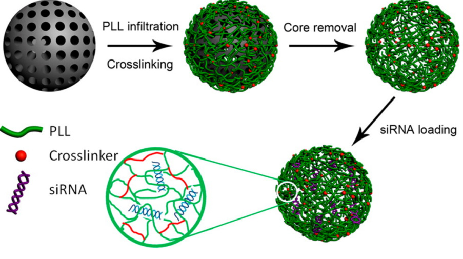
    [41] Lu Y, Low PS (2002) Folate-mediated delivery of macromolecular anticancer therapeutic agents. Adv Drug Deliv Rev 54: 675–693. doi: 10.1016/S0169-409X(02)00042-X
    [42] Nistor MT, Chiriac AP, Nita LE, et al. (2013) Semi-interpenetrated polymer networks of hyaluronic acid modified with poly(aspartic acid). J Polym Res 20: 86. doi: 10.1007/s10965-013-0086-8
    [43] Mackay JA, Chilkoti A (2008) Temperature sensitive peptides: engineering hyperthermia-directed therapeutics. Int J Hypertherm 24: 483–495. doi: 10.1080/02656730802149570
    [44] Aluri S, Janib SM, Mackay JA (2009) Environmentally responsive peptides as anticancer drug carriers. Adv Drug Deliver Rev 61: 940–952. doi: 10.1016/j.addr.2009.07.002
    [45] Ruszczak Z (2003) Effect of collagen matrices on dermal wound healing. Adv Drug Deliv Rev 55: 1595–1611. doi: 10.1016/j.addr.2003.08.003
    [46] Lupas A (1996) Coiled coils: new structures and new functions. Trends Biochem Sci 21: 375–382. doi: 10.1016/S0968-0004(96)10052-9
    [47] Petka WA, Harden JL, McGrath KP, et al. (1998) Reversible hydrogels from self-assembling artificial proteins. Science 281: 389–392.
    [48] Al-Ahmady ZS, Al-Jamal WT, Bossche JV, et al. (2012) Lipid–Peptide Vesicle Nanoscale Hybrids for Triggered Drug Release by Mild Hyperthermia in Vitro and in Vivo. ACS Nano 6: 9335–9346. doi: 10.1021/nn302148p
    [49] Banta S, Wheeldon IR, Blenner M (2010) Protein engineering in the development of functional hydrogels. Annu Rev Biomed Eng 12:167–186. doi: 10.1146/annurev-bioeng-070909-105334
    [50] Huang HC, Koria P, Parker S, et al. (2008) Optically responsive gold nanorod-polypeptide assemblies. Langmuir 24: 14139–14144. doi: 10.1021/la802842k
    [51] Lin Y, Xia X, Wang M, et al. (2014) Genetically programmable thermoresponsive plasmonic gold/silk-elastin protein core/shell nanoparticles. Langmuir 30: 4406–4414.
    [52] Chilkoti A, Dreher MR, Meyer DE, et al. (2002) Targeted drug delivery by thermally responsive polymers. Adv Drug Deliver Rev 54: 613–630. doi: 10.1016/S0169-409X(02)00041-8
    [53] Maeda H, Seymour LW, Miyamoto Y (1992) Conjugates of anticancer agents and polymers: advantages of macromolecular therapeutics in vivo. Bioconjug Chem 3: 351–362. doi: 10.1021/bc00017a001
    [54] Schmaljohann D (2006) Thermo- and pH-responsive polymers in drug delivery. Adv Drug Deliver Rev 58: 1655–1670.
    [55] Raucher D, Massodi I, Bidwell GL (2008) Thermally targeted delivery of chemotherapeutics and anti-cancer peptides by elastin like polypeptide. Expert Opin Drug Deliver 5: 353–369. doi: 10.1517/17425247.5.3.353
    [56] Massodi I, Thomas E, Raucher D (2009) Application of thermally responsive elastin-like polypeptide fused to a lactoferrin-derived peptide for treatment of pancreatic cancer. Molecules 14: 1999–2015. doi: 10.3390/molecules14061999
    [57] MacEwan SR, Chilkoti A (2010) Elastin-like polypeptides: biomedical applications of tunable biopolymers. Biopolymers 94: 60–77. doi: 10.1002/bip.21327
    [58] Liu W, Dreher MR, Furgeson DY, et al (2006) Tumor accumulation, degradation and pharmacokinetics of elastin-like polypeptides in nude mice. J Control Release 116: 170–178. doi: 10.1016/j.jconrel.2006.06.026
    [59] McDaniel JR, Callahan DJ, Chilkoti A, et al. (2010) Drug delivery to solid tumors by elastin-like polypeptides. Adv Drug Deliver Rev 62: 1456–1467. doi: 10.1016/j.addr.2010.05.004
    [60] 
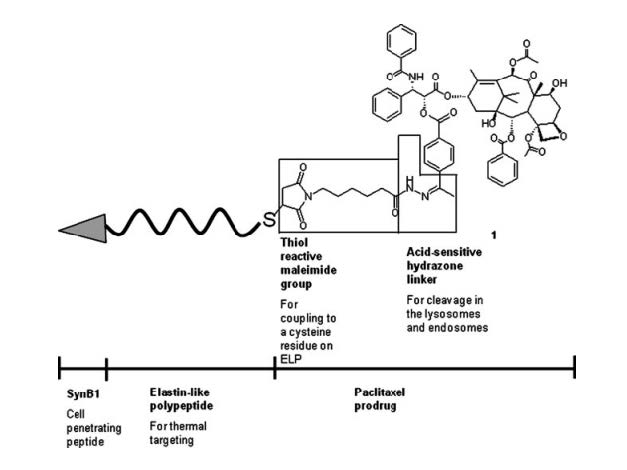
Moktan S, Ryppa C, Kratz F, et al. (2012) A thermally responsive biopolymer conjugated to an acid-sensitive derivative of paclitaxel stabilizes microtubules, arrests cell cycle, and induces apoptosis. Invest New Drugs 30: 236–248. doi: 10.1007/s10637-010-9560-x
    [61] Rousselle C, Smirnova M, Clair P, et al. (2001) Enhanced delivery of doxorubicin into the brain via a peptide-vector-mediated strategy: saturation kinetics and specificity. J Pharmacol Exp Ther 296: 124–13.
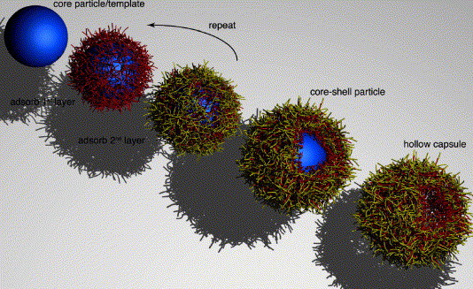
    [62] Jiang C, Tsukruk VV, et al. (2006) Freestanding Nanostructures via Layer-by-Layer Assembly. Adv Mater 18: 829–840.
    [63] Johnston APR, Cortez C, Angelatos AS, et al. (2006) Layer-by-layer engineered capsules and their applications. Curr Opin Colloid Interface Sci 11: 203–209.
    [64] Wattendorf U, Kreft O, Textor M, et al. (2008) Stable stealth function for hollow polyelectrolyte microcapsules through a poly(ethylene glycol) grafted polyelectrolyte adlayer. Biomacromolecules 9: 100–108.
    [65] Kamphuis MM, Johnston AP, Such GK, et al. (2010) Targeting of cancer cells using click-functionalized polymer capsules. J Am Chem Soc 132: 15881–15883.
    [66] Golonka M, Bulwan M, Nowakowska M, et al. (2011) Thermoresponsive multilayer films based on ionic elastin-like recombinamers. Soft Matter 7: 9402–9409. doi: 10.1039/c1sm06276j
    [67] Chen X, Zhang W, Li K, et al. (2012) Thermoresponsive oligoprolines. Soft Matter 8: 4869–4872. doi: 10.1039/c2sm25451d
    [68] Chen C, Wang Z, Li Z (2011) Thermoresponsive Polypeptides from Pegylated Poly-l-glutamate. Biomacromolecules 12: 2859–2863. doi: 10.1021/bm200849m
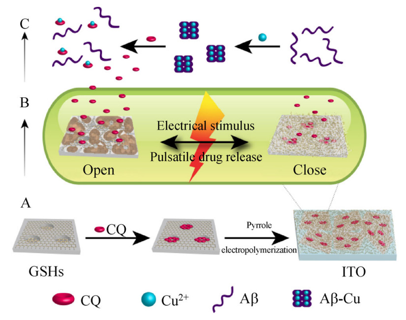
    [69] Fu X, Shen Y, Fu W (2013) Thermoresponsive Oligo(ethylene glycol) Functionalized Poly-l-cysteine. Macromolecules46: 3753–3760.
    [70] 
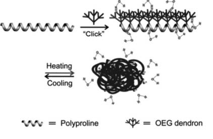
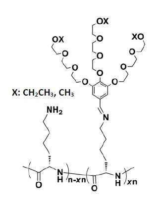
Zhang X, Li W, Zhao X, et al. (2013) Thermoresponsive Dendronized Polyprolines via the “Grafting to” Route. Macromol Rapid Commun 34: 1701–1707. doi: 10.1002/marc.201300605
    [71] Yan J, Liu K, Zhang X, at al. (2015) Dynamic covalent polypeptides showing tunable secondary structures and thermoresponsiveness. J Polym Sci A 53: 33–41. doi: 10.1002/pola.27433
    [72] Lam RTS, Belenguer A, Roberts SL (2005) Amplification of Acetylcholine-Binding Catenanes from Dynamic Combinatorial Libraries. Science 308: 667–669. doi: 10.1126/science.1109999
    [73] Carnall JMA, Waudby CA, Belenguer AM, et al (2010) Mechanosensitive Self-Replication Driven by Self-Organization. Science 327: 1502–1506. doi: 10.1126/science.1182767
    [74] Li J, Nowak P, Otto S (2013) Dynamic Combinatorial Libraries: From Exploring Molecular Recognition to Systems Chemistry. J Am Chem Soc 135: 9222–9239. doi: 10.1021/ja402586c
    [75] Matanović MR, Kristl J, Grabnar PA (2014) Thermoresponsive polymers: Insights into decisive hydrogel characteristics, mechanisms of gelation, and promising biomedical applications. Int J Pharm 472: 262–275.
    [76] Joly-Duhamel C, Hellio D, Djabourov M (2002) All gelatin networks: 1. Biodiversity and physical chemistry. Langmuir 18: 7208–7217.
    [77] Yang H, Kao W (2006) Thermoresponsive gelatin/monomethoxy poly(ethylene glycol)–poly(d,l-lactide) hydrogels: formulation, characterization, and antibacterial drug delivery. Pharm Res 23: 205–214.
    [78] Ohya S, Matsuda T (2005) Poly(N-isopropylacrylamide) (PNIPAM)-grafted gelatin as thermoresponsive three-dimensional artificial extracellular matrix: molecular and formulation parameters vs. cell proliferation potential. J Biomat Sci-Polym 16: 809–827.
    [79] Huang J, Hastings CL, Duffy GP, et al. (2013) Supramolecular Hydrogels with Reverse Thermal Gelation Properties from (Oligo)tyrosine Containing Block Copolymers. Biomacromolecules 14: 200–206. doi: 10.1021/bm301629f
    [80] Cheng Y, He C, Xiao C, et al. (2012) Decisive Role of Hydrophobic Side Groups of Polypeptides in Thermosensitive Gelation. Biomacromolecules 13: 2053–2059. doi: 10.1021/bm3004308
    [81] Park MH, Joo MK, Choi BG, et al. (2012) Biodegradable Thermogels. Acc Chem Res 45: 424–433.
    [82] Oh HJ, Joo MK, Sohn YS, et al. (2008) Secondary Structure Effect of Polypeptide on Reverse Thermal Gelation and Degradation of l/dl-Poly(alanine)–Poloxamer–l/dl-Poly(alanine) Copolymers. Macromolecules 41:8204–8209.
    [83] Zhang S, Fu W, Li Z (2014) Supramolecular hydrogels assembled from nonionic poly(ethylene glycol)-b-polypeptide diblocks containing OEGylated poly-L-glutamate. Polym Chem 5: 3346–3351.
    [84] Zhang DJ, Alvarez MV, Sofroniew TJ (2015) Deming. Design and Synthesis of Nonionic Copolypeptide Hydrogels with Reversible Thermoresponsive and Tunable Physical Properties. Biomacromolecules16: 1331–1340.
    [85] Huang J, Bonduelle C, Thévenot J, et al. (2012) Biologically Active polymersomes from Amphiphilic Glycopeptides. J Am Chem Soc 134: 119–122. doi: 10.1021/ja209676p
    [86] Schatz C, Louguet S, Meins JFL, et al. (2009) Polysaccharide block polypeptide copolymers vesicles towards synthetic viral capsids. Angew Chem Int Ed 48: 2572–2575. doi: 10.1002/anie.200805895
    [87] Zhang A, Zhang Z, Shi F, et al. (2013) Redox-Sensitive Shell-Crosslinked Polypeptide-block-Polysaccharide Micelles for Efficient Intracellular Anticancer Drug Delivery. Macromol Biosci 13: 1249–1258. doi: 10.1002/mabi.201300175
    [88] Liu L, Liu P (2015) Synthesis strategies for disulfide bond-containing polymer-based drug delivery system for reduction-responsive controlled release. Front Mater Sc 9: 211–226. doi: 10.1007/s11706-015-0283-y
    [89] Wang K, Luo GF, Liu Y, et al. (2012) Redox-sensitive shell cross-linked PEG-polypeptidehybrid micelles for controlled drug release. Polym Chem 3: 1084–1090. doi: 10.1039/c2py00600f
    [90] Beloor J, Ramakrishna S, Nam K, et al. (2015) Effective Gene Delivery into Human Stem Cells with a Cell-Targeting Peptide-Modified Bioreducible Polymer. Small 11: 2069–2079. doi: 10.1002/smll.201402933
    [91] Burnett JC, Rossi JJ (2012) RNA-Based Therapeutics: Current Progress and Future Prospects. Chem Biol 19: 60–71. doi: 10.1016/j.chembiol.2011.12.008
    [92] Cavalieri F, Beretta G, Cui J, et al. (2015) Redox-Sensitive PEG–Polypeptide Nanoporous Particles for Survivin Silencing in Prostate Cancer Cells. Biomacromolecules 16: 2168–2178. doi: 10.1021/acs.biomac.5b00562
    [93] Wang Y, Yu A, Caruso F (2005) Nanoporous Polyelectrolyte Spheres Prepared by Sequentially Coating Sacrificial Mesoporous Silica Spheres. Angew Chem Int Ed 44: 2888–2892. doi: 10.1002/anie.200462135
    [94] Manickam DS, Oupický D (2006) Multiblock Reducible Copolypeptides Containing Histidine-Rich and Nuclear Localization Sequences for Gene Delivery. Bioconjugate Chem17: 1395–1403.
    [95] Rosengart AJ, Kaminski MD, Chen HT, et al. (2005) Magnetizable implants and functionalized magnetic carriers: a novel approach for noninvasive yet targeted drug delivery. J Magn Magn Mater 293: 633–638
    [96] Mornet S, Vasseur S, Grasset F, et al. (2006) Magnetic nanoparticle design for medical applications. Prog Solid State Ch 34: 237–47. doi: 10.1016/j.progsolidstchem.2005.11.010
    [97] Lee H, Shao HP, Huang YQ, et al. (2005) Synthesis of MRI contrast agent by coating superparamagnetic iron oxide with chitosan. IEEE T Magn 41:4102–4.
    [98] Liu XQ, Kaminski MD, Riffle JS, et al. (2007) Preparation and characterization of biodegradable magnetic carriers by single emulsion-solvent evaporation. J Magn Magn Mater 311: 84–7. doi: 10.1016/j.jmmm.2006.10.1170
    [99] Ju XJ, Xie R, Yang L (2009) Biodegradable ‘intelligent’ materials in response to physical stimuli for biomedical applications. Expert Opin Ther Pat 19: 493–507. doi: 10.1517/13543770902771282
    [100] Aili D, Stevens MM (2010) Bioresponsive peptide-inorganic hybrid nanomaterials. Chem Soc Rev 39: 3358–3370. doi: 10.1039/b919461b
    [101] Pan BF, Cui DX, Sheng Y, et al. (2007) Dendrimer-modified magnetic nanoparticles enhance efficiency of gene delivery system. Cancer Res 67: 8156–8163. doi: 10.1158/0008-5472.CAN-06-4762
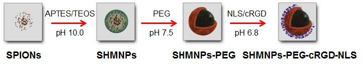
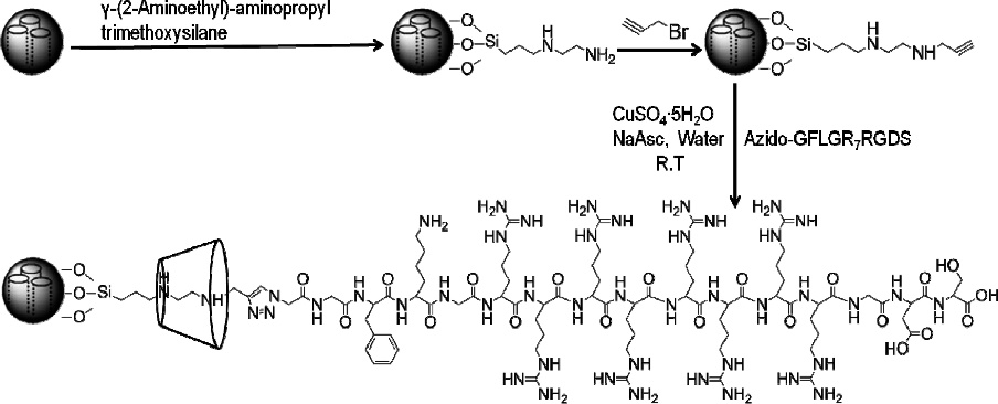
    [102] Digigow RG, Dechézelles JF, Dietsch H, et al. (2014) Preparation and characterization of functional silica hybrid magnetic nanoparticles. J Magn Magn Mater 362: 72–79.
    [103] Digigow RG, Vanhecke D, Rothen-Rutishauser B, et al. (2015)Uptake and Intracellular Fate of Peptide Surface-Functionalized Silica Hybrid Magnetic Nanoparticles In Vitro. Part Part Syst Char 33: 188–196.
    [104] Lerche MH, Jensen PR, Karlsson M, et al. (2015) NMR Insights into the Inner Workings of Living Cells. Anal Chem 87: 119–132.
    [105] Boutin C, Desvaux H, Carrière M, et al. (2011) Hyperpolarized129Xe NMR signature of living biological cells. NMR Biomed 24: 1264–1269. doi: 10.1002/nbm.1686
    [106] Kotera N, Dubost E, Milanole G, et al. (2015) A doubly responsive probe for the detection of Cys4-tagged proteins. Chem Commun 51: 11482–11484. doi: 10.1039/C5CC04721H
    [107] Kwon S, Kim BJ, Lim HK, et al. (2015) Magnetotactic molecular architectures from self-assembly of β-peptide foldamers. Nat Commun 6: 8747. doi: 10.1038/ncomms9747
    [108] Goodman CM, Choi S, Shandler S, et al. (2007) Foldamers as versatile frameworks for the design and evolution of function. Nat Chem Biol 3: 252–262. doi: 10.1038/nchembio876
    [109] Kwon S, Jeon A, Yoo S, et al. (2010) Unprecedented Molecular Architectures by the Controlled Self-Assembly of a β-Peptide Foldamer. Angew Chem Int Ed 49: 8232–8236. doi: 10.1002/anie.201003302
    [110] Lefèvre CT, Abreu F, Lins U, et al. (2011) A bacterial backbone: magnetosomes in magnetotactic bacteria. M. Rai, N. Duran (Eds.) in Metal nanoparticles in microbiology, Springer-Verlag, Berlin (2011), 75–102.
    [111] Unger E, Metzger P, Krupinski E, et al. (2000) The use of a thrombus-specific ultrasound contrast agent to detect thrombus in arteriovenous fistulae. Invest Radiol 35: 86–89. doi: 10.1097/00004424-200001000-00010
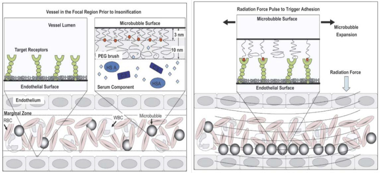
    [112] Weller GER, Wong MKK, Modzelewski RA, et al. (2005) Ultrasonic imaging of tumor angiogenesis using contrast microbubbles targeted via the tumor-binding peptide arginine-arginine-leucine. Cancer Res 65: 533–539.
    [113] Linker RA, Reinhardt M, Bendszus M, et al. (2005) In vivo molecular imaging of adhesion molecules in experimental autoimmune encephalomyelitis (EAE). J Autoimmun 25:199–205.
    [114] Sirsi S, Borden M (2009) Microbubble compositions, properties and biomedical applications. Bubble Sci Eng Technol 1: 3–17. doi: 10.1179/175889709X446507
    [115] Cochran MC, Eisenbrey J, Ouma RO, et al. (2011) Doxorubicin and paclitaxel loaded microbubbles for ultrasound triggered drug delivery. Int J Pharm 414:161–170. doi: 10.1016/j.ijpharm.2011.05.030
    [116] Borden MA, Sarantos MR, Stieger SM (2006) Ultrasound radiation force modulates ligand availability on targeted contrast agents. Mol Imag 5:139–147.
    [117] Lum AFH, Borden MA, Dayton PA (2006) Ultrasound radiation force enables targeted deposition of model drug carriers loaded on microbubbles. J Control Release 111: 128–134. doi: 10.1016/j.jconrel.2005.11.006
    [118] Borden MA,Zhang H, Gillies RJ, et al. (2008) A stimulus-responsive contrast agent for ultrasound molecular imaging. Biomaterials 29: 597–606. doi: 10.1016/j.biomaterials.2007.10.011
    [119] Bloch M, Jablonowski L, Yavin E, et al. (2015) Multi-modal detection of colon malignancy by NIR-tagged recognition polymers and ultrasound contrast agents. Int J Pharm 478: 504–516. doi: 10.1016/j.ijpharm.2014.11.066
    [120] Eisenbrey JR, Burstein OM, Kambhampati R, et al. (2010) Development and optimization of a doxorubicin loaded poly(lactic acid) contrast agent for ultrasound directed drug delivery. J Control Release 143: 38–44. doi: 10.1016/j.jconrel.2009.12.021
    [121] Eisenbrey JR, Soulen MC, Wheatley MA (2010) Delivery of encapsulated doxorubicin by ultrasound-mediated size reduction of drug-loaded polymer contrast agents. IEEE Trans Biomed Eng 57: 24–28. doi: 10.1109/TBME.2009.2030497
    [122] Duncan R, Gac-Breton S, Keane R (2001) Polymer-drug conjugates, PDEPT and PELT: basic principles for design and tranfer from the laboratory to clinic. J Control Release 74: 135–46. doi: 10.1016/S0168-3659(01)00328-5
    [123] Terada T, Iwai M, Kawakami S (2006) Novel PEG-matrix metalloproteinase-2 cleavable peptide-lipid containing galactosylated liposomes for hepatocellular carcinoma-selective targeting. J Control Release 111: 333–42.
    [124] Lee SJ, Jeong YI, Park HK, et al. (2015) Enzyme-responsive doxorubicin release from dendrimer nanoparticles for anticancerdrug delivery. Int J Nano 10: 5489–550.
    [125] Secret E, Kelly SJ, Crannell KE, et al. (2014) Enzyme-Responsive Hydrogel Microparticles for Pulmonary Drug Delivery. ACS Appl Mater Interfaces 6: 10313–10321. doi: 10.1021/am501754s
    [126] Secret E, Crannell KE, Kelly SJ, et al. (2015) Matrix metalloproteinase-sensitive hydrogel microparticles for pulmonary drug delivery of small molecule drugs or proteins. J Mater Chem B 3: 5629–5634. doi: 10.1039/C5TB00443H
    [127] Angelos S, Khashab NM, Yang YW, et al. (2009) pH clock-operated mechanized nanoparticles. J Am Chem Soc 131: 12912–12914.
    [128] Luo Z, Ding X, Hu Y, et al. (2013) Engineering a hollow nanocontainer platform with multifunctional molecular machines for tumor-targeted therapy in vitro and in vivo. ACS Nano 7: 10271–10284. doi: 10.1021/nn404676w
    [129] Li J, Liu F, Shao Q, et al. (2014) Enzyme-Responsive Cell-Penetrating Peptides Conjugated Mesoporous Silica Quantum Dots Nanocarriers for Controlled Release of Nucleus-Targeted Drug Molecules and Real-Time Intracellular Fluorescence Imaging of Tumor Cells. Adv Healthcare Mater 3: 1230–123. doi: 10.1002/adhm.201300613
    [130] Cheng YJ, Luo GF, Zhu JY, et al. (2015) Enzyme-Induced and Tumor-Targeted Drug Delivery System Based on Multifunctional Mesoporous Silica Nanoparticles. ACS Appl Mater Interfaces7: 9078–9087.
    [131] Liu J, Zhang B, Luo Z, et al. (2015) Enzyme responsive mesoporous silica nanoparticles for targeted tumor therapy in vitro and in vivo. Nanoscale 7: 3614–3626. doi: 10.1039/C5NR00072F
    [132] del Mercato LL, Ferraro MM, Baldassarre F, et al. (2014) Biological applications of LbL multilayer capsules: From drug delivery to sensing. Adv Colloid Interface Sci 207: 139–154. doi: 10.1016/j.cis.2014.02.014
    [133] De Geest BG, Vandenbroucke RE, Guenther AM, et al. (2006) Intracellularly degradable polyelectrolyte microcapsules. Adv Mater 18: 1005–1009. doi: 10.1002/adma.200502128
    [134] Ochs CJ, Such GK, Yan Y, et al. (2010) Biodegradable click capsules with engineered drug-loaded multilayers. Acs Nano 4: 1653–1663. doi: 10.1021/nn9014278
    [135] Aller SG, Yu J, Ward A, et al. (2009) Structure of P-Glycoprotein Reveals a Molecular Basis for Poly-Specific Drug Binding. Science 323: 1718–1722. doi: 10.1126/science.1168750
    [136] Yan Y, Ochs C, Such G, et al. (2010) Bypassing multidrug resistance in cancer cells with biodegradable polymer capsules. Adv Mater 22: 5398–5403. doi: 10.1002/adma.201003162
    [137] Zhao Y (2007) Rational design of light-controllable polymer micelles. Chem Rec 7: 286–294. doi: 10.1002/tcr.20127
    [138] Alatorre-Meda M, Alvarez-Lorenzo C, Concheiro A, et al. (2013) UV and Near-IR Triggered Release from Polymeric Micelles and Nanoparticles, in Smart Materials for Drug Delivery (Eds.: C. Alvarez-Lorenzo, A. Concheiro), RSC Publishing,Cambridge 304–348.
    [139] Hernanz D, Nunez V, Sancho A, et al. (2001) Hydroxycinnamic acids and ferulic acid dehydrodimers in barley and processed barley. J Agric Food Chem 49: 4884–4888. doi: 10.1021/jf010530u
    [140] Hoff WD, Dux P, Hard K, et al. (1994) Thiol ester-linked p-coumaric acid as a new photoactive prosthetic group in a protein with rhodopsin-like photochemistry. Biochemistry 33: 13959–13962. doi: 10.1021/bi00251a001
    [141] Wang G, Tong X, Zhao Y (2004) Preparation of Azobenzene-Containing Amphiphilic Diblock Copolymers for Light-Responsive Micellar Aggregates. Macromolecules 37: 8911–8917. doi: 10.1021/ma048416a
    [142] Shi D, Matsusaki M, Kaneko T, et al. (2008) Photo-Cross-Linking and Cleavage Induced Reversible Size Change of Bio-Based Nanoparticles. Macromolecules 41: 8167–8172. doi: 10.1021/ma800648e
    [143] Zhao Y (2012) Light-Responsive Block Copolymer Micelles. Macromolecules 45: 3647–3657. doi: 10.1021/ma300094t
    [144] Kotharangannagari VK, Sánchez-Ferrer A, Ruokolainen J, et al. (2011) Photo-Responsive Reversible Aggregation and Dissolution of Rod-Coil Polypeptide Diblock Copolymers. Macromolecules 44: 4569–4573. doi: 10.1021/ma2008145
    [145] Kumar S, Allard JF, Morris D, et al. (2012) Near-infrared light sensitive polypeptide block copolymer micelles for drug delivery. J Mater Chem 22: 7252–7257. doi: 10.1039/c2jm16380b
    [146] Li Y, Qian Y, Liu T, et al. (2012) Light-triggered concomitant enhancement of magnetic resonance imaging contrast performance and drug release rate of functionalized amphiphilic diblock copolymer micelles. Biomacromolecules 13: 3877–3886. doi: 10.1021/bm301425j
    [147] Liu G, Liu N, Zhou L, et al. (2015) NIR-responsive polypeptide copolymer upconversion composite nanoparticles for triggered drug release and enhanced cytotoxicity. Polym Chem 6: 4030–4039
    [148] Martinez-Cuezva A, Valero-Moya S, Alajarin M, et al. (2015) Light-responsive peptide [2]rotaxanes as gatekeepers of mechanised nanocontainers. Chem Commun 51: 14501–14504. doi: 10.1039/C5CC04365D
    [149] Stoddart JF (ed) (2001) Special issue on molecular machines. Acc Chem Res 34: 410 (2001).
    [150] Cavallini M, Biscarini F, Leon S (2003) Information Storage Using Supramolecular Surface Patterns.Science 299: 531–531. doi: 10.1126/science.1078012
    [151] Bottari G, Leigh DA, Pérez EM (2003) Chiroptical Switching in a Bistable Molecular Shuttle. J Am Chem.Soc125: 13360–13361.
    [152] Pernites RB, Santos CM, Maldonado M, et al. (2012) Tunable Protein and Bacterial Cell Adsorption on Colloidally Templated Superhydrophobic Polythiophene Films. Chem Mater 24: 870–880. doi: 10.1021/cm2007044
    [153] Gomez N, Schmidt CE (2007) Nerve growth factor-immobilized polypyrrole: Bioactive electrically conducting polymer for enhanced neurite extension. J Biomed Mater Res 81: 135–149.
    [154] Green RA, Lovell NH, Poole-Warren LA (2009) Cell attachment functionality of bioactive conducting polymers for neural interfaces. Biomaterials 30: 3637–3644. doi: 10.1016/j.biomaterials.2009.03.043
    [155] Zhong Y, Yu X, Gilbert R, et al (2001) Stabilizing electrode-host interfaces: a tissue engineering approach. J Rehabil Res Dev 38: 627–632.
    [156] Fabregat G, Ballano G, Armelin E (2013) An electroactive and biologically responsive hybrid conjugate based on chemical similarity. Polym Chem 4: 1412–1424. doi: 10.1039/C2PY20894F
    [157] Maione S, Gil AM, Fabregat G, et al. (2015) Electroactive polymer–peptide conjugates for adhesive biointerfaces. Biomater Sci 3: 1395–1405. doi: 10.1039/C5BM00160A
    [158] Jeon G, Yang SY, Byun J, et al. (2011) Electrically actuatable smart nanoporous membrane for pulsatile drug release. Nano Lett 11: 1284–1288. doi: 10.1021/nl104329y
    [159] Ge J, Neofytou E, Cahill TJ, et al. (2012) Drug release from electric-field-responsive nanoparticles. ACS Nano 6: 227–233. doi: 10.1021/nn203430m
    [160] Wu L, Wang J, Gao N, et al. (2015) Electrically pulsatile responsive drug delivery platform for treatment of Alzheimer’s disease. Nano Res 8: 2400–2414. doi: 10.1007/s12274-015-0750-x
    [161] Santos JL, Li Y, Culver HR, et al. (2014) Conducting polymer nanoparticles decorated with collagen mimetic peptides for collagen targeting. Chem Commun 50: 15045–15048.
    [162] 
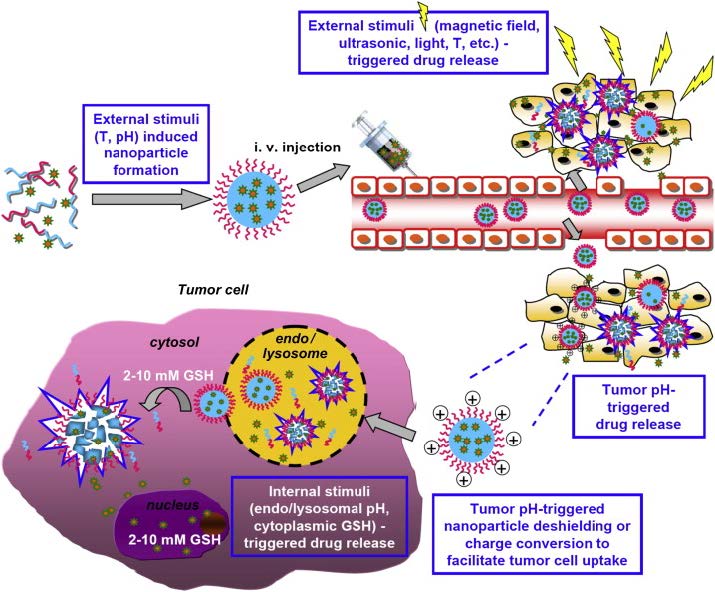
Cheng R, Meng F, Deng C, et al. (2013) Dual and multi-stimuli responsive polymeric nanoparticles for programmed site-specific drug delivery. Biomaterials 34: 3647–3657. doi: 10.1016/j.biomaterials.2013.01.084
    [163] Dai J, Lin SD, Cheng D, et al. (2011) Interlayer-crosslinked micelle with partially hydrated core showing reduction and pH dual sensitivity for pinpointed intracellular drug release. Angew Chem Int Ed 50: 9404–9408. doi: 10.1002/anie.201103806
    [164] Yu S, Wu G, Gu X, et al. (2013) Magnetic and pH-sensitive nanoparticles for antitumor drug deliver. Colloids Surf B 103: 15–22. doi: 10.1016/j.colsurfb.2012.10.041
    [165] Remant BKC, Thapa B, Xu P (2012) pH and Redox Dual Responsive Nanoparticle for Nuclear Targeted Drug Delivery. Mol Pharmaceutics 9: 2719–2729.
    [166] Gupta MK, Lee SH, Crowder SW, et al. (2015) Oligoproline-derived nanocarrier for dual stimuli-responsive gene delivery. J Mater Chem 3: 7271–7280. doi: 10.1039/C5TB00988J
  • This article has been cited by:

    1. Nguyen Thi Thu Thuy, Tran Thanh Tung, Strong convergence of one-step inertial algorithm for a class of bilevel variational inequalities, 2025, 0233-1934, 1, 10.1080/02331934.2024.2448737
    2. Nguyen Thi Thu Thuy, Tran Thanh Tung, Le Xuan Ly, Strongly convergent two-step inertial algorithm for a class of bilevel variational inequalities, 2025, 44, 2238-3603, 10.1007/s40314-024-03078-7
    3. Le Xuan Ly, Nguyen Thi Thu Thuy, Nguyen Quoc Anh, Relaxed two-step inertial method for solving a class of split variational inequality problems with application to traffic analysis, 2025, 0233-1934, 1, 10.1080/02331934.2025.2459191
  • Reader Comments
  • © 2016 the Author(s), licensee AIMS Press. This is an open access article distributed under the terms of the Creative Commons Attribution License (http://creativecommons.org/licenses/by/4.0)
通讯作者: 陈斌, bchen63@163.com
  • 1. 

    沈阳化工大学材料科学与工程学院 沈阳 110142

  1. 本站搜索
  2. 百度学术搜索
  3. 万方数据库搜索
  4. CNKI搜索

Metrics

Article views(11427) PDF downloads(1818) Cited by(7)

Figures and Tables

Figures(19)

/

DownLoad:  Full-Size Img  PowerPoint
Return
Return

Catalog